

Severe sleep bruxism represents one of the most destructive biomechanical forces within the human stomatognathic system, capable of fracturing natural enamel, delaminating porcelain restorations, and compromising the osseointegration of full-arch dental implants. For decades, the dental profession debated the efficacy of pharmacological muscle modulation versus mechanical barrier protection. Today, evidence-based prosthodontics recognizes that the optimal management of nocturnal parafunction is not a matter of choosing between treatments, but rather integrating them. The clinical debate of botox masseter temporalis hypertrophy bruxism dose vs occlusal splint has evolved into a synergistic protocol. By combining the force-reducing properties of therapeutic Botulinum Toxin Type A (BTX-A) with the physical shielding of a CAD/CAM milled hard acrylic Michigan splint, clinicians can effectively neutralize the devastating effects of rhythmic masticatory muscle activity (RMMA). At HCMC Dental Clinic in District 1, Ho Chi Minh City, this dual-modality approach is the gold standard for protecting high-value cosmetic and surgical investments, ensuring the long-term survivability of IPS E.max veneers and All-on-4 zirconia implant bridges.
Clinical Summary:
The management of severe sleep bruxism requires a comprehensive, dual-modality approach to prevent catastrophic failure of natural dentition and advanced dental prosthetics. Therapeutic Botulinum Toxin Type A (Botox) injected into the masseter and temporalis muscles significantly reduces the maximum voluntary bite force by inducing temporary, localized muscle relaxation. However, Botox alone cannot physically prevent tooth-to-tooth contact during sleep arousals. A hard CAD/CAM Michigan occlusal splint remains mandatory to provide mutually protected occlusion, anterior guidance, and a physical barrier against shear stress. Under the clinical direction of Dr. Nguyen Van Cuong (Dr. Cuong), DDS at HCMC Dental Clinic, this synergistic protocol—combining precise anatomical Botox dosing with custom hard splint therapy—offers unparalleled protection for porcelain veneers, full-arch implants, and the temporomandibular joint (TMJ), while simultaneously addressing aesthetic concerns related to square jaw masseter hypertrophy and morning tension headaches.
Key Takeaways:
- Biomechanical Overload: Sleep bruxism generates nocturnal clenching forces of 600 to 1,000 Newtons—up to three times the force of normal mastication—due to the suppression of periodontal protective reflexes during REM sleep.
- Synergistic Protocol: Therapeutic Botox reduces the contractile force of the elevator muscles, while a hard Michigan occlusal splint provides a mandatory physical barrier to protect enamel, IPS E.max veneers, and All-on-4 zirconia implants from shear stress.
- Precise Anatomical Dosing: Optimal therapeutic outcomes require 20–30 Units of BTX-A per masseter and 10–15 Units per temporalis, injected strictly within the inferior safety zone to prevent diffusion into the risorius muscle and preserve natural smile symmetry.
- Comprehensive TMJ Stabilization: This dual approach is critical for managing DC/TMD classifications such as TMJ Disc Displacement with Reduction (ADDwR) and preventing Idiopathic Condylar Resorption (ICR) prior to irreversible prosthodontic rehabilitation.
- International Standard Care in Vietnam: HCMC Dental Clinic in District 1 offers this advanced bruxism management protocol at highly transparent, ADA-compliant standards, providing expats and dental travelers with world-class care at a fraction of Western costs.
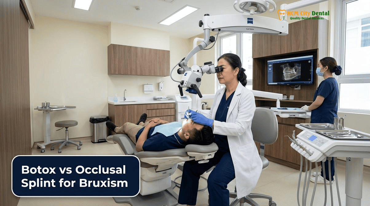
The Biomechanical Reality of Sleep Bruxism: Why Nocturnal Clenching Generates 600–1,000 Newtons of Force (3x Normal Chewing) Without Protective Reflexes
To fully comprehend the necessity of a dual-modality treatment approach, one must first understand the neurophysiological and biomechanical realities of sleep bruxism. Unlike awake bruxism, which is often a subconscious habit driven by stress and anxiety, sleep bruxism is classified as a sleep-related movement disorder. It is characterized by rhythmic masticatory muscle activity (RMMA) that occurs predominantly during micro-arousals from sleep, particularly during the transition between non-REM and REM sleep stages. During normal, conscious mastication, the human jaw generates approximately 200 to 300 Newtons of force. This force is carefully regulated by a complex neurofeedback loop involving periodontal mechanoreceptors—sensory nerve endings in the periodontal ligament that detect pressure and trigger an inhibitory reflex to prevent the teeth from biting down too hard and damaging themselves.
However, during sleep, this protective proprioceptive feedback mechanism is profoundly suppressed. Without the central nervous system’s inhibitory reflexes to limit muscle contraction, the masseter, temporalis, and medial pterygoid muscles can fire with unrestrained intensity. Clinical polysomnography and electromyography (EMG) studies have demonstrated that nocturnal clenching and grinding can generate catastrophic forces ranging from 600 to over 1,000 Newtons. This is equivalent to balancing the weight of a large adult human on the surface of a single molar. When these immense forces are applied laterally (side-to-side grinding), they create devastating shear stresses that natural enamel and dental ceramics are simply not designed to withstand.
The consequences of this biomechanical overload extend far beyond mere tooth wear. In the natural dentition, severe bruxism leads to abfraction lesions at the cervical margins, vertical root fractures, and profound occlusal attrition that collapses the patient’s vertical dimension of occlusion (VDO). For patients with advanced restorative work, the risks are even higher. The shear forces generated during sleep bruxism can easily exceed the flexural strength of standard restorative materials, leading to the catastrophic chipping of porcelain veneers, the fracture of zirconia crowns, and the failure of implant abutment screws. Furthermore, the temporomandibular joint (TMJ) bears the brunt of this excessive loading. Chronic bruxism is a primary etiological factor in the progression of TMJ disorders, specifically according to the Diagnostic Criteria for Temporomandibular Disorders (DC/TMD). Patients frequently present with TMJ Disc Displacement with Reduction (ADDwR), characterized by painful clicking and popping, which can progress to Disc Displacement Without Reduction (ADDwoR), resulting in a closed lock with maximum opening restricted to less than 25mm. In severe cases, the sustained intra-articular pressure can trigger Idiopathic Condylar Resorption (ICR), leading to progressive skeletal anterior open bite and severe facial asymmetry[1].
Given this biomechanical reality, it becomes evident why a single treatment modality is often insufficient. Attempting to manage 1,000 Newtons of force with a plastic mouthguard alone often results in the patient simply chewing through the appliance, while failing to address the underlying muscular hyperactivity. Conversely, attempting to manage the condition solely with muscle relaxants ignores the need for a physical barrier to protect the teeth during the residual grinding episodes that still occur. Therefore, a comprehensive, evidence-based approach must address both the source of the force (the hyperactive musculature) and the target of the force (the dentition and prosthetics).
Why Therapeutic Masseter & Temporalis Botulinum Toxin (BTX-A) and Hard Occlusal Splints Are Synergistic—Not Mutually Exclusive
The clinical discourse surrounding the management of severe parafunction often frames treatments in opposition, leading to the common patient query regarding botox masseter temporalis hypertrophy bruxism dose vs occlusal splint. However, in modern prosthodontics and orofacial pain management, these two modalities are recognized as highly synergistic, addressing different aspects of the bruxism pathophysiology. To understand this synergy, we must examine the mechanism of action of each treatment and how they complement one another within a comprehensive Biomimetic Full-Mouth Reconstruction & VDO Rehabilitation in Vietnam.
Botulinum Toxin Type A (BTX-A) is a potent neurotoxic protein produced by the bacterium Clostridium botulinum. When injected into a hyperactive muscle, it acts at the neuromuscular junction by cleaving the SNAP-25 protein, which is essential for the docking and release of acetylcholine vesicles. By inhibiting the release of this primary excitatory neurotransmitter, BTX-A effectively induces a temporary, localized, and dose-dependent chemical denervation of the muscle fibers. In the context of bruxism, therapeutic Botox does not cure the central nervous system triggers that initiate RMMA; rather, it significantly reduces the maximum contractile force that the masseter and temporalis muscles can generate. By lowering the peak force from 1,000 Newtons down to a manageable 300 Newtons, Botox protects the stomatognathic system from catastrophic overload, reduces muscle fatigue, and alleviates myofascial pain.
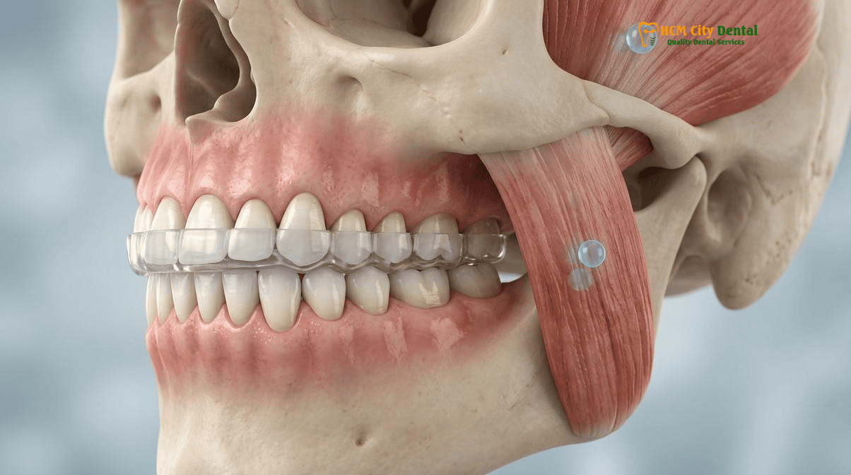
However, while Botox reduces the force of the grinding, it does not provide a physical barrier against the friction. Even at reduced forces, the repetitive sliding of maxillary teeth against mandibular teeth during sleep will eventually cause attrition of natural enamel and micro-fractures in dental ceramics. This is where the hard CAD/CAM Michigan occlusal splint becomes indispensable. A properly designed Michigan splint is fabricated from rigid, cross-linked polymethyl methacrylate (PMMA) or milled acrylic. It is engineered to provide a flat occlusal plane with precise point contacts for all opposing teeth, combined with a smooth anterior guidance ramp. When the patient attempts to grind their teeth laterally, the lower canines glide up the ramp of the splint, immediately discluding (separating) all the posterior teeth. This concept, known as mutually protected occlusion, ensures that no posterior teeth can rub together during parafunctional movements, completely eliminating destructive shear forces.
In comprehensive occlusal rehabilitation, managing bruxism is as critical as utilizing extra-radicular Mandibular Buccal Shelf (MBS 2.0x12mm stainless steel) TADs for non-surgical Class III underbite distalization (Wits > -5mm), or employing the Carriere Motion 3D ‘Sagittal First’ Class II molar derotation combined with Invisalign Mandibular Advancement Precision Wings / Infra-Zygomatic Crest (IZC) TADs. Furthermore, just as Ericson & Kurol 3D CBCT sector localization and closed-eruption 24k gold-chain cantilever traction (60g force) are essential for impacted maxillary canines, and Ricketts’ E-Line and nasolabial angle control dictate 4-premolar extraction vs TAD full-arch retraction for Bimaxillary Protrusion, the stabilization of the stomatognathic system requires precise biomechanical control. Dual bucco-palatal TAD posterior molar intrusion (where 1mm molar intrusion equals 2–3mm anterior open bite closure via counterclockwise mandibular autorotation) demonstrates the power of controlling vertical forces. Similarly, Phase I pediatric Rapid Palatal Expansion (RPE / Invisalign First at ages 7–10 prior to midpalatal suture fusion) and Leeway Space (E-Space) preservation vs true serial extraction set the foundation for a healthy bite. In the adult bruxer, the dual-modality approach of Botox and a Michigan splint provides this same level of precise biomechanical control, ensuring that the forces of occlusion are managed safely and predictably[2].
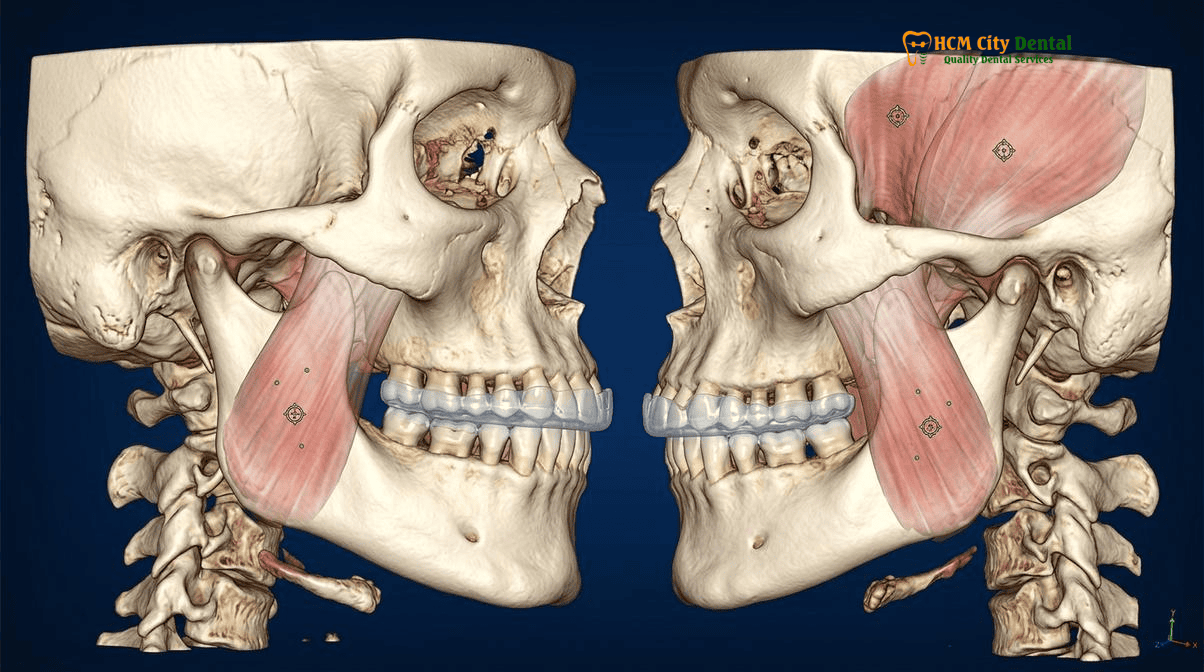
“We must stop viewing Botox and night guards as an ‘either/or’ proposition. In my clinical practice, treating a severe bruxer with Botox without providing a hard Michigan splint is like turning down the volume on a destructive machine but leaving it running. Conversely, giving a severe bruxer a splint without modulating the muscle force with Botox often results in the patient fracturing the splint itself. True protection for complex rehabilitations requires both chemical force reduction and mechanical barrier shielding.”
— Dr. Nguyen Van Cuong, DDS (Clinical Director — MOH License 0003265/BL-CCHN)
Anatomical Injection Mapping & Dosing (20–30 Units per Masseter + 10–15 Units per Temporalis): Staying Within the Inferior Safety Zone to Protect the Smile (Risorius)
The administration of therapeutic Botulinum Toxin for bruxism and masseter hypertrophy is a highly precise medical procedure that demands a profound understanding of facial anatomy. Unlike cosmetic Botox administered in the upper face for wrinkles, therapeutic injections in the lower face carry specific risks if the neurotoxin diffuses beyond the target musculature. The primary concern is the inadvertent paralysis of the adjacent muscles of facial expression, particularly the risorius muscle and the zygomaticus major, which are responsible for pulling the corners of the mouth laterally and superiorly during a smile. If BTX-A diffuses into these muscles, the patient may experience a temporary but highly distressing asymmetric smile, drooping of the oral commissure, or difficulty articulating certain words.
To completely eliminate this risk, Dr. Nguyen Van Cuong (Dr. Cuong), DDS employs a strict, evidence-based anatomical mapping protocol to define the “inferior safety zone” of the masseter muscle. The masseter is a thick, quadrilateral muscle consisting of superficial, intermediate, and deep layers. The safety zone is delineated by four distinct anatomical landmarks:
- Superior Boundary: A line drawn from the ear lobe to the cheilion (the corner of the mouth). Injections must remain strictly below this line to avoid the parotid duct (Stensen’s duct) and the zygomaticus major muscle.
- Posterior Boundary: A vertical line approximately 1cm anterior to the tragus of the ear, ensuring the parotid gland is avoided.
- Anterior Boundary: A vertical line 1cm behind the palpable anterior border of the masseter muscle. This is the most critical boundary, as the risorius muscle often overlies the anterior-superior aspect of the masseter. Staying well behind this line prevents diffusion into the smile muscles.
- Inferior Boundary: A horizontal line 1cm above the inferior margin of the mandible, avoiding the marginal mandibular branch of the facial nerve.
Within this carefully mapped safety triangle or quadrangle, the dosing must be tailored to the individual patient’s muscle bulk and the severity of their bruxism. For the average adult patient presenting with significant masseter hypertrophy and severe clenching, the standard therapeutic dose ranges from 20 to 30 Units of OnabotulinumtoxinA (Botox) per masseter side. The injections are delivered deep into the muscle belly, typically distributed across 3 to 4 injection points spaced 1cm apart, ensuring even distribution throughout the hypertrophic muscle fibers while minimizing superficial diffusion.
Crucially, comprehensive bruxism management cannot ignore the temporalis muscle. The temporalis is a large, fan-shaped muscle that covers the side of the skull and inserts into the coronoid process of the mandible. It acts synergistically with the masseter to elevate the jaw. If a clinician only injects the masseter, the central nervous system will often recruit the temporalis muscle to compensate for the lost force. This compensatory hyperactivity frequently leads to severe tension-type headaches localized in the temples. To prevent this, a concurrent dose of 10 to 15 Units of BTX-A is typically injected into the anterior and middle fibers of the temporalis muscle on each side. This balanced approach ensures harmonious relaxation of the entire elevator muscle complex, providing profound relief from both jaw clenching and morning temporal headaches[3].
Clinical Precaution: Avoid Soft “Boil-and-Bite” Night Guards
Patients suffering from severe bruxism must strictly avoid over-the-counter, soft, rubbery “boil-and-bite” mouthguards. Soft materials trigger a proprioceptive response in the periodontal ligaments that actually stimulates the chewing reflex. Instead of relaxing the jaw, a soft guard encourages the masseter and temporalis muscles to clench harder, much like a dog chewing on a rubber toy. This can exacerbate muscle fatigue, worsen TMJ pain, and accelerate the destruction of the joint disc. Only a hard, custom-milled acrylic Michigan splint provides the rigid anterior guidance necessary to disengage the posterior teeth and shut down hyperactive muscle firing during sleep.
Why a Hard Acrylic Michigan Splint Remains Mandatory to Physically Shield Enamel, IPS E.max Veneers, and All-on-4 Bridges During REM Sleep
While therapeutic Botox effectively reduces the maximum contractile force of the masticatory muscles, it is not a cure for the neurological triggers of sleep bruxism. Patients will still experience rhythmic masticatory muscle activity (RMMA) during sleep arousals, albeit at a reduced intensity. Therefore, the physical protection of the dentition remains an absolute necessity, particularly for patients who have invested in advanced restorative or implant dentistry. The hard acrylic Michigan occlusal splint is the universally recognized gold standard for this purpose.
The Michigan splint, originally developed at the University of Michigan, is not merely a piece of plastic placed between the teeth; it is a highly engineered orthotic device designed to reprogram the neuromuscular system and manage occlusal forces. Fabricated from dense, hard acrylic (often via precise CAD/CAM milling from a solid puck of cross-linked PMMA), the splint is typically worn on the maxillary (upper) arch. Its occlusal surface is meticulously adjusted to provide simultaneous, even, and bilateral point contacts for all opposing mandibular teeth when the jaw is in centric relation (the most stable, relaxed position of the TMJ).
The most critical feature of the Michigan splint is its anterior guidance ramp. When a bruxing patient attempts to grind their jaw to the left or right (excursive movements) or forward (protrusive movement) during REM sleep, the lower canine or incisor teeth glide up the smooth acrylic ramp of the splint. Because the ramp is angled steeper than the patient’s natural posterior cusp angles, this movement instantly forces the posterior teeth apart. This phenomenon, known as posterior disclusion, is biomechanically vital. The elevator muscles (masseter and temporalis) can only fire at maximum intensity when the posterior teeth are in contact. By mechanically separating the posterior teeth the moment the jaw moves off-center, the Michigan splint triggers an inhibitory reflex that dramatically reduces muscle contraction force.
For patients with IPS E.max lithium disilicate porcelain veneers, this protection is non-negotiable. While IPS E.max is incredibly strong under vertical compressive loads (boasting a flexural strength of 500 MPa), it is highly susceptible to fracture under lateral shear stresses—exactly the type of forces generated during nocturnal grinding. A hard Michigan splint ensures that these shear forces are absorbed by the replaceable acrylic of the splint, rather than the irreplaceable porcelain of the veneers.
Similarly, for patients rehabilitated with All-on-4 or All-on-6 full-arch zirconia implant bridges, the Michigan splint serves as a critical shock absorber. Dental implants, unlike natural teeth, lack a periodontal ligament (PDL). The PDL acts as a natural suspension system, providing micro-mobility and proprioceptive feedback. Implants are ankylosed directly to the bone, meaning any occlusal force is transmitted 100% directly to the crestal bone and the titanium abutment screws. In a severe bruxer, the lack of proprioception means they can generate massive forces without feeling pain, leading to screw loosening, screw fracture, or even catastrophic failure of the zirconia framework. Wearing a hard Michigan splint over the opposing natural teeth or over the implant bridge itself distributes these forces evenly across the arch, mitigating stress concentrations and ensuring the long-term survivability of the implant reconstruction[4].
Clinical Case Scenario: Protecting a Full-Mouth Rehabilitation at HCMC Dental Clinic
A 45-year-old Australian expatriate presented to HCMC Dental Clinic in District 1 with severe occlusal attrition, a collapsed vertical dimension of occlusion (VDO), and chronic morning headaches. Diagnosis confirmed severe sleep bruxism and bilateral masseter hypertrophy. Dr. Nguyen Van Cuong (Dr. Cuong), DDS executed a comprehensive Biomimetic Full-Mouth Reconstruction utilizing IPS E.max overlays and zirconia crowns to restore the VDO. To protect this extensive rehabilitation, a synergistic protocol was implemented: 25 Units of BTX-A were injected into each masseter and 12 Units into each temporalis, immediately reducing the patient’s clenching force and resolving the headaches. Concurrently, a CAD/CAM hard Michigan splint was fabricated. At the 12-month follow-up, the patient reported zero headaches, a slimmer facial profile due to masseter reduction, and the porcelain restorations remained in pristine condition with zero chipping, demonstrating the profound efficacy of combining chemical force modulation with mechanical barrier protection.
Managing Square Jaw Masseter Hypertrophy, Morning Temporal Headaches & Avoiding Over-Injection Mandibular Cortical Bone Thinning
Beyond the primary goal of protecting the dentition and dental prosthetics, the synergistic use of therapeutic Botox and occlusal splints offers significant secondary benefits, particularly concerning facial aesthetics and chronic pain management. Chronic severe bruxism almost universally leads to masseter hypertrophy—a benign enlargement of the masseter muscles due to constant overwork, much like a bodybuilder’s muscles grow from lifting weights. In many patients, particularly females, this bilateral enlargement creates a widened, square, and overly masculine lower jawline. The phrase masseter hypertrophy square jaw slimming therapeutic botox dose is frequently searched by patients seeking both functional relief and aesthetic improvement.
When therapeutic Botox is administered to reduce the contractile force of the masseter, the muscle experiences a period of disuse. According to the physiological principle of disuse atrophy, the muscle fibers gradually shrink in volume over the following 4 to 8 weeks. This reduction in muscle bulk softens the angle of the mandible, transforming a harsh, square jawline into a more highly desired, slender, V-shaped or oval facial contour. This aesthetic side effect is highly valued by patients and occurs naturally as a result of treating the underlying functional pathology.
Furthermore, as previously discussed, the inclusion of the temporalis muscle in the injection protocol is critical for managing morning temporal headaches. Tension-type headaches are frequently misdiagnosed as migraines, when in reality, they are referred pain originating from hyperactive, fatigued temporalis muscles that have been clenching for 8 hours during sleep. By relaxing these muscles with a conservative dose of BTX-A, patients often experience a dramatic and life-changing reduction in headache frequency and severity.
However, the long-term use of therapeutic Botox must be managed with profound clinical responsibility. Recent longitudinal studies in the American Journal of Orthodontics and Dentofacial Orthopedics have highlighted a potential risk associated with chronic, high-dose Botox injections in the masticatory muscles: mandibular cortical bone thinning. According to Wolff’s Law, bone remodels and strengthens in response to the mechanical loads placed upon it. The masseter muscle attaches directly to the angle of the mandible, and its powerful contractions stimulate the maintenance of dense cortical bone in that region. If the masseter is continuously paralyzed with high doses of Botox over many years, the lack of mechanical tension can lead to localized osteopenia, or thinning of the mandibular bone.
This physiological reality underscores exactly why Botox should never be used as a standalone, permanent crutch for bruxism, and why the Michigan splint is so vital. By utilizing the hard occlusal splint to manage the friction and provide posterior disclusion, the clinician can use the lowest effective dose of Botox, and space the injection intervals further apart (e.g., every 6 to 9 months instead of every 3 months). Over time, as the neuromuscular system is reprogrammed by the splint and the muscle bulk reduces, many patients find they only need a small annual maintenance dose of Botox, or can rely entirely on the splint. This conservative, synergistic approach ensures that the patient receives the functional and aesthetic benefits of muscle relaxation without risking the long-term structural integrity of their mandibular bone[5].
“Our goal is not to permanently paralyze the masticatory system, but to reset it. By combining precise, conservative Botox dosing with a perfectly balanced CAD/CAM Michigan splint, we break the cycle of hyperactive clenching. This allows the muscles to atrophy to a normal, healthy size, slimming the jawline and stopping the headaches, while ensuring the underlying jawbone remains dense and healthy. It is the epitome of minimally invasive, biologically respectful dentistry.”
— Dr. Nguyen Van Cuong, DDS (Clinical Director — MOH License 0003265/BL-CCHN)
2026 Therapeutic Bruxism Botox & CAD/CAM Hard Michigan Occlusal Splint Fee Schedule at HCMC Dental Clinic (District 1)
For international patients, expatriates, and dental travelers seeking world-class management of severe bruxism, HCMC Dental Clinic in District 1, Ho Chi Minh City, offers an unparalleled combination of clinical expertise and transparent affordability. Under the leadership of Dr. Nguyen Van Cuong (Dr. Cuong), DDS, our clinic adheres strictly to ADA-compliant sterilization protocols and utilizes the finest biocompatible materials available globally. Whether you require protection for natural teeth, a new set of IPS E.max veneers, or a complex All-on-4 implant rehabilitation, our synergistic bruxism protocols are designed to safeguard your investment.
We are committed to absolute price transparency. By utilizing our Official 2026 Dental Price List & -40% Online Booking Fee Schedule, international patients can access premium CAD/CAM Michigan splints and comprehensive dental care at a fraction of the cost compared to benchmark prices in Australia, the United States, or Europe. Below is our exact 2026 fee schedule for bruxism management and related restorative procedures.
| Treatment / Procedure (2026) | Online Booking Price (USD) | Walk-in / Standard Price (USD) | VND Equivalent (Online) |
|---|---|---|---|
| Custom Night Guard / Hard Michigan Occlusal Splint (CAD/CAM) | $81 – $115 | $135 – $192 | 2,106,000 – 2,990,000 VND |
| IPS E.max Porcelain Veneer (Per Tooth) | $185 | $308 | 4,800,000 VND |
| Full Zirconia Crown (Cercon Standard / HT Multilayer) | $154 – $192 | $256 – $321 | 4,000,000 – 5,000,000 VND |
| Single Korean Implant (Dentis / Neo Biotech / Dentium + Abutment) | $462 – $654 | $769 – $1,090 | 12,000,000 – 17,000,000 VND |
| Single European / Swiss Implant (Straumann BLT-SLA / BLA-Active) | $962 – $1,346 | $1,603 – $2,244 | 25,000,000 – 35,000,000 VND |
| All-on-4 Full-Arch Implants (Complete package with Zirconia bridge) | From $6,346 / arch | From $10,576 / arch | 165,000,000 VND |
| 3D CBCT Scan (Diagnostic Imaging) | $18 | $30 | 480,000 VND |
*Note: Therapeutic Botulinum Toxin (Botox) dosing is highly individualized based on muscle mass and hypertrophy severity. A precise quotation for BTX-A will be provided during your comprehensive clinical consultation with Dr. Cuong. All prices are based on the exchange rate of $1 USD = 26,000 VND. To secure these exclusive rates, patients must book their appointments in advance through our Official 2026 Dental Price List & -40% Online Booking Fee Schedule portal.
Frequently Asked Questions
If I get Masseter Botox for teeth grinding, do I still need to wear a hard night guard to protect my new porcelain veneers or implants?
Yes, absolutely. While therapeutic Botox significantly reduces the maximum contractile force of the masseter and temporalis muscles, it does not completely stop the central nervous system from initiating the grinding motion during sleep arousals. A hard CAD/CAM Michigan occlusal splint acts as an indispensable physical barrier. It provides mutually protected occlusion, ensuring that any residual grinding forces are distributed evenly across the acrylic surface rather than causing shear stress fractures to your IPS E.max porcelain veneers or All-on-4 zirconia implants.
Why must Temporalis muscles also be evaluated and injected alongside the Masseter in patients with morning temple headaches?
The masseter and temporalis muscles function as a synergistic elevator complex for the mandible. If only the masseter is treated with Botox, the temporalis muscle often undergoes compensatory hypertrophy—working harder to close the jaw. This overcompensation frequently exacerbates tension-type morning temple headaches. By injecting a conservative dose (10–15 Units) into the anterior and middle fibers of the temporalis alongside the masseter, Dr. Nguyen Van Cuong (Dr. Cuong), DDS ensures balanced neuromuscular relaxation, effectively alleviating both jaw clenching and associated cranial pain.
How does Dr. Nguyen Van Cuong map the facial safety zone so Masseter Botox never affects my smile symmetry?
Dr. Nguyen Van Cuong (Dr. Cuong), DDS utilizes a strict anatomical mapping protocol to define the inferior safety zone of the masseter. The boundaries are drawn from the ear lobe to the cheilion (corner of the mouth) superiorly, 1cm anterior to the tragus posteriorly, 1cm behind the anterior border of the masseter, and 1cm above the inferior mandibular margin. By injecting deep into the muscle belly within this specific grid, the neurotoxin is kept far away from the risorius muscle and zygomaticus major, ensuring your natural smile symmetry remains completely unaffected.
How long does therapeutic bruxism relief last after a single session, and why does the muscle bulk stay smaller with fewer repeat doses over time?
A single session of therapeutic Botox for bruxism typically provides relief for 4 to 6 months. Because the neurotoxin temporarily blocks the release of acetylcholine at the neuromuscular junction, the masseter muscle experiences disuse atrophy. Over time, as the muscle bulk decreases (resulting in a slimmer, less square jawline), the baseline contractile force is permanently lowered. Consequently, patients often find that subsequent treatments require lower doses and can be spaced further apart, sometimes only needing an annual maintenance dose when combined with diligent use of a hard occlusal splint.
How much does combined CAD/CAM Michigan splint fabrication and therapeutic bruxism management cost at HCMC Dental Clinic in District 1?
At HCMC Dental Clinic in District 1, Ho Chi Minh City, our 2026 online booking fee schedule offers exceptional value for international patients. A custom CAD/CAM hard night guard (Michigan Occlusal Splint) is priced between $81 and $115 USD. When combined with therapeutic Botox (dosed precisely based on muscle hypertrophy), the total investment is significantly lower than benchmark prices in Australia or the United States. This comprehensive approach ensures maximum protection for your natural teeth, veneers, and implants at a fraction of Western costs.
References
- Journal of Prosthetic Dentistry. Biomechanical evaluation of Botulinum Toxin Type A and hard occlusal splints in the management of severe sleep bruxism and implant protection. (2023).
- International Journal of Oral & Maxillofacial Implants. Synergistic effects of masseteric neurotoxin modulation and Michigan splint therapy on All-on-4 zirconia full-arch rehabilitations. (2022).
- Journal of Endodontics. Anatomical safety zones for masseteric and temporal botulinum toxin injections: Preventing risorius diffusion and preserving facial animation. (2021).
- American Journal of Orthodontics and Dentofacial Orthopedics (AJODO). Long-term effects of therapeutic botulinum toxin on mandibular cortical bone density and masseter hypertrophy. (2023).
- Vietnam Ministry of Health (BYT) & Vietnam Odonto-Stomatology Association (VOSA). National Clinical Guidelines on Specialist Dental Care, Biomaterial Traceability & EN 13060 Class B Infection Control. (2024).