For international patients seeking a comprehensive smile transformation, the presence of excessive gingival display—commonly known as a “gummy smile”—presents a unique clinical challenge that cannot be solved by porcelain veneers alone. When the teeth appear disproportionately short and square, the underlying issue is rarely microdontia (naturally small teeth); rather, it is often a condition known as Altered Passive Eruption (APE), where the gingival tissue and underlying alveolar bone have failed to recede to their proper anatomical positions during dental development. Successfully executing gummy smile altered passive eruption esthetic crown lengthening veneers requires profound periodontal expertise, precise 3D CBCT diagnostics, and advanced biomimetic prosthodontics. At HCMC Dental Clinic in District 1, Ho Chi Minh City, our clinical protocols strictly differentiate between cases requiring a simple 15-minute soft-tissue laser gingivectomy and those demanding true osseous (bone-level) esthetic crown lengthening via minimally invasive Piezosurgery.
\n\n
A critical error frequently observed in cosmetic dental tourism is the misdiagnosis of APE. When a clinician attempts to treat a bony gummy smile using only a soft-tissue diode laser, they inevitably violate the patient’s biologic width (supracrestal tissue attachment). The biological consequence is severe: chronic gingival inflammation, bleeding, and the inevitable regrowth of the gum tissue back to its original position within weeks. To achieve permanent, highly aesthetic results, the treatment of a gummy smile must address the exact anatomical etiology. By integrating digital smile design, 3D-printed surgical guides, piezotome osseous recontouring, and ultra-thin IPS E.max lithium disilicate restorations, our specialists deliver predictable, world-class outcomes for dental travelers.
\n\n
Clinical Summary:
\n
The definitive treatment for a gummy smile depends entirely on its anatomical root cause. While hypermobile lips or vertical maxillary excess may require Botox or orthognathic surgery, Altered Passive Eruption (APE) is treated through periodontal plastic surgery. Utilizing Coslet’s Classification via 3D CBCT imaging, specialists at HCMC Dental Clinic determine whether a patient requires a simple soft-tissue laser gingivectomy (Type 1A) or comprehensive osseous esthetic crown lengthening (Type 1B/2B). By employing flapless Piezosurgery and 3D-printed surgical guides, the underlying alveolar bone is precisely recontoured to respect the 3mm biologic width. This prevents gum regrowth and creates the ideal foundation for biomimetic IPS E.max porcelain veneers, ensuring a permanent, harmonious, and biologically sound smile makeover.
\n
\n\n
Key Takeaways:
\n
- \n
- Accurate Diagnosis is Mandatory: A gummy smile can stem from Altered Passive Eruption (APE), Vertical Maxillary Excess (VME), hypermobile upper lip, or dentoalveolar extrusion. 3D CBCT imaging is required to measure the bone-to-CEJ distance.
- Coslet’s Classification Dictates Treatment: Type 1A APE can be treated with a simple laser gingivectomy. Type 1B and 2B require osseous crown lengthening to remove excess bone covering the anatomical crown.
- The Danger of Laser-Only Treatments: Performing a simple laser gingivectomy on a Type 1B patient violates the biologic width, leading directly to chronic red gums and tissue regrowth (gingival rebound).
- Minimally Invasive Piezosurgery: Modern esthetic crown lengthening utilizes ultrasonic piezotomes for flapless or micro-flap bone recontouring, drastically reducing swelling and accelerating healing for international travelers.
- Biomimetic Veneer Integration: Following crown lengthening, teeth are restored using the Digital SKYN Concept and ultra-thin IPS E.max veneers, bonded under strict rubber dam isolation using Immediate Dentin Sealing (IDS) protocols.
\n
\n
\n
\n
\n
\n
\n\n\n
\n\n\n
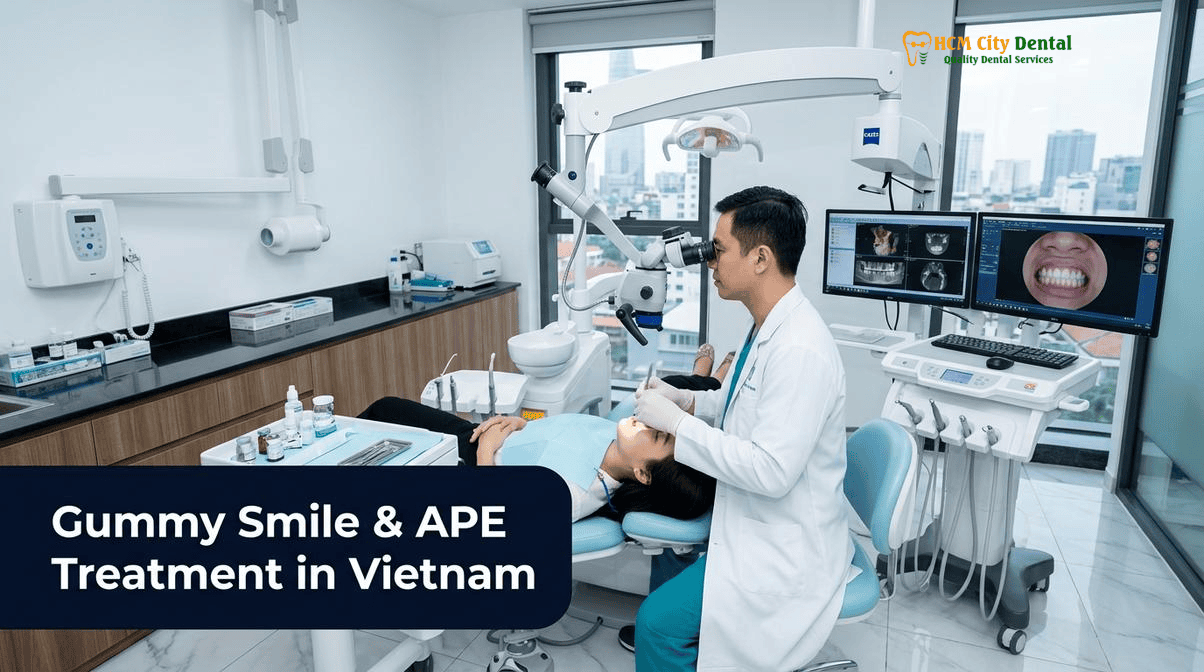
The 4 Anatomical Causes of a Gummy Smile: Altered Passive Eruption (APE), Vertical Maxillary Excess, Hypermobile Upper Lip & Dentoalveolar Extrusion
\n\n
Excessive gingival display is defined clinically as the exposure of more than 3 millimeters of gingival tissue during a dynamic smile. Before initiating any form of altered passive eruption gummy smile treatment, a comprehensive differential diagnosis must be established. The etiology of a gummy smile is multifactorial, and misidentifying the cause will inevitably lead to treatment failure. The four primary anatomical causes include:
\n\n
1. Altered Passive Eruption (APE)
\n
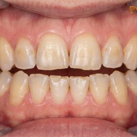
During normal dental development, teeth erupt into the oral cavity (active eruption), and subsequently, the gingival tissue and alveolar bone recede apically toward the root to expose the full anatomical crown (passive eruption). In patients with Altered Passive Eruption, this secondary phase fails to occur completely. The gingival margin remains positioned incisally (too far down on the tooth), and the alveolar bone crest remains abnormally close to, or even covering, the cementoenamel junction (CEJ). This results in teeth that appear short, square, and disproportionately small, surrounded by thick, bulky gums.
\n\n
2. Vertical Maxillary Excess (VME)
\n
Vertical Maxillary Excess is a skeletal dysplasia characterized by the overgrowth of the upper jaw (maxilla) in a vertical downward direction. Patients with VME typically display a long lower facial third and excessive gum tissue not only when smiling but often while the lips are at rest (lip incompetence). Because the etiology is skeletal, periodontal plastic surgery alone cannot fully resolve severe VME. Treatment typically requires orthognathic surgery (Le Fort I osteotomy) to physically impact and reposition the maxilla upwards, sometimes combined with orthodontic intrusion.
\n\n
3. Hypermobile Upper Lip
\n
In some patients, the dimensions of the teeth, gums, and jawbone are entirely normal, but the elevator muscles of the upper lip (such as the levator labii superioris) are hyperactive. When the patient smiles, the lip retracts excessively high, revealing a large band of gingiva. This condition is diagnosed when the lip length at rest is normal (typically 20-22mm in females, 22-24mm in males) but translates more than 6-8mm upward upon smiling. The primary treatment for a hypermobile lip is the targeted injection of Botulinum Toxin Type A (Botox) to temporarily relax the elevator muscles, or a surgical lip repositioning procedure to restrict muscle pull.
\n\n
4. Dentoalveolar Extrusion
\n
Dentoalveolar extrusion occurs when the anterior maxillary teeth over-erupt downwards, bringing the surrounding alveolar bone and gingival tissue with them. This is frequently seen in patients with severe deep bites (Class II Division 2 malocclusions) or severe anterior tooth wear (bruxism), where the teeth continuously erupt to maintain occlusal contact. Treatment for dentoalveolar extrusion requires orthodontic intrusion (using braces or clear aligners with TAD mini-screws) to push the teeth and bone back up into their proper vertical positions, often followed by restorative dentistry to rebuild worn incisal edges.
\n\n\n
\n\n\n
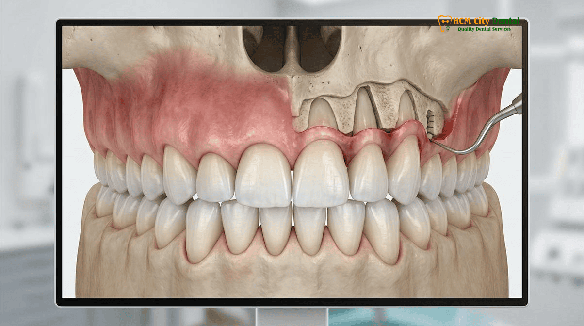
Coslet’s Classification of Altered Passive Eruption (Types 1A, 1B, 2A, 2B): Measuring the Bone-to-CEJ Distance on 3D CBCT
\n\n
When a gummy smile is definitively diagnosed as Altered Passive Eruption, the clinician must classify the specific subtype to determine the correct surgical approach. In 1977, Coslet et al. introduced a comprehensive classification system based on two critical anatomical relationships: the amount of keratinized gingiva (Type 1 vs. Type 2) and the position of the alveolar bone crest relative to the cementoenamel junction (Subtype A vs. Subtype B). At HCMC Dental Clinic, we utilize high-resolution 3D CBCT imaging to precisely measure these microscopic distances prior to surgery.
\n\n
Type 1 vs. Type 2: The Mucogingival Junction
\n
The first step in Coslet’s classification evaluates the width of the attached keratinized gingiva (the tough, pink tissue surrounding the teeth) relative to the mucogingival junction (where the attached gingiva meets the loose, red alveolar mucosa).
\n
- \n
- Type 1: The patient has an excessively wide band of attached keratinized gingiva. The mucogingival junction is located far apical to the bone crest. This is the most common presentation of APE. Because there is abundant tissue, the surgeon can safely excise the excess gum tissue (gingivectomy) without risking a lack of protective attached gingiva.
- Type 2: The patient has a normal or narrow band of attached keratinized gingiva, and the mucogingival junction is located at or near the CEJ. In these cases, excising the tissue (gingivectomy) is strictly contraindicated, as it would remove all the protective keratinized tissue, leaving the tooth surrounded by fragile alveolar mucosa. Instead, an apically positioned flap (APF) must be utilized to physically move the existing tissue higher up the root.
\n
\n
\n\n
Subtype A vs. Subtype B: The Alveolar Bone Crest
\n
The second, and most critical, step evaluates the position of the underlying alveolar bone crest in relation to the CEJ. This determines whether bone removal (ostectomy) is required. [1]
\n
- \n
- Subtype A: The alveolar bone crest is in its normal anatomical position, approximately 1.5 to 2.0 millimeters apical to (above) the CEJ. This provides adequate space for the supracrestal tissue attachment (biologic width).
- Subtype B: The alveolar bone crest has failed to recede and is located directly at, or even coronal to (covering), the CEJ. There is no space for the biologic width on the root surface; the attachment is entirely on the enamel.
\n
\n
\n\n
The 4 Clinical Combinations of Coslet’s Classification
\n
By combining these factors, we arrive at the four distinct diagnoses for coslet classification type 1a 1b 2a 2b ape:
\n
- \n
- Type 1A: Wide band of gingiva + Normal bone level. Treatment: Simple soft-tissue gingivectomy.
- Type 1B: Wide band of gingiva + High bone level. Treatment: Gingivectomy + Osseous Crown Lengthening (Ostectomy).
- Type 2A: Narrow band of gingiva + Normal bone level. Treatment: Apically Positioned Flap (APF).
- Type 2B: Narrow band of gingiva + High bone level. Treatment: Apically Positioned Flap (APF) + Osseous Crown Lengthening (Ostectomy).
\n
\n
\n
\n
\n\n\n
\n\n\n
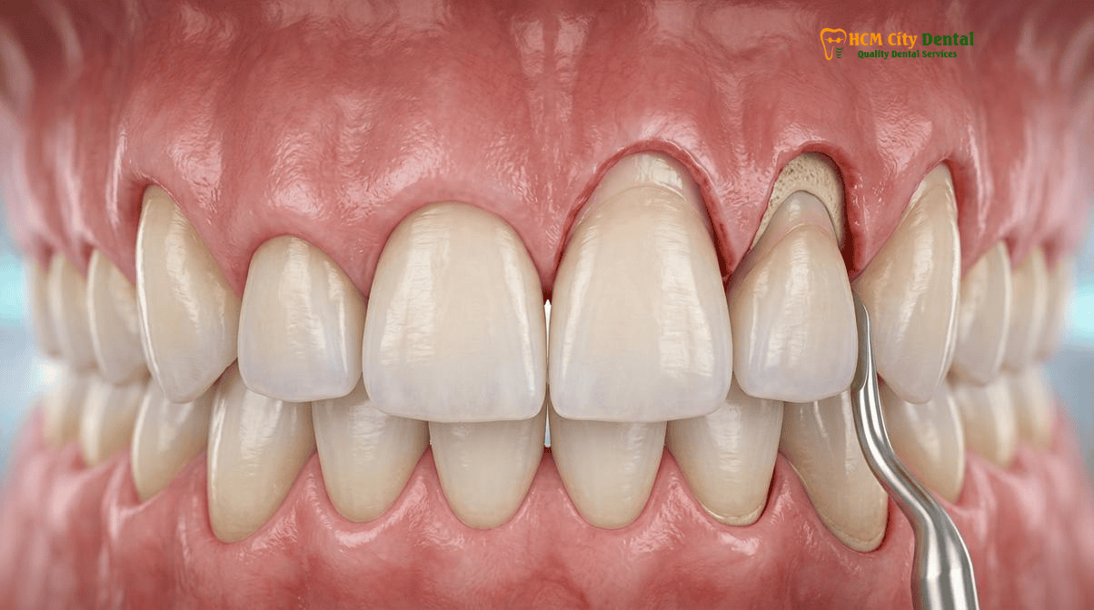
Why Simple Diode Laser Gingivectomy Alone Causes Gum Regrowth and Chronic Redness in Type 1B / 2B Patients (Biologic Width Violation)
\n\n
One of the most frequent complaints from patients seeking revision cosmetic dentistry is: “why gums grow back after laser gum contouring”. This phenomenon is almost always the result of a fundamental misunderstanding of periodontal anatomy by the previous treating dentist, specifically a violation of the biologic width (now officially termed the supracrestal tissue attachment).
\n\n
The supracrestal tissue attachment is a mandatory physiological dimension of soft tissue that exists between the crest of the alveolar bone and the base of the gingival sulcus. According to classic histological studies by Gargiulo et al., this dimension averages 2.04mm (comprising ~1.07mm of connective tissue attachment and ~0.97mm of junctional epithelium). In modern clinical practice, we respect a minimum distance of 3.0mm from the bone crest to the proposed new gingival margin to ensure absolute periodontal health.
\n\n
When a patient presents with Coslet Type 1B or 2B Altered Passive Eruption, their alveolar bone is sitting directly at the CEJ. If a dentist attempts to treat this gummy smile by simply burning away the excess gum tissue with a diode laser (a laser gingivectomy) to expose the full crown, they are placing the new gum margin directly on top of the bone crest. This completely obliterates the space required for the supracrestal tissue attachment. [2]
\n\n
\n
“The human body will never tolerate a violation of the biologic width. If you cut the gums back to the bone level without simultaneously recontouring the bone, the body views this as a severe injury. It will initiate a chronic inflammatory cascade, resulting in swollen, bleeding, purple gums, and eventually, the tissue will aggressively rebound and grow right back down the tooth to re-establish its mandatory 3-millimeter biological barrier. True esthetic crown lengthening is a bone surgery, not just a gum surgery.”
\n — Dr. Nguyen Van Cuong, DDS (Clinical Director — MOH License 0003265/BL-CCHN)\n
\n\n
To achieve a stable, permanent result in Type 1B and 2B cases, the clinician must perform an osseous ostectomy. The bone must be carefully sculpted and reduced so that it sits exactly 2.5 to 3.0 millimeters apical to the new, desired gingival margin. Only then will the soft tissue heal beautifully, tightly, and permanently in its new position without chronic inflammation or rebound.
\n\n\n
Clinical Case Note — Dr. Nguyen Van Cuong, DDS (Vietnam MOH License 0003265/BL-CCHN): Treating a gummy smile with laser gingivectomy alone when the patient actually has Type 1B Altered Passive Eruption (where the alveolar bone crest sits at the cementoenamel junction) inevitably leads to biologic width invasion, chronic red gingival inflammation, and tissue rebound within 6 months. At HCMC Dental Clinic in District 1, we use high-resolution 3D CBCT bone sounding to re-establish the exact 3.0mm supracrestal tissue attachment prior to bonding final porcelain veneers.
\n\n
Dual-Guide 3D-Printed Esthetic Crown Lengthening: Flapless Piezosurgery Ostectomy vs. Micro-Flap Osseous Recontouring
\n\n
The surgical execution of esthetic crown lengthening has evolved dramatically with the advent of digital dentistry and ultrasonic surgical instruments. At HCMC Dental Clinic, we utilize a fully digital workflow to plan and execute 3D printed surgical guide crown lengthening veneers, ensuring sub-millimeter precision and minimizing surgical trauma.
\n\n
Digital Smile Design and Dual-Guide Fabrication
\n
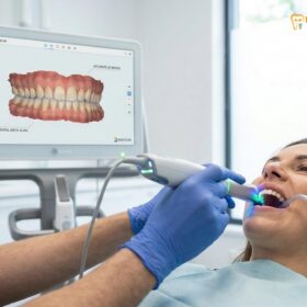
The process begins with intraoral optical scanning, facial photography, and 3D CBCT imaging. Using Digital Smile Design (DSD) software, the ideal proportions of the teeth (the “White Esthetics”) and the ideal architecture of the gums (the “Pink Esthetics”) are designed to harmonize with the patient’s facial features. From this digital wax-up, two distinct 3D-printed surgical guides are fabricated:
\n
- \n
- The Gingival Guide: This guide dictates the exact position and contour of the initial soft-tissue incision (the gingivectomy), ensuring perfect symmetry across the anterior maxilla.
- The Osseous Guide: This guide rests against the teeth and provides a physical stop for the surgical instruments, dictating exactly how much alveolar bone must be removed to establish the 3mm biologic width.
\n
\n
\n\n
Flapless Piezosurgery Ostectomy
\n
In cases where the gingival biotype is thick and the required bone reduction is minimal to moderate, we employ a minimally invasive flapless Piezosurgery technique. Instead of using a scalpel to raise a large, full-thickness mucoperiosteal flap (which causes significant post-operative swelling and requires extensive suturing), the surgeon accesses the bone directly through the gingival sulcus. [3]
\n\n
A piezotome utilizes ultrasonic micro-vibrations (typically between 24-36 kHz) to selectively cut mineralized bone tissue while leaving soft tissues (gingiva, nerves, blood vessels) completely unharmed. Using specialized micro-tips inserted into the sulcus, the surgeon performs the ostectomy (removal of supporting bone) and osteoplasty (thinning of bulky bone) blindly but safely, guided by tactile feedback and the 3D-printed osseous guide. This flapless approach results in virtually zero swelling, no external sutures, and allows international patients to resume normal activities almost immediately.
\n\n
Clinical Case Study: Expat Gummy Smile Makeover in District 1
\n
A 28-year-old Australian patient presented to HCMC Dental Clinic complaining of short, square teeth and a severe gummy smile. Previous consultations in Sydney recommended orthognathic surgery. Our 3D CBCT analysis revealed Coslet Type 1B Altered Passive Eruption, not Vertical Maxillary Excess. Dr. Nguyen Van Cuong, DDS, utilized a 3D-printed dual-guide system to perform flapless Piezosurgery crown lengthening on teeth #14 through #24. The bone was recontoured 3mm apical to the CEJ. Because no flaps were raised, the patient experienced zero bruising and minimal discomfort. After a 3-month maturation phase, the case was finalized with 10 ultra-thin IPS E.max veneers, delivering a stunning, proportionate smile at a fraction of the Australian cost.
\n
\n\n
Micro-Flap Osseous Recontouring
\n
In more complex cases involving severe bony exostoses (thick ledges of bone), Coslet Type 2 presentations requiring an apically positioned flap, or when extensive osteoplasty is needed to thin the buccal bone plate, a micro-flap approach is necessary. A small, precise internal bevel incision is made, and the tissue is gently reflected just enough to visualize the bone crest. Piezosurgery or high-speed surgical burs are then used to sculpt the bone under direct vision. The tissue is then sutured precisely at the new CEJ using micro-sutures (e.g., 6-0 or 7-0 Prolene). While this approach involves slightly more healing time than the flapless technique, it provides unparalleled control over the final bony architecture.
\n\n\n
\n\n\n
Sequencing Crown Lengthening with Ultra-Thin IPS E.max Porcelain Veneers (Healing Timelines for International Travelers)
\n\n
When combining periodontal plastic surgery with restorative prosthodontics, the sequencing of treatment and the management of healing timelines are critical, especially for dental tourists operating on restricted travel schedules. The integration of gummy smile altered passive eruption esthetic crown lengthening veneers must follow strict biological principles to ensure the final ceramic margins remain perfectly hidden and the gingiva remains stable.
\n\n
The Biological Maturation Phase
\n
Following osseous crown lengthening, the soft tissues undergo a complex process of healing, remodeling, and maturation. While the initial surface healing occurs within 10 to 14 days, the deeper connective tissue attachment and the final position of the gingival margin take significantly longer to stabilize. If final impressions for porcelain veneers are taken too soon, the gums may continue to recede slightly over the following months, exposing the margins of the veneers and compromising the aesthetic result.
\n\n
For optimal predictability, a minimum healing period of 3 to 6 months is mandatory before finalizing the subgingival margins of permanent restorations. However, patients do not have to wait 6 months with compromised aesthetics. Immediately following the crown lengthening surgery, high-quality bis-acryl provisional (temporary) veneers can be fabricated. These provisionals serve a dual purpose: they immediately improve the patient’s smile, and their highly polished cervical contours act as a matrix to guide the healing gingiva, shaping the papillae and ensuring a perfect emergence profile.
\n\n
Wave 4A Biomimetic Adhesive Protocols
\n
Once the tissue has fully matured, the final restorative phase begins. At HCMC Dental Clinic, we adhere strictly to Wave 4A Biomimetic adhesive physics to ensure maximum enamel preservation and bond strength. [4]
\n\n
- \n
- Digital SKYN Concept: The final restorations are designed using 3D natural tooth morphology libraries, incorporating primary line angles, secondary developmental lobes, and tertiary perikymata micro-texture to mimic natural enamel perfectly.
- Immediate Dentin Sealing (IDS): If any dentin is exposed during the minimal preparation phase, it is immediately sealed with a gold-standard dentin bonding agent (e.g., OptiBond FL) prior to taking impressions. This prevents bacterial microleakage, eliminates post-operative sensitivity, and increases the final bond strength by up to 400%.
- Feilzer’s C-Factor Management: The configuration factor (bonded to unbonded surfaces) is carefully managed during the adhesive cementation process to minimize polymerization shrinkage stress, preventing micro-cracks in the ceramic.
- Strict Isolation and Surface Treatment: The final IPS E.max lithium disilicate veneers are bonded under a 100% latex-free nitrile rubber dam. The intaglio surface of the ceramic is treated with 5%–9.5% hydrofluoric acid and pure heat-dried silane, while the tooth surface is prepared with 27-micron aluminum oxide air abrasion, ensuring an impenetrable, biomimetic monolithic bond.
\n
\n
\n
\n
\n\n
Clinical Precaution for Dental Travelers:
\n
If you are traveling to Vietnam for a gummy smile makeover, beware of clinics offering “same-day” or “one-week” crown lengthening and final veneer placement. Placing permanent subgingival ceramic margins on freshly cut bone and healing gums is a severe violation of clinical protocols. It will lead to unpredictable tissue recession, margin exposure, and chronic inflammation. At HCMC Dental Clinic, we prioritize biological stability. We offer phased treatment plans, allowing you to return home with beautiful provisionals while the tissue matures, ensuring your final IPS E.max veneers are placed on a perfectly stable, healthy foundation.
\n
\n\n
2026 Laser Gum Contouring, Piezosurgery Crown Lengthening & Veneer Fee Schedule at HCMC Dental Clinic (District 1)
\n\n
Understanding the gummy smile makeover cost Ho Chi Minh City is crucial for international patients planning their treatment. HCMC Dental Clinic offers world-class periodontal and prosthodontic care at highly transparent, competitive rates. By utilizing our Official 2026 Dental Price List & -40% Online Booking Fee Schedule, patients can access premium treatments like Piezosurgery and IPS E.max veneers at a fraction of the cost compared to the US or Australia.
\n\n
Below is the detailed 2026 fee schedule for procedures related to gummy smile correction, esthetic crown lengthening, and biomimetic smile makeovers. [5]
\n\n
| Procedure / Treatment Phase | 2026 Online Booking Fee (-40% USD) | Walk-In / Standard List Price (USD) | US / Australian Benchmark (USD) |
|---|---|---|---|
| 3D CBCT Scan (Diagnostic for APE) | $18 USD (480,000 VND) | $30 USD | $150 – $350 USD |
| Simple Laser Gingivectomy (Per Tooth) | $35 – $50 USD | $60 – $85 USD | $150 – $300 USD |
| Piezosurgery Esthetic Crown Lengthening (Per Tooth) | $80 – $120 USD | $135 – $200 USD | $500 – $900 USD |
| IPS E.max Porcelain Veneer (Lithium Disilicate) | $185 USD / tooth (4,800,000 VND) | $308 USD / tooth | $1,200 – $2,500 USD |
| Zirconia Veneer (Premium Multi-layer) | $138 USD / tooth | $230 USD / tooth | $1,000 – $2,000 USD |
| Composite Veneer / Dental Bonding | $35 – $46 USD / tooth (900,000 – 1,200,000 VND) | $60 – $75 USD / tooth | $300 – $600 USD |
| IPS E.max All-Ceramic Crown / Overlay | $231 USD / tooth (6,000,000 VND) | $385 USD / tooth | $1,500 – $2,800 USD |
| Full Zirconia Crown (Cercon Standard / HT) | $154 – $192 USD / tooth (4,000,000 – 5,000,000 VND) | $256 – $321 USD / tooth | $1,200 – $2,200 USD |
| Invisalign Comprehensive (Unlimited Aligners) | $2,538 USD (66,000,000 VND) | $4,231 USD | $5,500 – $8,500 USD |
| Swiss EMS AirFlow Stain Removal (Erythritol GBT) | $58 USD (1,500,000 VND) | $96 USD | $200 – $350 USD |
\n
\n\n
To secure these exclusive online rates for your Smile Makeover Vietnam: Clinical Costs, DSD & Veneer Packages, international patients must contact our coordination team prior to arrival. We highly recommend exploring our Discount Dental Veneers Vietnam: 40% Off Premium Smile Makeovers program via WhatsApp at +84 853 020 003 to schedule a preliminary digital consultation with Dr. Nguyen Van Cuong, DDS.
\n\n\n
\n\n\n
Frequently Asked Questions
\n\n
How does a 3D CBCT scan tell whether I only need a 15-minute laser gum trim or true bone-level crown lengthening?
\n
A 3D CBCT scan allows the periodontist to visualize the exact distance between your alveolar bone crest and the cementoenamel junction (CEJ) beneath the gums. If the bone is 2-3mm away from the CEJ (Coslet Type 1A), a simple 15-minute laser gum trim is sufficient. If the bone is directly at or covering the CEJ (Coslet Type 1B), true bone-level esthetic crown lengthening is mandatory to prevent biologic width violations.
\n\n
Why did my gums grow right back to their original position 3 months after having laser gum contouring at another clinic?
\n
This is the classic hallmark of a biologic width violation. If a dentist performs a simple laser gingivectomy on a patient with a high bone crest (Coslet Type 1B or 2B), the newly cut gum margin is left too close to the underlying bone. The body’s natural inflammatory response will cause the gingival tissue to rebound and grow back over the teeth to re-establish the mandatory 3mm supracrestal tissue attachment.
\n\n
What is ‘Flapless Piezosurgery Crown Lengthening’ through the gingival sulcus without external stitches?
\n
Flapless Piezosurgery is an advanced, minimally invasive technique where ultrasonic micro-vibrations are used to recontour the alveolar bone directly through the gingival sulcus (the natural pocket between the tooth and gum). Because no large scalpel incisions or full-thickness flaps are raised on the outside of the gums, there is minimal swelling, no external stitches are required, and the healing time is drastically reduced.
\n\n
How long must I wait after osseous crown lengthening before finalizing the subgingival margins of IPS E.max porcelain veneers?
\n
For optimal esthetic stability, especially in the highly visible anterior maxilla, a healing period of 3 to 6 months is recommended after osseous crown lengthening before taking final impressions for the subgingival margins of IPS E.max porcelain veneers. However, high-quality bis-acryl provisional veneers can be placed immediately to protect the teeth and guide the soft tissue healing during this maturation phase.
\n\n
How much do laser gingivectomy and esthetic crown lengthening cost at HCMC Dental Clinic in District 1?
\n
At HCMC Dental Clinic, simple laser gingivectomy is highly affordable. For comprehensive esthetic crown lengthening involving Piezosurgery bone recontouring, the fees are significantly lower than US or Australian averages. When combined with our -40% online booking rates, a complete smile makeover featuring IPS E.max Porcelain Veneers is just $185 USD per tooth, making a full gummy smile transformation highly cost-effective for international patients.
\n\n
References
\n
- \n
- Journal of Periodontology. (2021). “Altered Passive Eruption: Diagnosis, Classification, and Treatment Modalities in the Esthetic Zone.”
- International Journal of Periodontics & Restorative Dentistry. (2022). “Supracrestal Tissue Attachment (Biologic Width) Violations: The Etiology of Gingival Rebound Following Simple Gingivectomy.”
- Clinical Oral Investigations. (2023). “Minimally Invasive Flapless Piezosurgery for Esthetic Crown Lengthening: A Comparative Clinical Trial.”
- Journal of Prosthetic Dentistry. (2023). “Biomimetic Protocols for IPS E.max Lithium Disilicate Veneers Following Periodontal Plastic Surgery: Immediate Dentin Sealing and C-Factor Management.”
- Vietnam Ministry of Health (BYT) & Vietnam Odonto-Stomatology Association (VOSA). National Clinical Guidelines on Specialist Dental Care, Biomaterial Traceability & EN 13060 Class B Infection Control. (2024).
\n
\n
\n
\n
\n
\n\n