The clinical management of an ankylosed tooth or a submerged molar represents one of the most complex biomechanical challenges in modern restorative dentistry and oral implantology. When the periodontal ligament (PDL) is destroyed—whether due to severe dental trauma, delayed replantation of an avulsed tooth, or genetic predisposition—the tooth root fuses directly to the surrounding alveolar bone. This pathological dentoalveolar fusion halts the tooth’s natural eruptive potential. As the patient’s jaw continues to grow vertically, the fused tooth is left behind, sinking below the occlusal plane into a state of progressive infraocclusion. For international patients and dental travelers seeking definitive care, the decision between preserving the tooth with a biomimetic overlay, performing a surgical decoronation, or executing an atraumatic extraction followed by a 3D-guided dental implant requires precise, evidence-based diagnostics. At HCMC Dental Clinic in Ho Chi Minh City, our clinical protocols prioritize the preservation of the buccal bundle bone, the optimization of soft tissue architecture, and the long-term biomechanical stability of the final restoration.
Clinical Summary:
When evaluating an ankylosed tooth, submerged molar, decoronation vs. implant therapy, the patient’s skeletal maturity and the severity of infraocclusion dictate the treatment pathway. In growing adolescents, decoronation (removing the crown and submerging the root) preserves vertical alveolar bone growth for future implant site development. In adults with mild infraocclusion and intact roots, a biomimetic IPS E.max table-top overlay ($231 USD online) restores occlusal function without surgery. For adults with severe infraocclusion and progressive root replacement resorption, the gold standard is Piezosurgery atraumatic extraction combined with guided bone regeneration and the placement of a 3D-guided dental implant (starting at $462 USD online for a Korean fixture). Accurate 3D CBCT diagnosis is mandatory to prevent catastrophic orthodontic failures, as braces cannot move an ankylosed tooth.
Key Takeaways:
- Dentoalveolar Fusion: Ankylosis involves the complete loss of the periodontal ligament (PDL), resulting in the tooth root fusing directly to the jawbone, making orthodontic movement physically impossible.
- Adolescent Protocol (Decoronation): Placing dental implants in growing children leads to severe aesthetic and functional failures as the jaw grows around the static implant. Decoronation preserves the alveolar ridge until skeletal maturity is reached.
- Adult Conservative Protocol: A retained primary molar in mild infraocclusion can often be restored functionally and aesthetically using a minimally invasive IPS E.max ceramic overlay, avoiding extraction entirely.
- Adult Surgical Protocol: Extracting an ankylosed tooth with traditional forceps causes massive bone defects. Piezosurgery (ultrasonic micro-surgery) is required to safely separate the fused root from the bone prior to 3D-guided implant placement.
- Transparent 2026 Pricing: HCMC Dental Clinic offers a -40% online booking fee schedule, providing world-class implantology and biomimetic restorations at a fraction of US or Australian benchmark costs.
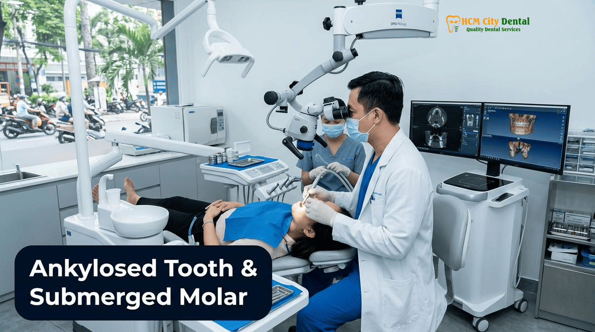
What Is Dental Ankylosis (Dentoalveolar Fusion) and Why Do Retained Primary Molars or Replanted Avulsed Teeth Sink Below the Occlusal Plane?
Dental ankylosis is a severe disruption of the normal tooth-to-bone interface. In a healthy dentition, every tooth is suspended in its bony socket by the periodontal ligament (PDL)—a complex network of collagen fibers, blood vessels, and mesenchymal stem cells. The PDL acts as a biological shock absorber during mastication and serves as a physical barrier that prevents the surrounding alveolar bone from directly contacting the tooth root. Furthermore, the PDL is the engine of tooth eruption, constantly remodeling to allow the tooth to move in harmony with the growing jaws.
When the PDL is irreversibly damaged, the protective barrier is lost. This damage most commonly occurs following severe dental trauma (such as intrusive luxation or the delayed replantation of an avulsed tooth) or due to idiopathic reasons in retained primary (baby) molars that lack a permanent successor. Without the PDL, osteoclasts begin to resorb the root dentin, and osteoblasts immediately deposit alveolar bone into the resorbed spaces. This process, known as replacement resorption, results in the direct, rigid fusion of the tooth root to the jawbone—a condition termed dentoalveolar fusion or ankylosis[1].
The most visible clinical consequence of ankylosis is infraocclusion. Because the ankylosed tooth is locked into the bone, it loses all eruptive capacity. Meanwhile, the adjacent healthy teeth continue to erupt, and the surrounding alveolar bone continues to grow vertically. Over time, the ankylosed tooth appears to “sink” below the occlusal plane (the biting surface). In reality, the ankylosed tooth is stationary; it is the rest of the jaw that is growing away from it. If left untreated, severe infraocclusion can lead to the tipping of adjacent teeth into the space, supra-eruption of the opposing teeth, severe localized periodontal defects, and a collapsed bite architecture.
In the context of modern biological surgery, managing these cases requires a deep understanding of alveolar bone dynamics. At HCMC Dental Clinic, Dr. Nguyen Van Cuong, DDS (Clinical Director — MOH License 0003265/BL-CCHN) utilizes advanced microsurgical techniques to address the unique challenges presented by ankylosed teeth, ensuring that any intervention—whether restorative or surgical—preserves the maximum amount of native bone and soft tissue.
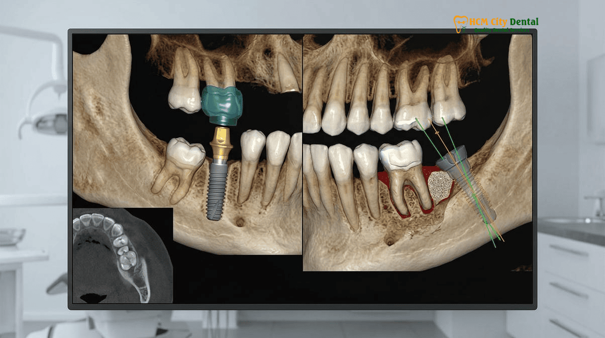
3D CBCT & Percussion Diagnosis: Why Braces and Invisalign Cannot Move an Ankylosed Tooth (And Instead Pull Adjacent Teeth Into Open Bite)
Accurate diagnosis of dental ankylosis is the critical first step in formulating a treatment plan. Clinically, an ankylosed tooth exhibits zero physiological mobility. When tapped with the handle of a dental mirror, it produces a characteristic high-pitched, metallic percussion sound, contrasting sharply with the dull, cushioned sound of a healthy tooth suspended by a PDL. However, clinical signs alone are insufficient for comprehensive treatment planning.
High-resolution 3D Cone Beam Computed Tomography (CBCT) is mandatory to visualize the extent of the dentoalveolar fusion. Traditional 2D panoramic or periapical X-rays often fail to show the fusion points due to anatomical superimposition. A 3D CBCT scan allows the specialist to examine the tooth in axial, coronal, and sagittal planes, identifying the exact locations where the PDL space has been obliterated and replaced by bone. This imaging is crucial for determining whether the tooth can be surgically luxated, decoronated, or if it requires complex piezosurgical extraction[2].
A catastrophic error frequently seen in general dental practice is the attempt to orthodontically move an ankylosed tooth. Because the tooth is fused to the bone, it possesses infinite anchorage value—it acts exactly like a titanium dental implant. According to Newton’s Third Law of Motion (for every action, there is an equal and opposite reaction), applying orthodontic force (via braces or clear aligners) to extrude an ankylosed tooth will not move the fused tooth a single millimeter. Instead, the reciprocal forces will intrude the adjacent healthy anchor teeth, driving them into the jawbone and creating a severe, iatrogenic open bite.
“Attempting to pull an ankylosed canine or incisor into the dental arch with orthodontics is a biomechanical impossibility. The fused tooth will not yield; instead, it will drag the adjacent healthy teeth out of alignment, destroying the patient’s occlusion. This is why we mandate in-house 3D CBCT imaging for every suspected case of infraocclusion before any treatment is initiated.”
— Dr. Nguyen Van Cuong, DDS
For patients seeking Safe Dental Implants Ho Chi Minh City: 3D CBCT & Guided Surgery, our facility in District 1, Ho Chi Minh City, is equipped with state-of-the-art digital imaging technology to ensure precise, risk-free diagnostics.
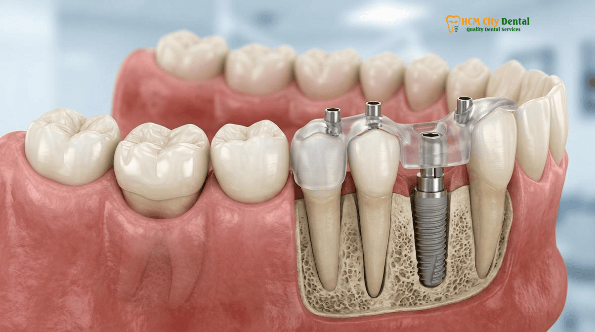
Scenario A (Mild Infraocclusion of a Retained Baby Molar with Intact Roots): Biomimetic IPS E.max Table-Top Overlay ($231 USD)
Not all ankylosed teeth require surgical extraction. A common clinical scenario involves an adult patient with a retained primary (baby) second molar. Often, the permanent second premolar is congenitally missing, and the primary molar has become ankylosed. If the 3D CBCT scan reveals that the primary molar has long, intact roots with minimal replacement resorption, no periapical infection, and is only in mild infraocclusion (1–3 mm below the occlusal plane), the most biologically conservative approach is to retain the tooth.
In these cases, preparing the tooth for a traditional full-coverage dental crown is highly destructive and contraindicated, as primary teeth have large pulp chambers and thin enamel. Instead, Dr. Nguyen Van Cuong, DDS employs a biomimetic approach using an IPS E.max lithium disilicate table-top overlay. This minimally invasive ceramic onlay is bonded directly to the occlusal surface of the submerged molar, effectively building up its height to meet the opposing tooth.
The biomimetic protocol involves Immediate Dentin Sealing (IDS) and resin coating to protect the pulp and maximize bond strength. The IPS E.max overlay is etched with hydrofluoric acid, treated with a silane coupling agent, and bonded using heated composite resin under strict rubber dam isolation. This restores the patient’s chewing function, prevents the opposing teeth from supra-erupting, and maintains the alveolar bone volume—all without a single surgical incision.
At HCMC Dental Clinic, an IPS E.max All-Ceramic Overlay is priced at just $231 USD per tooth online (6,000,000 VND), representing a highly cost-effective and biologically sound alternative to extraction and implant placement for mild cases.
Scenario B (Progressive Infraocclusion & Root Replacement Resorption): Piezosurgery Atraumatic Extraction + Guided Bone Regeneration + Implant
When an ankylosed tooth in an adult patient exhibits severe infraocclusion, extensive root replacement resorption, or structural failure, preservation is no longer viable. The tooth must be extracted and replaced. However, extracting an ankylosed tooth is notoriously difficult. Because the root is fused to the bone, the use of traditional extraction forceps or elevators will inevitably result in the fracture of the buccal (outer) bone plate. This creates a massive, three-dimensional alveolar ridge defect that makes subsequent implant placement highly unpredictable and aesthetically compromised.
To circumvent this, HCMC Dental Clinic utilizes Piezosurgery for atraumatic bone micro-surgery. Piezosurgery utilizes ultrasonic micro-vibrations (operating at a frequency of 25–30 kHz) that selectively cut mineralized tissue (bone and tooth) while completely sparing soft tissues (nerves, blood vessels, and the periosteum). Using specialized micro-tips, Dr. Cuong precisely severs the ankylotic fusion lines between the root and the bone, allowing the root fragments to be removed without expanding the socket or damaging the fragile buccal bundle bone.
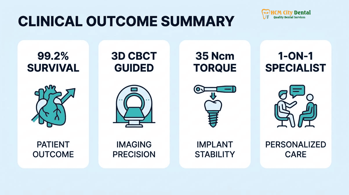
Following the atraumatic extraction, the site is often grafted using particulate xenograft material and a resorbable collagen membrane (Guided Bone Regeneration) to reconstruct the ridge. Depending on the primary stability achieved, a 3D-guided dental implant may be placed immediately or after a healing period of 3 to 4 months. Our implant protocols strictly adhere to advanced biomechanical metrics, including Osstell ISQ Resonance Frequency Analysis (requiring an ISQ ≥70 and insertion torque ≥35 Ncm for immediate loading) and the use of Conical Morse Taper Platform Switching implants to ensure <0.2mm crestal bone loss at 36 months[3].
Patients have the option of Grade 4 pure titanium implants or 100% metal-free Y-TZP Zirconia implants ($1,154 USD online) for optimal soft tissue integration and aesthetics. For patients with complex medical backgrounds, we also offer specialized protocols, such as our Dental Implants on Blood Thinners Protocol | HCMC Dental.
Clinical Precaution: Never allow a general dentist to attempt a forceful extraction of an ankylosed tooth using standard forceps. The resulting bone trauma can necessitate extensive, costly block bone grafting procedures. Always seek a specialist equipped with Piezosurgery and 3D CBCT capabilities. If you have a submerged tooth, contact Dr. Nguyen Van Cuong, DDS via WhatsApp at +84 853 020 003 for a comprehensive digital assessment.
Scenario C (Growing Adolescents & Severe Labial Bone Fusion): Decoronation Ridge-Preservation Technique vs. Root Submergence Therapy
The management of an ankylosed permanent tooth (often a central incisor following trauma) in a growing child or adolescent presents a unique dilemma. Dental implants are strictly contraindicated in growing patients. Because an implant lacks a PDL, it acts exactly like an ankylosed tooth; it will not erupt as the child’s jaw grows. Placing an implant in a 12-year-old will result in the implant being left high up in the bone by the time the patient is 18, leading to a disastrous aesthetic failure and a severe vertical bone defect.
To manage this, the gold standard treatment is the Decoronation Technique (originally described by Malmgren). Decoronation is a surgical ridge-preservation strategy designed to maintain the width and height of the alveolar bone until the patient reaches skeletal maturity.
The protocol involves reflecting a mucoperiosteal flap and amputating the crown of the ankylosed tooth approximately 1.5 to 2.0 mm below the crest of the alveolar bone. Any endodontic filling material (such as gutta-percha) inside the root canal is completely removed. The empty root canal is then intentionally allowed to fill with blood from the surrounding tissues. The mucoperiosteal flap is sutured completely over the submerged root. Over the next several years, the submerged root undergoes continuous replacement resorption, acting as a biological scaffold. As the adjacent teeth erupt and the jaw grows, the periosteum lays down new vertical alveolar bone over the resorbing root[4].
Once the patient reaches skeletal maturity (typically age 18-21, confirmed via cervical vertebral maturation or serial cephalometric radiographs), the site will have adequate bone volume to receive a definitive 3D-guided dental implant. In adult patients where the root is healthy but the crown is destroyed, a similar concept known as Root Submergence Therapy or the Socket Shield Technique (Partial Extraction Therapy) for Front Tooth Implants in Vietnam: Protocol & Cost can be utilized. This involves leaving a 1.5mm buccal root fragment to preserve the periodontal ligament blood supply and the labial bundle bone, preventing the natural post-extraction resorption of the anterior maxilla.
Clinical Case Study: District 1, Ho Chi Minh City
A 14-year-old international patient presented to HCMC Dental Clinic with a severely ankylosed and infraoccluded maxillary right central incisor, the result of an avulsion injury sustained at age 9. The tooth was 4mm below the occlusal plane, causing a severe aesthetic deformity and localized bone arrest. Because the patient was still actively growing, Dr. Nguyen Van Cuong, DDS performed a surgical decoronation under local anesthesia. The crown was removed, and the root was submerged to act as a biological scaffold. A temporary Essix retainer with a pontic was provided for aesthetics. Over the next 4 years, the alveolar bone grew vertically over the resorbing root, perfectly matching the adjacent healthy incisor. At age 19, following confirmation of skeletal maturity, a 3D-guided Straumann BLT-SLA implant was placed into the preserved ridge without the need for complex bone grafting, yielding a flawless, highly aesthetic emergence profile.
2026 Surgical & Prosthetic Treatment Costs at HCMC Dental Clinic (District 1, Ho Chi Minh City)
International patients, expats, and dental travelers seeking definitive care for ankylosed teeth and complex implantology often face prohibitive costs in their home countries. HCMC Dental Clinic provides world-class, evidence-based surgical and restorative care at highly accessible price points. By utilizing our Official 2026 Dental Price List & -40% Online Booking Fee Schedule, patients can access premium European and Korean implant systems, Piezosurgery, and biomimetic ceramics at a fraction of US or Australian benchmark costs.
Below is the official 2026 fee schedule for procedures related to the management of ankylosed teeth, decoronation, and 3D-guided implantology under the clinical direction of Dr. Nguyen Van Cuong, DDS[5].
| Clinical Procedure (Ankylosis & Implantology) | 2026 Online Booking Price (-40% USD) | Walk-In / Standard Price (USD) | VND Equivalent (Online) |
|---|---|---|---|
| Surgical Wisdom/Ankylosed Tooth Extraction (Piezotome) | $58 – $115 USD | $96 – $192 USD | 1,500,000 – 3,000,000 VND |
| IPS E.max All-Ceramic Overlay (Biomimetic Onlay) | $231 USD / tooth | $385 USD | 6,000,000 VND |
| Single Korean Implant (Dentis / Neo Biotech / Dentium) | $462 – $654 USD | $769 – $1,090 USD | 12,000,000 – 17,000,000 VND |
| Single European/Swiss Implant (Straumann BLT-SLA / BLA-Active) | $962 – $1,346 USD | $1,603 – $2,244 USD | 25,000,000 – 35,000,000 VND |
| 100% Metal-Free Zirconia Ceramic Implant | $1,154 USD | $1,923 USD | 30,000,000 VND |
| Implant Zirconia HT Crown + Custom Abutment | $154 USD | $256 USD | 4,000,000 VND |
| 3D CBCT Scan (In-House Diagnostic Imaging) | $18 USD | $30 USD | 480,000 VND |
“Our commitment at HCMC Dental Clinic is to provide uncompromising clinical excellence. By integrating Piezosurgery, 3D CBCT diagnostics, and Custom CAD/CAM Hybrid Zirconia-TiBase abutments, we ensure that every implant placed into a previously ankylosed site achieves optimal osseointegration and long-term biomechanical stability, all while maintaining absolute transparency in our fee structure.”
— Dr. Nguyen Van Cuong, DDS
Frequently Asked Questions
Why did my orthodontist try to pull my ankylosed canine or incisor down with braces, only for my neighboring healthy teeth to tilt inward?
An ankylosed tooth is fused directly to the jawbone, meaning it has zero periodontal ligament (PDL) to allow for movement. In orthodontic biomechanics, it acts as an absolute anchor. According to Newton’s Third Law, the force applied to pull the ankylosed tooth down will instead cause the adjacent healthy teeth to intrude or tilt inward, creating a severe open bite. This is why 3D CBCT diagnosis is mandatory before attempting to move a submerged tooth.
If my retained baby second molar (with no adult premolar underneath) is slightly low, can an IPS E.max onlay build up its height without pulling it?
Yes. If the retained baby molar has intact roots, no infection, and is only in mild infraocclusion (slightly below the bite plane), a biomimetic IPS E.max table-top overlay ($231 USD online) is the ideal conservative treatment. This ceramic onlay builds up the submerged molar to the correct occlusal height, restoring chewing function and preventing opposing teeth from over-erupting, all without the need for surgical extraction.
Why does extracting a long-ankylosed tooth with traditional forceps cause large jawbone defects—and how does Piezosurgery prevent this?
Because an ankylosed root is fused to the surrounding bone, traditional forceps extraction often fractures the fragile buccal (outer) bone plate, leaving a massive defect that makes future implant placement difficult. Piezosurgery uses ultrasonic micro-vibrations (25–30 kHz) to selectively cut the mineralized fusion line while preserving the surrounding bone and soft tissue, allowing for an atraumatic extraction and immediate bone grafting.
What is ‘decoronation’ and when is it performed before placing a dental implant?
Decoronation is a ridge-preservation technique used primarily in growing adolescents. The crown of the ankylosed tooth is removed below the gumline, and the root is left in the bone to undergo natural replacement resorption. As the child grows, the surrounding alveolar bone continues to develop vertically over the submerged root. Once skeletal maturity is reached, a 3D-guided dental implant can be placed into the preserved, fully developed bone ridge.
How much does Piezosurgery extraction, bone grafting, and a 3D-guided dental implant cost at HCMC Dental Clinic?
At HCMC Dental Clinic, a surgical extraction using Piezotome costs $58 – $115 USD online. A single Korean Implant (Dentis, Neo Biotech, or Dentium) ranges from $462 – $654 USD online, while a premium European/Swiss Implant (like Straumann BLT-SLA) ranges from $962 – $1,346 USD online. A 100% metal-free Zirconia implant is $1,154 USD online. These transparent 2026 online booking rates represent a -40% savings compared to standard walk-in prices.
References
- Malmgren, B., & Malmgren, O. (2002). Rate of infraposition of reimplanted ankylosed incisors related to age and growth in children and adolescents. Dental Traumatology.
- Filippi, A., Pohl, Y., & von Arx, T. (2001). Decoronation of an ankylosed tooth for preservation of alveolar bone prior to implant placement. Dental Traumatology.
- Buser, D., Sennerby, L., & De Bruyn, H. (2017). Modern implant dentistry based on osseointegration: 50 years of progress, current trends and open questions. Periodontology 2000.
- Magne, P., & Belser, U. (2002). Bonded Porcelain Restorations in the Anterior Dentition: A Biomimetic Approach. Quintessence Publishing.
- Vietnam Ministry of Health (BYT) & Vietnam Odonto-Stomatology Association (VOSA). National Clinical Guidelines on Specialist Dental Care, Biomaterial Traceability & EN 13060 Class B Infection Control. (2024).