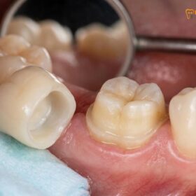
A zirconia crown is a highly durable, biocompatible dental restoration milled from yttria-stabilized zirconium dioxide. It effectively restores severely damaged, decayed, or root canal-treated teeth. Known for exceptional fracture resistance and natural aesthetics, this metal-free cap provides long-lasting functional and structural support for both anterior and posterior teeth.
Clinical Summary:
Zirconia crowns represent the gold standard in modern restorative dentistry, utilizing yttria-stabilized zirconium dioxide to achieve unparalleled flexural strength (up to 1200+ MPa). Available in 3Y, 4Y, and 5Y formulations, these metal-free restorations balance extreme durability with lifelike translucency. Clinical success relies heavily on precise conservative tooth preparation, advanced digital CAD/CAM fabrication, and strict MDP-based cementation protocols to prevent debonding. By eliminating the metallic substructure found in traditional crowns, zirconia provides a highly biocompatible, aesthetically superior solution that resists chipping, minimizes wear on opposing dentition, and promotes optimal gingival health.
Key Takeaways:
- Zirconium dioxide is a bio-inert, metal-free crystalline oxide offering superior fracture toughness and biocompatibility.
- Modern dental zirconia is categorized into 3Y (maximum strength), 4Y (balanced), and 5Y (high aesthetic translucency) generations.
- Unlike traditional porcelain-fused-to-metal (PFM) restorations, monolithic zirconia eliminates the risk of dark gingival margins.
- Successful long-term retention requires specialized MDP-containing primers and strict isolation to prevent saliva contamination.
- With proper clinical execution, precise occlusal management, and routine oral hygiene, these restorations routinely last 10 to 15 years or longer.
The Clinical Anatomy and Material Science of a Zirconia Crown
Zirconia crowns are fabricated from yttria-stabilized zirconium dioxide, a crystalline lattice structure that delivers extreme mechanical toughness, phase-transformation capabilities, and complete biocompatibility.
To fully appreciate the clinical superiority of a zirconia crown, one must understand the underlying material science. Zirconium dioxide (ZrO₂) is a polymorphic material that exists in three distinct crystalline phases depending on the temperature: monoclinic (room temperature), tetragonal (elevated temperatures), and cubic (extremely high temperatures). In its pure form, cooling zirconia from the tetragonal to the monoclinic phase results in a massive volumetric expansion, which would cause the material to shatter. To utilize this material in dentistry, manufacturers add yttrium oxide (Y₂O₃) to stabilize the tetragonal and cubic phases at room temperature[1].
This yttria stabilization creates a phenomenon known as “transformation toughening.” When a micro-crack begins to propagate through the material due to heavy occlusal forces, the stress at the tip of the crack triggers a localized phase transformation from tetragonal to monoclinic. This transformation is accompanied by a 3% to 5% localized volume expansion, which effectively compresses the crack and halts its progression. This self-healing mechanism is what gives traditional zirconia its legendary fracture toughness, making it an ideal material for a dental crown in high-stress areas.
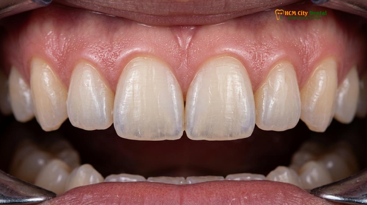
In clinical practice, zirconia is utilized in two primary forms: monolithic and layered. Monolithic zirconia is milled from a single, solid block of material. It offers maximum strength and eliminates the risk of porcelain chipping—a common failure point in older restorations. Layered zirconia, on the other hand, consists of a high-strength zirconia coping overlaid with highly aesthetic feldspathic porcelain. While layered restorations provide exceptional optical depth for anterior teeth, the monolithic approach has become the dominant standard of care due to rapid advancements in the translucency of the base material itself.
Types of Zirconia Used in Modern Dentistry (3Y, 4Y, 5Y)
Dental professionals classify zirconia based on its yttria content—3Y, 4Y, and 5Y—which dictates the precise balance between flexural strength and optical translucency for specific clinical applications.
The evolution of dental zirconia has been driven by the desire to combine the strength of metal with the beauty of glass. By manipulating the percentage of yttria in the composition, material scientists have created different generations of zirconia, each tailored for specific clinical indications. Understanding these classifications is crucial for achieving predictable restorative outcomes, as the optical properties and flexural strength vary significantly between generations[4].
3Y Zirconia (Tetragonal Zirconia Polycrystal)
Containing approximately 3 mol% yttria, 3Y zirconia is the original “workhorse” of metal-free dentistry. It consists almost entirely of the tetragonal phase, providing maximum transformation toughening. With a flexural strength ranging from 1000 to 1200 MPa, it is highly resistant to normal human bite forces. However, its dense crystalline structure scatters light, resulting in a highly opaque appearance. It is primarily indicated for posterior single crowns, multi-unit bridges, and masking severely discolored underlying tooth structures or titanium implant abutments.
4Y Zirconia (High Translucency)
By increasing the yttria content to 4 mol%, manufacturers introduce a small percentage of the cubic phase into the microstructure. The cubic phase is isotropic, meaning light passes through it more linearly, significantly improving translucency. 4Y zirconia offers a flexural strength of 800 to 1000 MPa, making it an incredibly versatile material. It provides an excellent balance, offering enough strength for posterior restorations while delivering sufficient aesthetics for premolars and many anterior applications.
5Y Zirconia (Ultra-Translucent)
Containing 5 mol% yttria, 5Y zirconia features a microstructure with up to 50% cubic phase. This dramatically enhances light transmission, allowing the material to mimic the natural opalescence and translucency of human enamel closely. The trade-off for this aesthetic brilliance is a reduction in flexural strength, which drops to approximately 600 to 800 MPa, and a loss of transformation toughening. 5Y zirconia is strictly indicated for highly demanding anterior aesthetic cases, such as Porcelain Veneers and single anterior crowns, where occlusal forces are lower.
| Zirconia Type | Yttria Content | Flexural Strength | Primary Clinical Indication | Aesthetic Profile |
|---|---|---|---|---|
| 3Y (TZP) | 3 mol% | 1000 – 1200+ MPa | Posterior crowns, long-span bridges, bruxism cases | High opacity, excellent masking ability |
| 4Y (HT) | 4 mol% | 800 – 1000 MPa | Universal use, premolars, short-span bridges | Balanced translucency and strength |
| 5Y (UT) | 5 mol% | 600 – 800 MPa | Anterior crowns, aesthetic veneers | Enamel-like translucency, lifelike optics |
“Selecting the appropriate yttria concentration is the most critical diagnostic decision when planning a restoration. We must evaluate the patient’s occlusal forces, the location of the tooth, and the aesthetic demands before prescribing the specific zirconia generation.”
— Clinical Material Science Review
Clinical Indications and Contraindications
While highly versatile, zirconia restorations are specifically indicated for high-stress posterior areas and bruxism cases, but require careful evaluation regarding interocclusal space and extreme anterior aesthetic demands.
The decision to utilize a zirconia crown is based on a comprehensive clinical assessment. Monolithic zirconia is the material of choice for patients exhibiting parafunctional habits, such as severe bruxism (teeth grinding) or clenching. Unlike layered porcelain, which is prone to cohesive fracture under extreme shear forces, a polished monolithic zirconia surface can withstand immense pressure without catastrophic failure. Clinical outcomes and survival rates of monolithic zirconia crowns in posterior dentition demonstrate exceptional long-term success[3]. Furthermore, highly polished zirconia is remarkably kind to opposing natural dentition, causing less enamel wear than traditional feldspathic porcelain or even natural enamel against enamel.
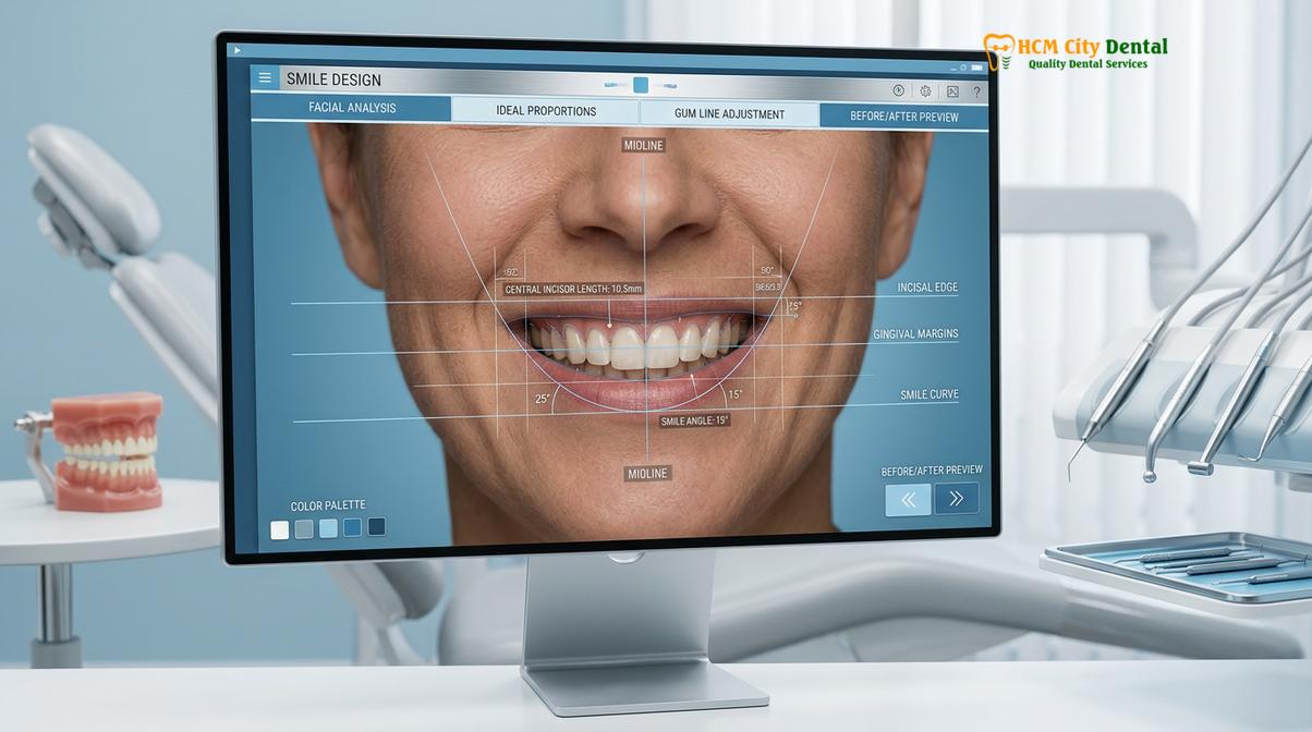
Zirconia is also highly indicated for masking dark substrates. When a patient presents with a severely discolored non-vital tooth, or when restoring over a dark titanium implant abutment, the inherent opacity of 3Y or 4Y zirconia effectively blocks the underlying darkness, preventing the grayish hue that often plagues other all-ceramic systems. For patients requiring a crown after root canal therapy, zirconia provides the necessary structural reinforcement to protect the brittle, endodontically treated tooth from vertical root fractures.
Despite its vast applications, there are specific contraindications. Zirconia requires a minimum material thickness to maintain its structural integrity (typically 0.8mm to 1.0mm axially and 1.0mm to 1.5mm occlusally). In cases where interocclusal space is severely limited and cannot be corrected, a cast gold restoration might be the only viable option. Additionally, while 5Y zirconia is highly aesthetic, restoring a single central incisor to perfectly match an adjacent natural tooth remains one of the most challenging tasks in dentistry. In cases of extreme aesthetic demand, a lithium disilicate (E.max) crown or a layered zirconia approach may be preferred over a purely monolithic restoration.
Clinical Warning: Saliva Contamination
During the try-in phase, salivary phospholipids rapidly bind to the zirconia intaglio surface, permanently occupying the chemical bonding sites. Rinsing with water or alcohol is insufficient to remove this contamination. Failure to decontaminate the crown with a specialized alkaline cleaning agent prior to cementation will result in a catastrophic loss of bond strength and premature crown debonding.
The Zirconia Crown Procedure: A Step-by-Step Clinical Workflow
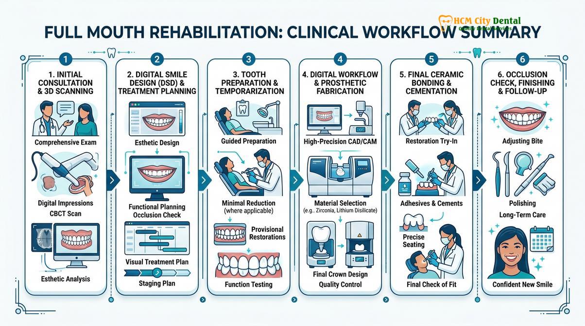
The clinical workflow involves conservative tooth preparation, precise intraoral digital scanning, CAD/CAM milling in the green state, and high-temperature sintering to achieve the final crystalline structure.
The fabrication and delivery of a zirconia crown represent a seamless integration of clinical precision and advanced digital engineering. The process begins with tooth preparation. Unlike traditional metal crowns that can tolerate feather-edge margins, zirconia requires a distinct, well-defined margin—typically a heavy chamfer or a rounded shoulder. The clinician must ensure that all internal line angles are smooth and rounded; sharp angles act as stress concentrators that can initiate micro-cracks within the ceramic material under occlusal loading.
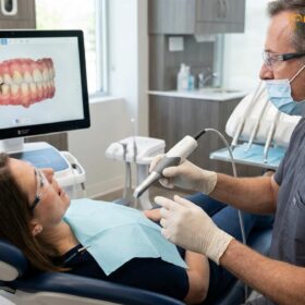
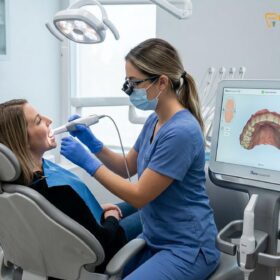
Once the tooth is optimally prepared and the gingival tissues are managed (often using retraction cord or soft tissue lasers), the clinician captures the impression. Modern practices utilize intraoral 3D scanners, which project structured light or utilize confocal microscopy to capture the exact geometry of the preparation, adjacent teeth, and the opposing arch. This digital file is instantly transmitted to the dental laboratory, eliminating the distortions, gag reflexes, and material shrinkage associated with traditional polyvinyl siloxane (PVS) impression materials.
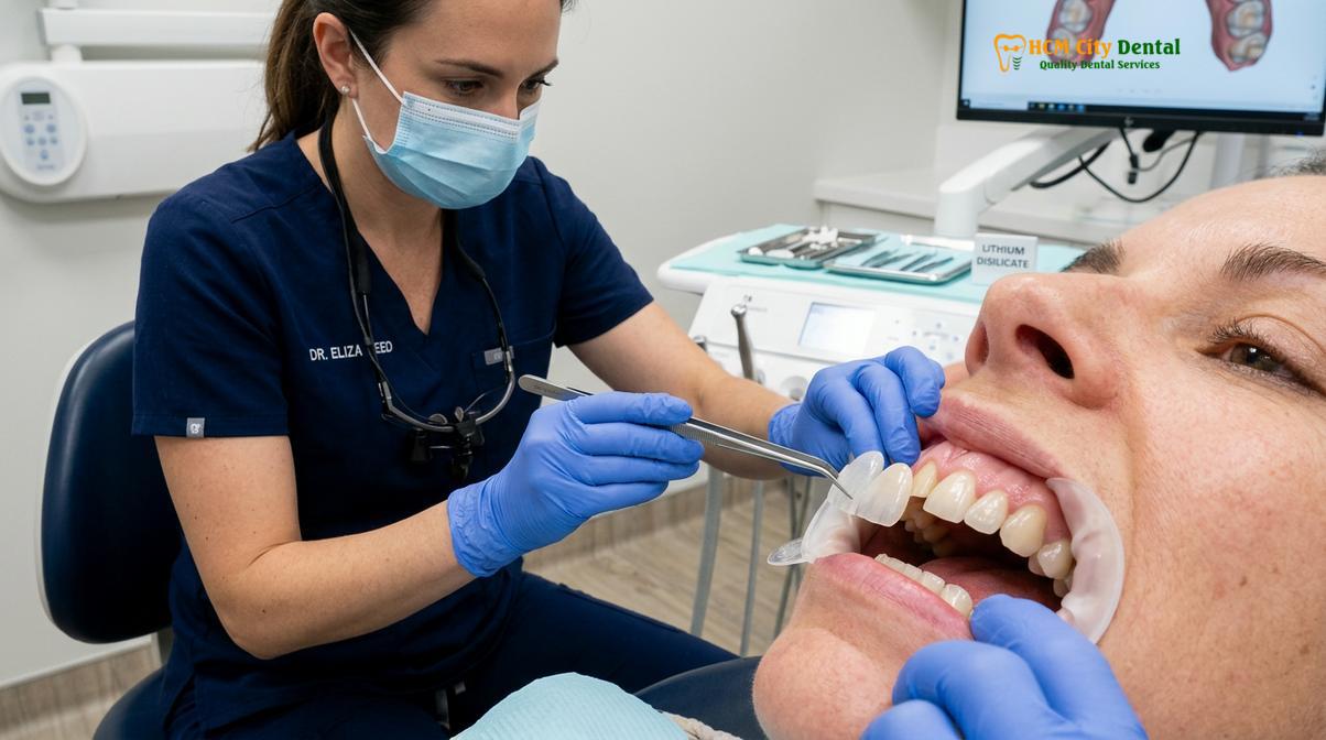
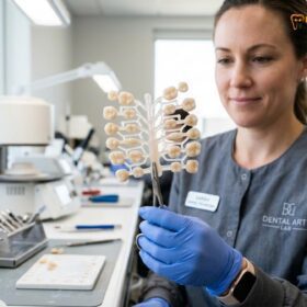
In the laboratory, a technician uses Computer-Aided Design (CAD) software to digitally sculpt the restoration, precisely defining the occlusal contacts, proximal contours, and cement space. The design is then sent to a Computer-Aided Manufacturing (CAM) milling machine. The crown is milled from a “green state” (pre-sintered) zirconia puck. In this chalk-like state, the material is soft and easily machined. Because zirconia shrinks by approximately 20% to 25% during the final firing process, the CAM software automatically mills the crown oversized to compensate.
The milled restoration is then placed into a high-temperature sintering furnace, where it is heated to temperatures between 1450°C and 1550°C for several hours. This sintering process densifies the material, eliminates porosity, and triggers the final crystalline phase, resulting in the restoration’s ultimate strength and precise final dimensions. After sintering, the crown is characterized with specialized stains and glazes to match the patient’s natural dentition perfectly.
Advanced Cementation Protocols for Zirconia
Long-term retention of zirconia relies on chemical bonding using 10-MDP monomers, requiring meticulous decontamination and surface activation of the intaglio surface prior to final cementation.
One of the most critical phases of zirconia restorative therapy is the cementation protocol. Unlike silica-based ceramics (such as feldspathic porcelain or lithium disilicate), zirconia lacks a glass matrix. Therefore, traditional acid etching with hydrofluoric acid and silanization is completely ineffective. Zirconia requires a specific chemical approach to achieve a durable bond to the underlying tooth structure, primarily utilizing 10-MDP based primers to maximize shear bond strength[2].
The gold standard for bonding a dental crown cap made of zirconia involves the use of 10-MDP (10-methacryloyloxydecyl dihydrogen phosphate). This unique monomer contains a hydrophilic phosphate group that chemically bonds to the hydroxyl groups on the zirconia surface, and a hydrophobic methacrylate group that copolymerizes with the resin cement. To facilitate this bond, the intaglio (internal) surface of the crown must first be sandblasted with aluminum oxide (Al₂O₃) particles at low pressure (typically 1-2 bar). This micro-roughening increases the surface area and surface energy, preparing it for chemical interaction.
“The success of a zirconia restoration is dictated in the final five minutes of the appointment. Strict isolation, meticulous decontamination of salivary proteins, and the precise application of MDP-based primers are non-negotiable steps for ensuring decades of clinical stability.”
— Advanced Prosthodontic Guidelines
After the crown is tried in the patient’s mouth to verify fit and aesthetics, it must be thoroughly decontaminated. As noted previously, saliva instantly neutralizes the bonding sites. The clinician applies a specialized zirconia cleaner, rinses it thoroughly, and air-dries the surface. An MDP-containing ceramic primer is then applied to the internal surface. Simultaneously, the prepared tooth is isolated, cleaned, and treated with a compatible bonding agent. A dual-cure resin cement is then extruded into the crown, which is seated with firm pressure. Excess cement is carefully removed, and the margins are light-cured to finalize the polymerization process.
Zirconia vs. Porcelain Fused to Metal (PFM) and Lithium Disilicate
Compared to traditional PFM and lithium disilicate, monolithic zirconia offers superior fracture resistance, eliminates the risk of metal oxidation at the gumline, and provides exceptional biocompatibility.
For decades, traditional porcelain-fused-to-metal bridges and crowns were the standard of care. However, PFM restorations possess inherent flaws. The metallic substructure blocks light transmission, resulting in an opaque, unnatural appearance. More problematically, as gingival tissues naturally recede over time, the dark metal collar of the PFM crown becomes visible, creating an unsightly gray line at the gum margin. Furthermore, the bond between the porcelain and the metal framework is susceptible to cohesive failure, leading to porcelain chipping.
Zirconia eliminates these issues entirely. Being 100% metal-free, there is no dark substructure to hide and no risk of a gray margin appearing over time. The monolithic nature of modern zirconia also eradicates the risk of porcelain delamination. When compared to lithium disilicate (E.max), another popular all-ceramic material, zirconia offers significantly higher flexural strength. While lithium disilicate is often preferred for highly aesthetic single anterior crowns due to its superior glass-like translucency, zirconia remains the undisputed champion for posterior restorations, long-span bridges, and patients with heavy bruxism.
Clinical Case Study: Full-Mouth Rehabilitation in Ho Chi Minh City
A comprehensive restorative case demonstrates the transformative functional and aesthetic benefits of utilizing monolithic zirconia for full-arch rehabilitation.
A recent patient visited HCMC Dental Clinic in Ho Chi Minh City presenting with severe generalized attrition, loss of vertical dimension, and multiple failing amalgam restorations. The clinical objective was to restore proper occlusal function while delivering a highly aesthetic, natural-looking smile. After a thorough digital smile design and occlusal analysis, a full-mouth rehabilitation utilizing monolithic 4Y and 5Y zirconia crowns was prescribed.
Dr. Nguyen Van Cuong, a leading restorative specialist at HCMC Dental Clinic, emphasizes a conservative approach when designing these restorations. He routinely utilizes advanced digital scanning to ensure optimal marginal fit, which is crucial for long-lasting dental aesthetics. Under his guidance, patients receive highly customized treatment plans that prioritize both structural integrity and natural beauty.
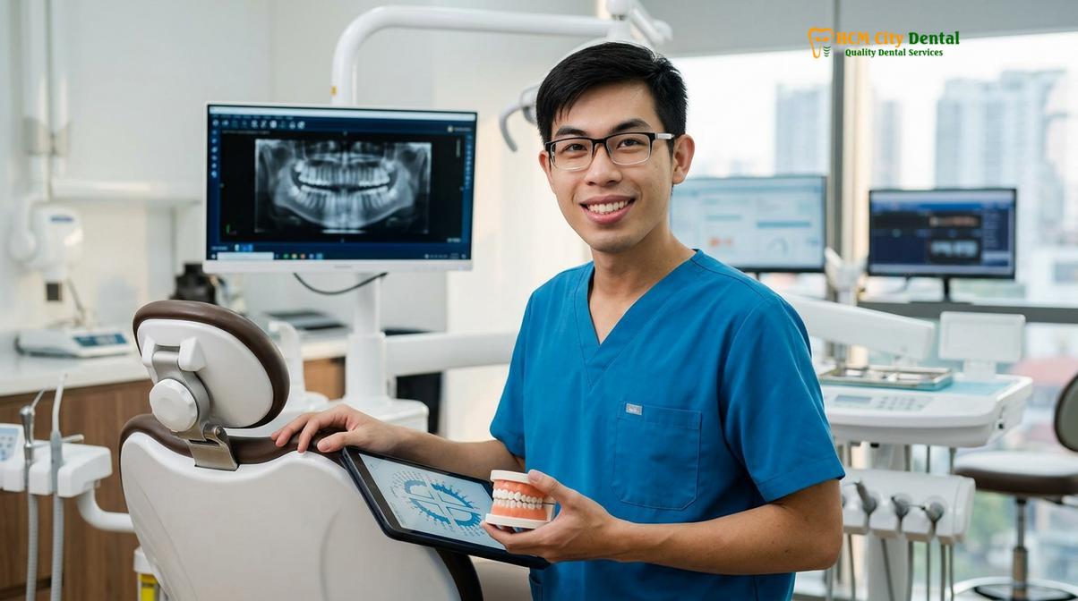
The posterior segments were restored with 4Y zirconia to withstand heavy masticatory forces, while the anterior teeth were restored with ultra-translucent 5Y zirconia to mimic natural enamel opalescence. The restorations were milled using state-of-the-art CAD/CAM technology and cemented using a strict MDP-based adhesive protocol. The final result provided the patient with a harmonious, functional, and highly durable smile, highlighting the exceptional capabilities of modern zirconia materials when executed with precision.
When to Consult a Dentist for Your Restoration
Timely professional evaluation is critical if you experience persistent sensitivity, notice structural damage to an existing crown, or require a comprehensive assessment for new restorations.
While zirconia is incredibly resilient, the underlying tooth structure and surrounding gingival tissues require ongoing care. Patients should schedule a clinical evaluation if they experience localized pain upon biting, which may indicate an occlusal discrepancy or underlying endodontic complication. Additionally, any signs of gingival inflammation or bleeding around the crown margin warrant immediate attention, as this can lead to periodontal attachment loss or secondary caries beneath the restoration. Following the guidelines on the clinical application of monolithic zirconia restorations ensures that potential issues are identified and managed proactively[5].
Routine dental prophylaxis and radiographic examinations are essential for monitoring the long-term health of the restoration. For patients considering zirconia crowns in Vietnam, a comprehensive consultation will determine the most appropriate material generation (3Y, 4Y, or 5Y) based on individual functional requirements and aesthetic goals.
References
- Journal of Prosthetic Dentistry. Phase transformation toughening and mechanical properties of yttria-stabilized zirconia.
- Dental Materials. The effect of 10-MDP based primers on the shear bond strength of zirconia restorations.
- Journal of the American Dental Association. Clinical outcomes and survival rates of monolithic zirconia crowns in posterior dentition.
- International Journal of Prosthodontics. Optical properties and flexural strength of 3Y, 4Y, and 5Y highly translucent zirconia.
- Vietnam Odonto-Stomatology Association (VOSA). Guidelines on the clinical application of monolithic zirconia restorations.
Schedule Your Consultation Today
If you are experiencing dental discomfort or wish to explore the aesthetic and functional benefits of advanced metal-free restorations, expert care is available. Contact HCMC Dental Clinic in Ho Chi Minh City to schedule a comprehensive evaluation and discover how precision-milled zirconia can restore your smile’s natural strength and beauty.