Neuromuscular dentistry full mouth rehabilitation is an advanced clinical approach that aims to restore the entire dentition while aligning the temporomandibular joint (TMJ) and masticatory muscles. By establishing a physiologic rest jaw position, this comprehensive treatment seeks to resolve chronic pain, correct bite collapse, and rebuild optimal chewing function and aesthetics.
Clinical Summary:
Full mouth rehabilitation guided by neuromuscular principles goes beyond traditional tooth restoration by prioritizing the biomechanical harmony of muscles, joints, and teeth. Utilizing diagnostic splints, ultra-low frequency TENS therapy, and advanced digital workflows, clinicians can accurately determine the optimal vertical dimension of occlusion. This comprehensive strategy is designed to treat TMJ disorders, alleviate chronic facial pain, and promote the long-term structural stability of permanent restorations, such as monolithic zirconia crowns or implant-supported prostheses, aiming to provide patients with a functional and comfortable stomatognathic system.
Key Takeaways:
- Neuromuscular dentistry integrates TMJ alignment, muscle relaxation, and dental occlusion to predictably manage complex bite collapse.
- TENS therapy is utilized to help achieve a true physiologic rest jaw position before any irreversible permanent restorations are placed.
- Diagnostic splints temporarily stabilize the bite, allowing patients to test the new vertical dimension of occlusion for comfort and function.
- Advanced CAD/CAM precision and intraoral 3D scans assist in ensuring that final restorations fit with micrometer accuracy.
- Comprehensive rehabilitation often incorporates IV conscious sedation and PRF to support patient comfort and aid in tissue healing.
The Relationship Between Masticatory Muscles, TMJ, and Dental Occlusion
The stomatognathic system relies on a delicate balance between the temporomandibular joint, masticatory muscles, and dental occlusion, where any disruption can contribute to biomechanical instability and chronic pain.
To fully comprehend the scope of neuromuscular dentistry, one must first understand the intricate triad that governs oral function: the teeth (occlusion), the temporomandibular joints (TMJ), and the masticatory musculature. In a healthy stomatognathic system, these three components operate in synchrony. When the jaw closes, the teeth should meet evenly without forcing the TMJ out of its socket or requiring the facial muscles to strain to accommodate the bite. However, when teeth are lost, severely worn down, or improperly restored, this delicate balance is often compromised[1].
This disruption frequently manifests as bite collapse, a condition where the loss of posterior tooth support causes the lower jaw to over-close. As the vertical dimension of occlusion decreases, the mandible may be forced backward and upward into the glenoid fossa. This unnatural trajectory can compress the highly innervated retrodiscal tissues of the TMJ, potentially leading to inflammation, clicking, and popping sounds during function. Furthermore, the masticatory muscles—particularly the masseter, temporalis, and lateral pterygoid—are often forced into a state of chronic hyperactivity as they constantly struggle to find a stable resting position.
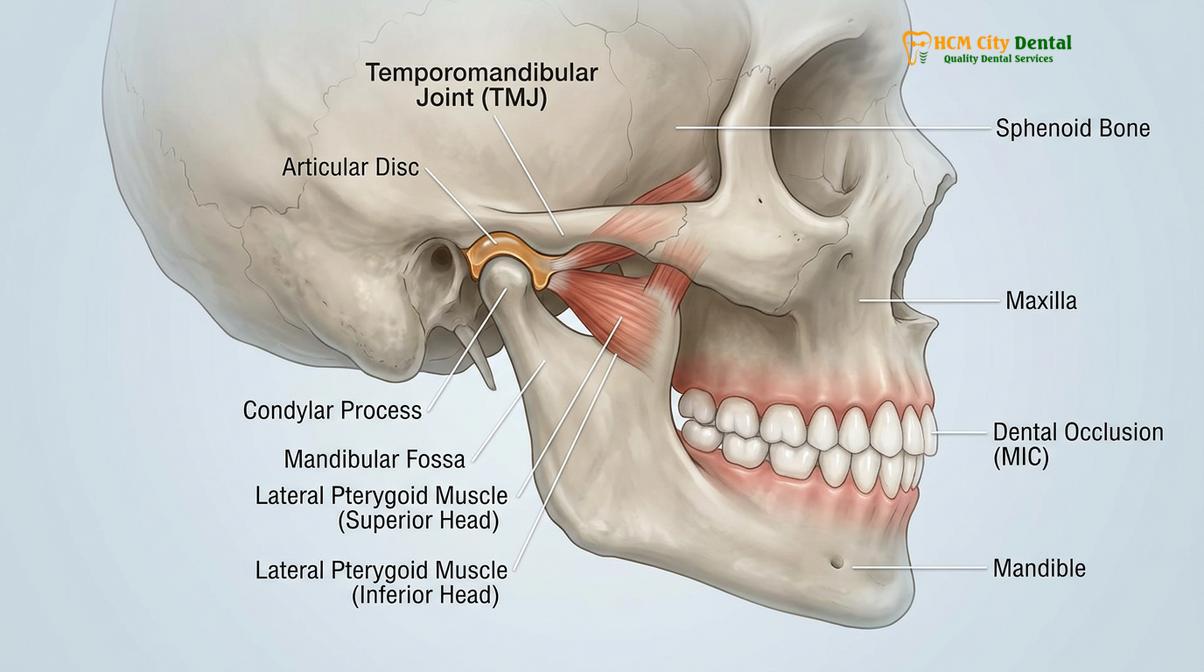
Addressing this complex dysfunction requires more than simply placing new crowns or fillings. A comprehensive approach must first aim to deprogram the strained musculature and decompress the joint space. By focusing on bite collapse restoration through neuromuscular principles, clinicians can rebuild the dentition to support the jaw in a position that is dictated by relaxed muscles rather than worn or misaligned teeth. This foundational step is critical; placing new restorations on a pathological bite trajectory can increase the risk of dental work failure and the continuation of the patient’s discomfort.
TENS Therapy (Transcutaneous Electrical Nerve Stimulation) in Dental Triage
TENS therapy utilizes ultra-low frequency electrical impulses to involuntarily contract and relax facial muscles, helping to erase pathological muscle memory and facilitate accurate bite registration.
One of the primary diagnostic and therapeutic tools in neuromuscular dentistry is the use of Ultra-Low Frequency Transcutaneous Electrical Nerve Stimulation (ULF-TENS). Unlike standard medical TENS units designed primarily for pain masking via sensory nerve stimulation, the ultra-low frequency units used in dentistry specifically target the motor nerves that control the muscles of mastication and the cervical region[2].
When a patient suffers from a misaligned bite, their muscles can develop a “pathological engram”—a learned, defensive muscle memory that guides the jaw into a compromised position to avoid painful tooth interferences. This chronic muscle tension may lead to a buildup of lactic acid and a depletion of ATP, resulting in fatigue and spasm. TENS therapy in dentistry aims to break this cycle by delivering mild, rhythmic electrical impulses that cause the muscles to involuntarily contract and relax.
“The application of ultra-low frequency TENS is a cornerstone of neuromuscular diagnostics, allowing the clinician to bypass the patient’s defensive muscle reflexes and capture a bite registration based on physiological relaxation rather than habitual accommodation.”
Over a standard clinical session, this rhythmic pulsing acts as a muscular massage. It helps flush out accumulated metabolic waste products, supports normal blood flow, and effectively erases the pathological muscle memory. Once the muscles are relaxed and deprogrammed, the mandible naturally drops into a more comfortable, unstrained posture. This state of jaw muscle relaxation is a vital prerequisite for accurately diagnosing the true extent of the occlusal discrepancy and planning the subsequent rehabilitation.
Finding the Physiologic Rest Position of the Mandible
Identifying the physiologic rest jaw position is critical for establishing a new vertical dimension of occlusion that supports both muscular relaxation and joint health.
The cornerstone of neuromuscular dentistry is the identification and capture of the physiologic rest jaw position. Traditional dentistry often relies on “centric relation,” a mechanically determined position where the condyles are seated in the uppermost, anterior position within the glenoid fossa. While useful for many restorative procedures, centric relation does not always account for the dynamic state of the surrounding musculature.
In contrast, the physiologic rest position is a muscle-determined trajectory. It is the three-dimensional spatial relationship of the mandible to the maxilla where the masticatory and cervical muscles are at their lowest state of electrical activity. Finding this position is essential for determining the correct vertical dimension of occlusion (VDO)—the optimal height of the bite that allows the muscles to rest comfortably when the teeth are slightly apart, and to contract efficiently during chewing and swallowing.
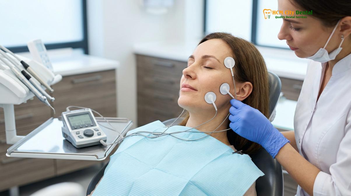
To precisely locate this position, neuromuscular dentists employ advanced computerized diagnostics. Surface electromyography (sEMG) may be used to measure the electrical activity of the muscles before, during, and after TENS therapy, providing objective data on muscle relaxation. Concurrently, Computerized Mandibular Scanning (CMS) tracks the three-dimensional movements of the jaw. By analyzing this data, the clinician can pinpoint the trajectory where the muscles are most relaxed and the joint is decompressed. This precise coordinate becomes the blueprint for the entire rehabilitation process.
How Misaligned Restorations Cause Chronic Headaches and Neck Pain
Improperly contoured dental restorations can force the mandible into a strained trajectory, potentially triggering chronic muscle spasms, tension headaches, and cervical neck pain.
The impact of dental occlusion extends far beyond the oral cavity. The teeth, jaw joints, and muscles are intimately connected to the central nervous system via the trigeminal nerve, which is responsible for the majority of sensory input from the face and motor control of the chewing muscles. When dental restorations—such as crowns, bridges, or fillings—are placed without regard for the neuromuscular trajectory, they can introduce premature contacts or occlusal interferences.
Even a minor discrepancy can trigger a cascade of neuromuscular dysfunction. The proprioceptors in the periodontal ligaments detect the uneven bite and signal the brain to alter the jaw’s closing path to avoid the high spot. This constant, subconscious muscular accommodation often leads to severe muscle fatigue. The overworked masseter and temporalis muscles frequently refer pain to the temples, forehead, and behind the eyes, which can manifest as severe tension headaches.
Furthermore, the stomatognathic system is biomechanically linked to the cervical spine. To maintain an open airway and a level line of sight when the jaw is misaligned, the body often compensates by altering head posture, typically resulting in a forward head posture. This places immense strain on the cervical and shoulder muscles, potentially leading to chronic neck pain and stiffness. Achieving TMJ headache relief therefore requires a holistic approach that corrects the dental occlusion to support proper head and neck posture.
Constructing Orthotic Splints for Bite Stabilization
A diagnostic splint acts as a reversible therapeutic device, stabilizing the mandible in its newly determined neuromuscular trajectory before committing to irreversible dental preparations.
Once the physiologic rest position has been identified, the next critical step is to test and verify this new bite position clinically. This is achieved through the fabrication of a neuromuscular orthotic, commonly referred to as a diagnostic splint. Unlike a standard night guard, which is typically flat and designed merely to protect teeth from bruxism, a neuromuscular orthotic is an anatomically contoured appliance worn over the lower teeth.
The orthotic is precisely milled or 3D printed to feature specific indentations that guide the upper teeth into the newly established vertical dimension of occlusion. By wearing this appliance, the patient’s mandible is supported in the optimal, relaxed trajectory. This phase, often lasting three to six months, serves multiple vital purposes.
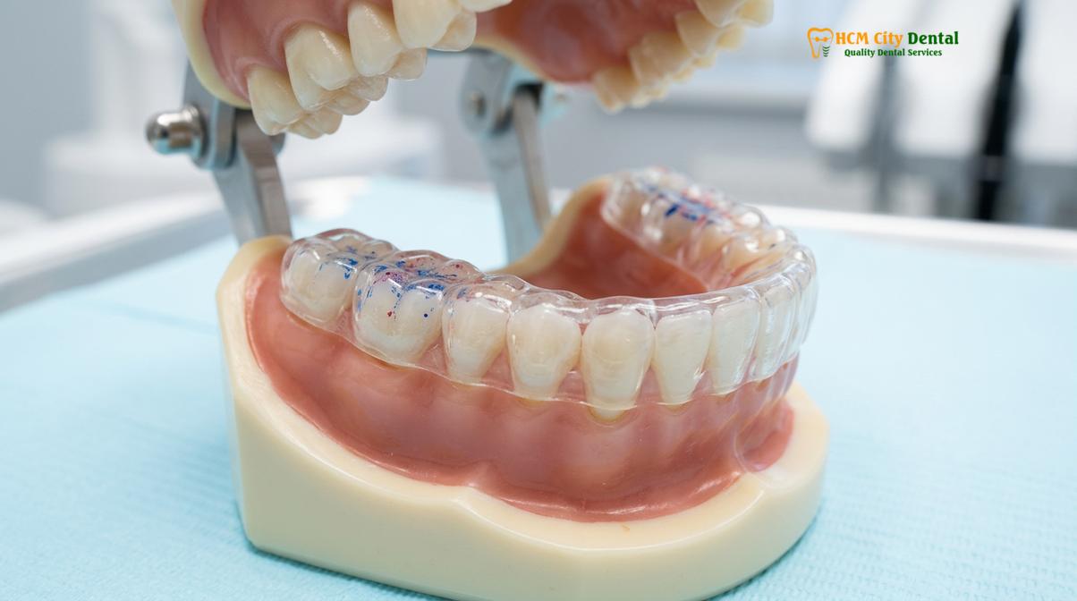
First, it acts as a reversible trial period. It allows the clinician to confirm that the new bite position effectively helps resolve the patient’s TMJ symptoms, muscle pain, and headaches. Second, it provides the patient with an opportunity to adapt to the new vertical dimension phonetically and functionally. If adjustments are needed, the acrylic orthotic can be easily modified. Only after the patient is asymptomatic and comfortable with the diagnostic splint does the clinician proceed to the irreversible phase of full mouth rehabilitation.
Comprehensive Full Mouth Rehabilitation Workflows
Modern full mouth rehabilitation combines neuromuscular diagnostics with digital smile design and biocompatible materials to predictably reconstruct the entire dentition.
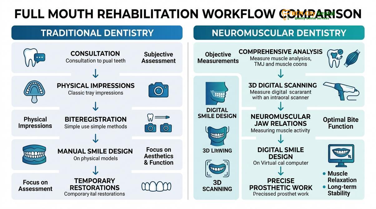
Transitioning a patient from a stabilized diagnostic splint to a permanent, functional dentition requires a meticulously planned clinical workflow. This phase integrates advanced material science, digital dentistry, and surgical precision to rebuild the mouth.
Digital Diagnostics and Treatment Planning
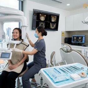
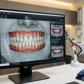
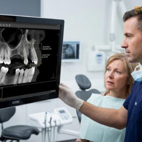
The process begins with comprehensive data acquisition. An intraoral 3D scan captures the exact topography of the teeth and the stabilized bite provided by the orthotic. This digital impression is combined with Cone Beam Computed Tomography (CBCT) to evaluate bone volume and joint anatomy. Utilizing Digital Smile Design (DSD) software, the clinical team creates a 3D digital mock-up of the proposed final restorations, allowing the dentist to evaluate phonetic compatibility and aesthetic proportions before a single tooth is prepared[5].
Surgical Phase and Patient Comfort
For patients requiring extractions, bone grafting, or dental implants as part of their rehabilitation, the surgical phase is executed with a focus on patient comfort and biological healing. Complex procedures are routinely performed under IV conscious sedation, administered by a qualified professional, to help ensure a safe and anxiety-free experience.
To optimize healing, clinicians frequently utilize Platelet-Rich Fibrin (PRF). By drawing a small amount of the patient’s blood and centrifuging it, a matrix rich in autologous growth factors is created. When applied to extraction sites or bone grafts, PRF can significantly aid in soft tissue healing, reduce post-operative swelling, and support the integration of dental implants[4].
Clinical Case Review: A 55-year-old expatriate presented to HCMC Dental Clinic in Ho Chi Minh City with severe bite collapse, chronic TMJ pain, and failing dentition. Following a thorough evaluation, the patient underwent neuromuscular deprogramming followed by a full arch implant-supported rehabilitation. Utilizing IV sedation and PRF protocols, the surgical phase was completed smoothly. The final restoration, utilizing CAD/CAM precision milled monolithic zirconia, restored the patient’s vertical dimension, significantly alleviating their chronic headaches while delivering an excellent aesthetic outcome.
Provisionalization and Final Restorations
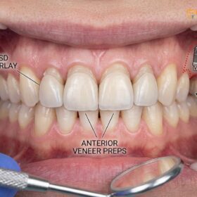
Following any necessary surgical healing, the teeth are prepared, and provisional (temporary) restorations are placed. These provisionals are replicas of the digital design and serve as a final test drive for aesthetics and function. Once approved, the final restorations are fabricated, taking into account the biomechanical load distribution required for long-term success, especially in complex implant cases like All-on-4 restorations[3].
The material of choice for modern neuromuscular rehabilitation is often biocompatible monolithic zirconia. This metal-free material offers high fracture toughness and aesthetic translucency. Utilizing CAD/CAM precision milling, the final crowns or implant bridges achieve an exceptional margin fit, supporting optimal gingival health and long-term biomechanical stability.
“The transition from a diagnostic orthotic to permanent monolithic zirconia restorations requires absolute precision. Digital workflows help ensure that the exact neuromuscular trajectory, proven to relieve pain and restore function, is accurately replicated in the final ceramics.”
Comparing Dental Approaches
Understanding the differences in treatment philosophy can help patients make informed decisions regarding their restorative care.
| Feature | Traditional Dentistry | Neuromuscular Dentistry |
|---|---|---|
| Primary Focus | Restoring individual teeth and mechanical bite relationships. | Harmonizing teeth, TMJ, and masticatory muscles. |
| Bite Registration | Often relies on habitual bite or mechanical centric relation. | Utilizes TENS therapy to find the physiologic rest position. |
| Diagnostic Tools | Standard X-rays, physical models, visual inspection. | sEMG, computerized mandibular scanning, 3D CBCT. |
| Treatment Phasing | Direct progression to permanent restorations. | Mandatory stabilization phase using a diagnostic splint. |
When to Consult a Neuromuscular Specialist
Recognizing the early signs of occlusal dysfunction and TMJ disorders is crucial for preventing severe bite collapse and chronic facial pain.
Patients experiencing unexplained chronic headaches, jaw clicking, limited mouth opening, or excessive tooth wear should seek a comprehensive evaluation from a dental professional trained in neuromuscular principles. Early intervention can often prevent the need for extensive full mouth reconstruction. According to guidelines supported by the Vietnam Odonto-Stomatology Association (VOSA), a multidisciplinary approach to temporomandibular joint disorders and prosthodontic rehabilitation is highly recommended to achieve optimal patient outcomes and prevent further deterioration of the stomatognathic system[6].
Important Note: Ignoring chronic jaw pain, frequent tension headaches, or noticeable changes in how your teeth fit together can lead to progressive joint damage and severe tooth wear. If you experience these symptoms, it is highly advisable to consult a qualified dental professional for a comprehensive neuromuscular evaluation before pursuing any irreversible cosmetic or restorative dental procedures.
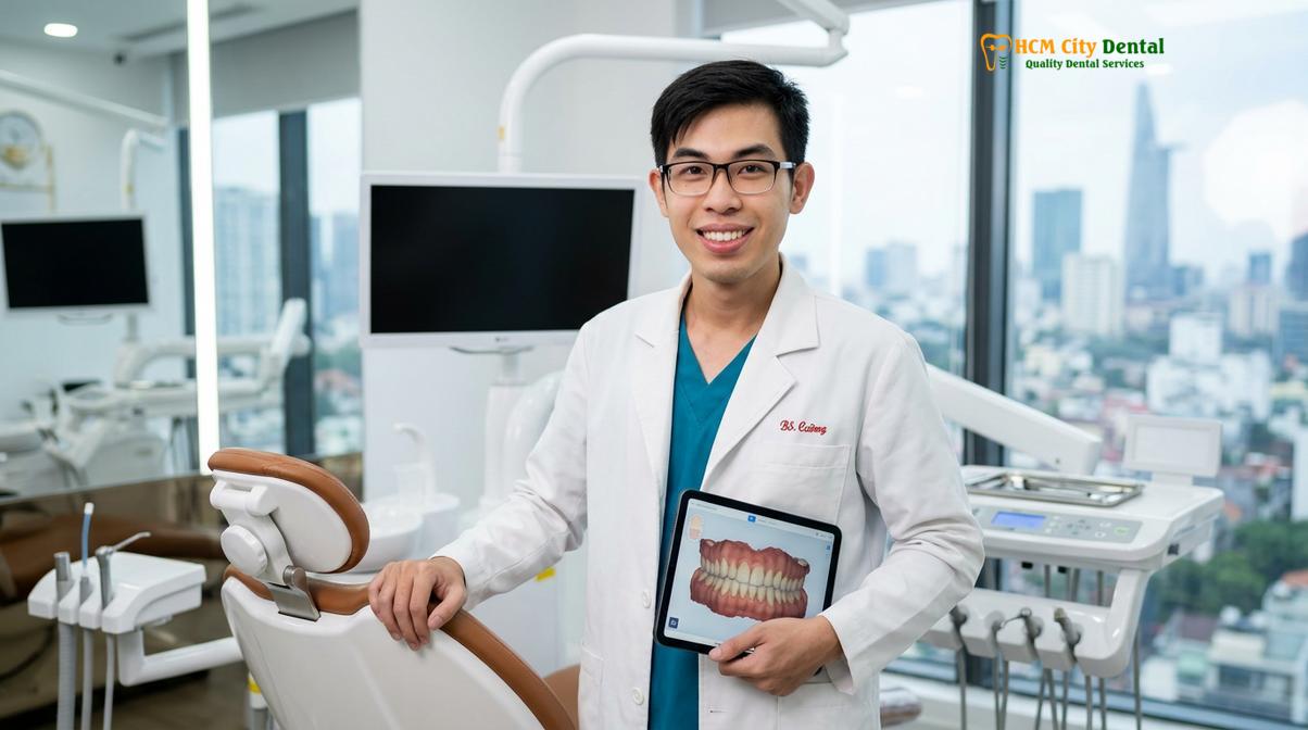
Dr. Nguyen Van Cuong, a leading specialist, emphasizes that successful full mouth rehabilitation requires a deep understanding of both biomechanics and facial harmony. With extensive clinical experience in neuromuscular dentistry, Dr. Cuong meticulously evaluates each patient’s unique jaw trajectory to help ensure that final restorations provide lasting comfort, functional efficiency, and structural stability.
Conclusion and Next Steps
Neuromuscular dentistry offers a scientifically grounded pathway to restoring not just the teeth, but the entire functional harmony of the jaw, muscles, and joints.
By prioritizing the physiologic rest position of the mandible, clinicians can predictably manage complex cases of bite collapse and chronic facial pain. Through the careful integration of TENS therapy, diagnostic splinting, and advanced digital restorative workflows, patients can achieve a stable, comfortable, and aesthetically pleasing smile. If you are experiencing chronic jaw pain or considering comprehensive restorative care, contact HCMC Dental Clinic in Ho Chi Minh City to explore how full mouth rehabilitation can help restore your oral health and improve your quality of life.
Frequently Asked Questions
What is neuromuscular dentistry?
Neuromuscular dentistry is an advanced dental approach that evaluates and treats the complex relationship between teeth, temporomandibular joints (TMJ), and masticatory muscles. By establishing a harmonious physiologic rest position for the jaw, this methodology aims to alleviate chronic pain and support the long-term stability of comprehensive dental restorations.
Can misaligned teeth contribute to headaches and neck pain?
Yes, misaligned teeth can force the jaw muscles into a constant state of tension, which may lead to chronic headaches and cervical neck pain. This muscular hyperactivity, often triggered by an improper bite trajectory, can refer pain through the trigeminal nerve pathways to surrounding facial and cervical structures.
How long do I wear a physiologic bite splint?
A physiologic bite splint is typically worn for three to six months during the diagnostic and stabilization phase. This duration allows the masticatory muscles to deprogram and relax, helping the clinician confirm that the newly established vertical dimension of occlusion is comfortable before transitioning to permanent dental restorations.
How does a diagnostic splint differ from a standard night guard?
A diagnostic splint is actively designed to reposition the mandible into an optimal neuromuscular trajectory, whereas a standard night guard is generally intended to protect teeth from grinding friction. The neuromuscular orthotic alters the bite to help relieve joint compression and muscle fatigue, serving as a blueprint for full mouth reconstruction.
Is full mouth rehabilitation painful?
Full mouth rehabilitation is generally well-tolerated, as modern protocols utilize advanced anesthesia and IV conscious sedation to help maintain patient comfort. Post-operative discomfort is typically managed effectively with prescribed analgesics, and the use of techniques like PRF can aid in tissue healing and reduce inflammation.
References
- The Journal of Prosthetic Dentistry. Vertical dimension of occlusion restoration parameters. (2021).
- Journal of Craniomandibular & Sleep Practice. TENS therapy in neuromuscular dentistry. (2020).
- International Journal of Prosthodontics. Biomechanical load distribution in All-on-4 restorations. (2019).
- Clinical Oral Implants Research. Platelet-rich fibrin in guided bone regeneration. (2022).
- Journal of Esthetic and Restorative Dentistry. Digital smile design functional protocols. (2018).
- Vietnam Odonto-Stomatology Association (VOSA). Guidelines on Temporomandibular Joint Disorders and Prosthodontic Rehabilitation. (2023).
For customized treatment planning, transparent cost estimates, and direct consultation with Dr. Cuong, visit our comprehensive Full Mouth Rehabilitation service page or message our team directly via WhatsApp to receive an instant assessment.