Rehab for congenital missing teeth involves a highly specialized, multidisciplinary clinical approach designed to restore both structural function and facial aesthetics. By integrating advanced orthodontics, precision bone grafting, and biocompatible dental implants, clinicians can correct bite collapse and permanently replace missing dentition for patients suffering from developmental dental anomalies.
Clinical Summary:
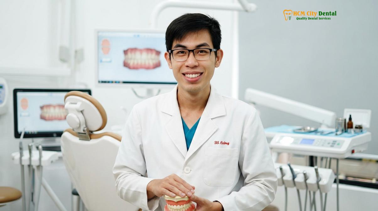
The congenital absence of teeth, ranging from localized hypodontia to severe ectodermal dysplasia, necessitates comprehensive Full Mouth Rehabilitation. Dr. Nguyen Van Cuong emphasizes that successful outcomes rely on meticulous phased interventions, beginning with early diagnosis and transitional prosthetics. The definitive reconstructive protocol integrates Digital Smile Design (DSD) for aesthetic planning, 3D CBCT diagnostics for precise anatomical mapping, and biocompatible monolithic zirconia for the final restorations. For complex cases presenting with severe bone resorption, advanced surgical techniques such as guided bone regeneration utilizing Platelet-Rich Fibrin (PRF) and the administration of IV conscious sedation ensure predictable, anxiety-free outcomes while successfully restoring the physiologic rest jaw position and optimal chewing function.
Key Takeaways:
- Congenital hypodontia and ectodermal dysplasia require a sequenced, multidisciplinary treatment approach spanning several years.
- Orthodontic space optimization and root paralleling are critical prerequisites before any surgical implant placement.
- Restoring the Vertical Dimension of Occlusion (VDO) is essential to prevent neuromuscular fatigue and temporomandibular joint (TMJ) disorders.
- Premium monolithic zirconia restorations provide superior biocompatibility, exceptional fracture toughness, and a margin fit under 50 micrometers.
- IV conscious sedation protocols ensure a highly safe, anxiety-free, and comfortable surgical experience for complex rehabilitations.
What is Ectodermal Dysplasia and Congenital Hypodontia?
Congenital hypodontia is the developmental absence of one to five permanent teeth, while ectodermal dysplasia is a genetic disorder causing multiple missing teeth and abnormal tissue development. Both conditions require specialized prosthetic and surgical interventions to restore oral function.
The development of the human dentition is a highly complex biological process regulated by a precise sequence of genetic expressions and epithelial-mesenchymal interactions. When this delicate process is disrupted, it can result in tooth agenesis, the most common developmental anomaly in humans. Clinically, this condition is categorized based on severity: hypodontia refers to the absence of one to five permanent teeth (excluding third molars), oligodontia denotes the absence of six or more teeth, and anodontia is the extremely rare complete absence of all primary and permanent teeth.
The etiology of congenital missing teeth is primarily rooted in genetic mutations. Specific gene defects, particularly involving the MSX1, PAX9, and AXIN2 genes, have been strongly correlated with non-syndromic tooth agenesis. These genetic anomalies disrupt the formation of the dental lamina during early embryogenesis, preventing the tooth buds from developing. In many instances, this genetic hypodontia dysplasia is inherited in an autosomal dominant pattern, meaning a thorough family dental history is a crucial component of the initial diagnostic workup.
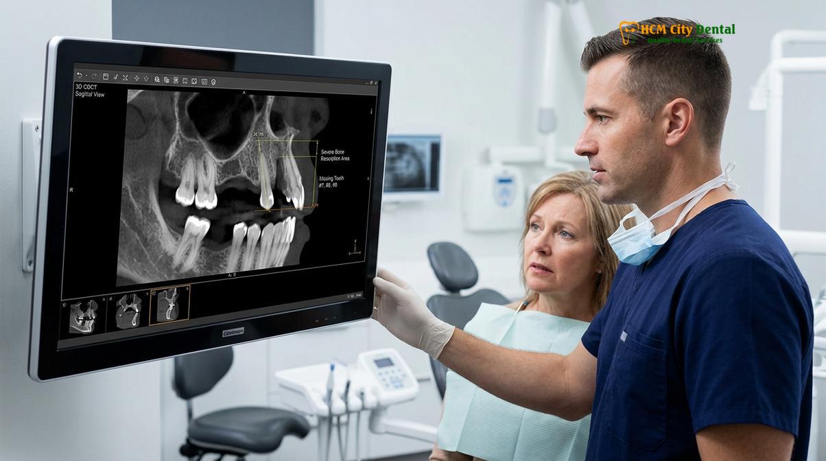
Beyond isolated missing teeth, tooth agenesis is frequently a hallmark symptom of broader systemic conditions, the most notable being ectodermal dysplasia. This is a heterogeneous group of inherited disorders characterized by abnormalities in structures derived from the embryonic ectoderm. Patients presenting with ectodermal dysplasia typically exhibit not only severe oligodontia or anodontia but also sparse hair (hypotrichosis), abnormal nail development, and a reduced ability to sweat (hypohidrosis) due to malformed eccrine glands. The dental manifestations are particularly severe; the few teeth that do erupt are often malformed, presenting as conical shape teeth, and the alveolar bone fails to develop normally due to the lack of intraosseous tooth buds.
The clinical implications of these conditions extend far beyond aesthetics. The absence of multiple teeth leads to a cascade of functional and structural complications. Without a complete dental arch, patients suffer from a compromised ability to masticate food properly, which can impact nutritional intake and overall systemic health. Furthermore, the lack of occlusal support often leads to a gradual bite collapse, altering the Vertical Dimension of Occlusion (VDO) and placing abnormal stress on the temporomandibular joints and associated musculature. Addressing these profound deficits requires a comprehensive understanding of craniofacial growth and the application of advanced prosthodontic principles [1].
Pediatric and Young Adult Case Coordination: Timing the Reconstruction
Timing is critical in pediatric jaw development; definitive implant placement must wait until skeletal growth is complete to prevent infraocclusion, while interim prostheses maintain space and function.
Managing congenital missing teeth is uniquely challenging because the condition is typically diagnosed in childhood, yet the definitive surgical solutions—namely, dental implants—cannot be implemented until the patient reaches early adulthood. This necessitates a carefully coordinated, long-term treatment strategy that adapts to the patient’s ongoing pediatric jaw development. The primary goal during the developmental years is to preserve alveolar bone volume, maintain adequate spacing for future restorations, and provide the child with functional and aesthetically acceptable interim prostheses.
The fundamental contraindication for placing dental implants in a growing child or adolescent is the phenomenon of ankylosis. Unlike natural teeth, which are suspended in the socket by the periodontal ligament and can erupt in tandem with vertical jaw growth, a dental implant fuses directly to the bone (osseointegration). If an implant is placed before skeletal maturity, it will remain stationary while the surrounding natural teeth and alveolar bone continue to grow vertically and horizontally. This results in the implant appearing submerged or “infraoccluded,” leading to severe aesthetic discrepancies, periodontal defects, and complex restorative failures that are exceedingly difficult to correct.
To navigate this waiting period, clinicians employ a variety of transitional restorative techniques. In cases of mild hypodontia, such as missing maxillary lateral incisors, orthodontic treatment is often utilized to either close the space (substituting the canine for the lateral incisor) or open and maintain the space for a future implant. If the space is maintained, a removable Essix retainer with a pontic (a fake tooth) or a minimally invasive resin-bonded Maryland bridge can be bonded to the adjacent teeth to provide a temporary aesthetic solution without interfering with jaw growth.
Clinical Warning: Premature placement of dental implants in adolescents before the cessation of craniofacial growth can lead to irreversible aesthetic and functional complications, including severe infraocclusion and adjacent root damage. Skeletal maturity must be confirmed via serial cephalometric radiographs before surgical intervention.
For patients with severe oligodontia or ectodermal dysplasia, the transitional phase is more complex. These children often require pediatric removable partial dentures or even complete overdentures to facilitate normal speech development, chewing function, and social integration. These prostheses must be frequently adjusted and remade as the child grows to accommodate the expanding dental arches. Dr. Nguyen Van Cuong advocates for a highly empathetic approach during this phase, recognizing that the psychological burden of missing teeth during adolescence is significant. Continuous monitoring through serial cephalometric radiographs is employed to track skeletal maturation, ensuring that the transition to definitive implant therapy occurs at the precise optimal moment, typically between the ages of 18 and 21 [2].
Aesthetic Challenges: Conical-Shaped Teeth and Reduced Jawbone Volume
Patients often present with malformed, conical shape teeth and severe bone volume deficiency due to the lack of alveolar bone stimulation from missing tooth roots.
The anatomical landscape of a mouth affected by congenital tooth agenesis presents significant hurdles for the restorative dentist. The challenges are twofold: the morphological abnormalities of the teeth that are present, and the severe volumetric deficiencies of the supporting alveolar bone. Addressing these issues requires a meticulous blend of periodontal plastic surgery, advanced bone grafting, and precise prosthodontic design to achieve a harmonious, natural-looking result.
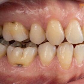
A frequent clinical finding in patients with hypodontia, particularly those with ectodermal dysplasia, is the presence of microdontia or malformed teeth. The most common manifestation is conical shape teeth, often referred to as “peg laterals” when affecting the maxillary lateral incisors. These teeth have a narrow, pointed appearance and lack the normal anatomical contours required for proper interproximal contact and aesthetic proportion. Before any implant therapy is initiated, these existing teeth must often be restored to their ideal dimensions. This is typically achieved through conservative composite resin bonding, porcelain veneers, or full-coverage crowns, depending on the structural integrity of the tooth. In cases where the tooth requires significant preparation, techniques like vital pulp capping using biocompatible materials (such as MTA or Biodentine) may be employed to preserve the vitality of the nerve.
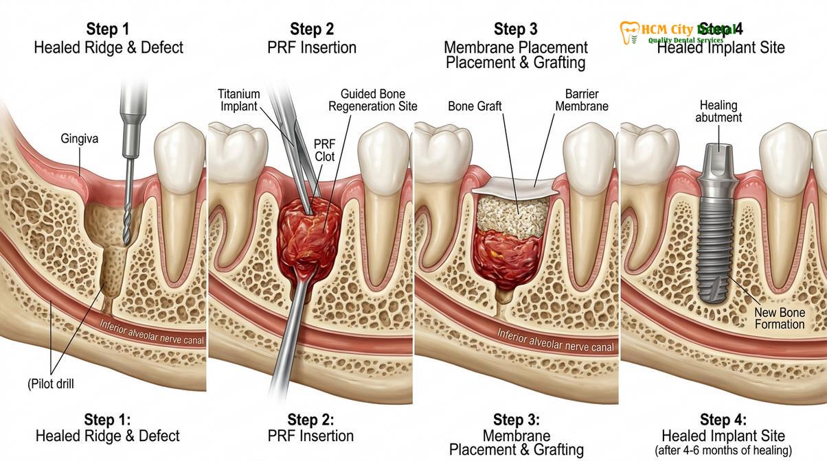
However, the most profound challenge in rehabilitating these patients is the severe bone volume deficiency. According to Wolff’s Law, bone remodels in response to the mechanical forces placed upon it. In a healthy mouth, the roots of the teeth transmit chewing forces to the surrounding alveolar bone, stimulating osteoblastic activity and maintaining bone volume. When tooth buds fail to develop, the alveolar ridge never forms to its full potential. Furthermore, if primary teeth are retained for prolonged periods and eventually lost, the subsequent bone resorption is often rapid and severe, leaving a knife-edge ridge that is entirely inadequate for housing a dental implant.
Reconstructing this deficient anatomy requires sophisticated hard and soft tissue engineering. The concept of biological width—the natural dimension of soft tissue attached to the tooth or implant above the crestal bone—must be respected to ensure long-term gingival health and prevent peri-implantitis. To create a robust foundation, clinicians utilize guided bone regeneration (GBR) techniques. This involves elevating a mucoperiosteal flap, placing particulate bone graft material (allografts, xenografts, or autografts) to build width and height, and covering the graft with a resorbable collagen membrane to exclude fast-growing epithelial cells, allowing the slower-growing bone cells to populate the defect. The integration of autologous blood concentrates, such as Platelet-Rich Fibrin (PRF), has revolutionized this process, accelerating angiogenesis and significantly improving soft tissue healing [3].
Multi-Disciplinary Rehabilitation: Orthodontics, Bone Grafting, and Implants
A sequenced approach utilizing orthodontics to align existing teeth, guided bone regeneration to build the foundation, and dental implants to anchor the final restorations ensures long-term biomechanical stability.
The definitive rehabilitation of congenital missing teeth is a highly orchestrated process that demands seamless collaboration between orthodontists, oral surgeons, and prosthodontists. This multi-disciplinary workflow is designed to optimize the biological foundation, ensure precise biomechanical load distribution, and deliver a final restoration that mimics the aesthetics and function of natural dentition.
Orthodontic Site Preparation and 3D Diagnostics
The rehabilitation journey almost always begins with comprehensive orthodontic therapy. The primary objective is not merely to straighten the existing teeth, but to strategically distribute them to create optimal, symmetrical spaces for the future implants. This involves precise root paralleling to ensure that the implant fixtures can be placed at the correct angulation without damaging adjacent tooth roots. Once the orthodontic phase nears completion, advanced 3D diagnostics become paramount. High-resolution Cone Beam Computed Tomography (CBCT) scans are utilized to assess the exact topography of the alveolar bone, identify vital anatomical structures (such as the inferior alveolar nerve and the maxillary sinus), and virtually plan the implant placement with sub-millimeter accuracy.
Surgical Phase: Implants, PRF, and IV Sedation
Following meticulous digital planning, the surgical phase commences. For patients requiring extensive bone grafting or the placement of multiple implants, managing surgical anxiety and ensuring absolute comfort is a clinical priority. At premier facilities like HCMC Dental Clinic in Ho Chi Minh City, these complex procedures are routinely performed under IV conscious sedation. Administered and monitored by board-certified anesthesiologists, protocols utilizing medications like Midazolam and Propofol, combined with continuous EKG and pulse oximetry monitoring, provide a safe, pain-free experience where the patient remains responsive but deeply relaxed and amnesic to the procedure.
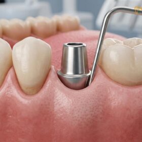
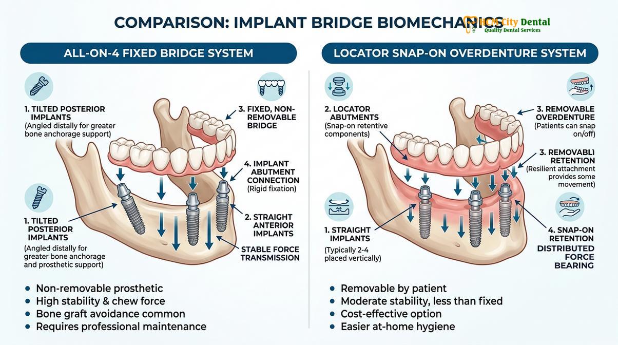
During the surgery, the implant fixtures—typically manufactured from high-grade titanium or titanium-zirconium alloys—are precisely torqued into the prepared osteotomies. In cases of severe oligodontia where the entire arch requires replacement, advanced structural biomechanics are employed. The All-on-4 or All-on-6 concepts utilize strategically angled implants to maximize contact with available dense bone, often bypassing the need for extensive sinus lifts or nerve repositioning. For patients opting for a removable but highly stable solution, implant-supported overdentures utilizing a locator snap-on attachment system provide excellent retention and improved chewing efficiency compared to traditional dentures.
Prosthetic Phase: Biocompatible Monolithic Zirconia
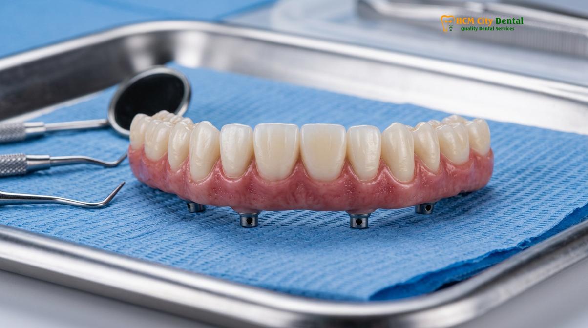
After a period of osseointegration—during which the bone fuses to the implant surface—the restorative phase begins. The current gold standard for full arch and multi-unit implant restorations is biocompatible monolithic zirconia. Sourced from premium Swiss and German manufacturers, this advanced ceramic material is entirely metal-free, highly bio-inert, and exhibits exceptional fracture toughness. Utilizing CAD/CAM technology, the final prostheses are milled from a single block of zirconia, ensuring a precise margin fit of under 50 micrometers. This microscopic precision is crucial for preventing bacterial microleakage and ensuring the long-term health of the peri-implant tissues.
Financial Investment and Global Support
Understanding the financial parameters of full mouth rehabilitation is essential for patients seeking care. International dental tourism has made premium treatments highly accessible. The pricing structure at leading clinics incorporates significant advantages for proactive patients, such as a 40% pre-arrival discount for WhatsApp bookings. According to current clinical fee schedules, the investment parameters are structured as follows:
| Implant Tier (Fixture & Abutment) | Standard Walk-in Rate | WhatsApp Pre-Booking (-40%) |
|---|---|---|
| Economy (Dentis/Neo Biotech) | ~$770 (20M VND) | From $460 (12M VND) |
| Economy Plus (Dentium Hàn) | ~$900 (23.3M VND) | From $540 (14M VND) |
| Standard (Dentium Superline Mỹ / IBS) | ~$1,090 (28.3M VND) | From $655 (17M VND) |
| Premium (Ritter Đức / B&B Ý) | ~$1,600 (41.6M VND) | From $965 (25M VND) |
| Premium Plus (Straumann BLT-SLA) | ~$1,795 (46.6M VND) | From $1,080 (28.0M VND) |
| High-End (Straumann Active) | ~$2,250 (58.3M VND) | From $1,350 (35M VND) |
For the restorative components, a Crown on Implant ranges from ~$260 to $385 for walk-ins, but reduces to From $155 to $230 per tooth with the WhatsApp pre-booking discount. Comprehensive Full Mouth Rehabilitation Packages also reflect this structure:
- Full Arch Zirconia Bridge on Implants (All-on-4 / All-on-6): Walk-in: ~$12,000 – $22,000 | WhatsApp pre-booking: From $7,200 – $13,200 per arch.
- Snap-On Overdenture (Implant-supported): Walk-in: ~$5,000 – $8,000 | WhatsApp pre-booking: From $3,000 – $4,800 per arch.
- Full Mouth Crowns (14 crowns per arch, Zirconia HT): Walk-in: ~$3,640 per arch | WhatsApp pre-booking: From $2,184 per arch.
Beyond the initial investment, reputable clinics provide robust global manufacturer warranty cards, offering 10-year to lifetime guarantees on implant fixtures. Furthermore, international patients benefit from remote post-op checkups via WhatsApp and support for financial planning, including 0% interest monthly installment plans and documentation for tax offsets (NMETO/IRS deductions) [4].
Restoring Normal Chewing Power and Psychological Confidence
Full mouth rehabilitation not only rebuilds the structural integrity of the bite but also profoundly impacts a patient’s self-esteem and social interactions by delivering a natural, functional smile.
The culmination of the rehabilitation process is the delivery of the final prostheses, a moment that often represents a profound life change for the patient. The restoration of normal chewing power is an immediate and tangible benefit. Patients who have spent years modifying their diets to accommodate missing teeth or unstable removable dentures can suddenly enjoy a full spectrum of nutritious, fibrous foods. The biomechanical stability provided by osseointegrated implants ensures that masticatory forces are efficiently transferred to the bone, mimicking the function of a healthy, natural dentition.
To ensure the final result meets the highest aesthetic and functional standards, clinicians employ Digital Smile Design (DSD) protocols. This involves creating 3D digital mock-ups and physical tooth wax-ups based on the patient’s unique facial proportions. Before the final monolithic zirconia restorations are milled, the patient wears a provisional acrylic prototype. This crucial step allows the clinician to verify phonetic compatibility—ensuring the patient can clearly pronounce fricative and sibilant sounds—and to confirm proper chewing guidance and occlusal harmony.
The psychological impact of this transformation cannot be overstated. Individuals with congenital missing teeth often develop deep-seated insecurities, leading to social withdrawal, a reluctance to smile, and diminished self-esteem. By providing a permanent, highly aesthetic solution, full mouth rehabilitation restores not just the teeth, but the patient’s psychological confidence. The ability to speak clearly, eat comfortably in public, and smile without hesitation fundamentally improves their overall quality of life, highlighting the profound intersection of dental science and human well-being [5].
Advanced Clinical Considerations for Complex Cases
Managing complex rehabilitations requires meticulous attention to the Vertical Dimension of Occlusion, neuromuscular harmony, and the patient’s underlying systemic health profile.
While the mechanical aspects of placing implants and fabricating crowns are critical, the true mastery of full mouth rehabilitation lies in managing the complex interplay between the teeth, muscles, and jaw joints. In patients with severe oligodontia, the prolonged absence of posterior teeth inevitably leads to a collapsed bite. This loss of the Vertical Dimension of Occlusion (VDO) forces the mandible to over-close, placing immense strain on the temporomandibular joints and the muscles of mastication.
To correct this, clinicians apply the principles of neuromuscular dentistry. The goal is to find the patient’s physiologic rest jaw position—the spatial orientation where the jaw muscles are at their most relaxed state. This is often achieved using diagnostic temporary splints or TENS (Transcutaneous Electrical Nerve Stimulation) therapy to deprogram the fatigued musculature. Once the optimal VDO is established and tested with provisional restorations, it is permanently captured in the final bio-inert zirconia prostheses. This meticulous approach prevents chronic headaches, neck pain, and premature wear of the new restorations.
Clinical Case Review: A 22-year-old patient presented with severe non-syndromic oligodontia, missing 14 permanent teeth, resulting in a collapsed VDO and compromised speech. Following a 24-month multidisciplinary protocol involving orthodontic space distribution, bilateral sinus lifts, and the placement of 8 dental implants under IV sedation, the patient’s bite was successfully reconstructed using full arch monolithic zirconia bridges. The restoration of the physiologic rest jaw position eliminated the patient’s chronic TMJ discomfort and dramatically improved facial aesthetics.
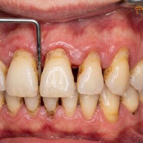
Furthermore, the patient’s broader systemic health must be rigorously evaluated before initiating surgical therapy. Conditions that affect bone metabolism require specific clinical protocols. For instance, in cases involving full mouth reconstruction seniors or patients with a history of dental osteoporosis, the density and healing capacity of the alveolar bone may be compromised. A particularly critical consideration is the patient’s medication history; the use of bisphosphonates or other antiresorptive drugs poses a significant risk for BRONJ bisphosphonate (Bisphosphonate-Related Osteonecrosis of the Jaw). In such scenarios, extensive surgical interventions may be contraindicated, and alternative, less invasive restorative strategies must be explored. Similarly, if the patient suffers from active periodontal disease bone loss around their remaining natural teeth, this infection must be completely eradicated through deep scaling and root planing before any implant surgery can safely proceed, ensuring a sterile environment for osseointegration.
When to See a Doctor
Early diagnosis is the cornerstone of successfully managing congenital missing teeth. Parents should schedule a comprehensive pediatric dental evaluation if they notice any delays in the eruption of their child’s primary or permanent teeth, or if there are visible gaps that do not close naturally. Specifically, if a child retains their baby teeth well beyond the normal shedding age (typically past age 12 or 13 for most teeth), it is a strong clinical indicator that the underlying permanent teeth may be absent or impacted.
Adults who have retained primary teeth or who have been functioning with missing teeth should seek a consultation with a prosthodontist or an implant specialist. Delaying treatment allows for progressive alveolar bone resorption and shifting of adjacent teeth, which exponentially increases the complexity and cost of future rehabilitation. A thorough clinical examination, including 3D CBCT imaging, is essential to determine the viability of the remaining bone and to formulate a personalized, evidence-based treatment plan.
Frequently Asked Questions
What are the options for people born with missing teeth?
Treatment options for congenital missing teeth include orthodontic space closure, resin-bonded bridges, removable partial dentures, and definitive implant-supported restorations. The optimal choice depends on the patient’s age, the number of missing teeth, jawbone volume, and overall facial skeletal development, requiring a customized multidisciplinary approach. In growing children, temporary solutions are utilized to maintain space and function until skeletal maturity is reached, at which point permanent dental implants can be safely placed.
At what age can someone with hypodontia get dental implants?
Dental implants are typically placed only after skeletal growth is complete, which is generally around age 18 to 21 for most individuals. Placing implants too early in a growing jaw can lead to infraocclusion, where the implant remains stationary while adjacent natural teeth continue to erupt. Clinicians use serial cephalometric radiographs to track bone growth and determine the precise, safe timing for surgical intervention.
How does ectodermal dysplasia affect jawbone growth?
Ectodermal dysplasia severely impacts jawbone growth because the absence of developing tooth buds deprives the alveolar ridge of the mechanical stimulation needed for normal bone apposition. This results in significant bone volume deficiency, often requiring extensive guided bone regeneration before any implant placement can occur. The lack of alveolar bone also contributes to a collapsed facial profile, making early prosthetic intervention crucial for both function and psychological development.
Is the full mouth rehabilitation process painful?
The surgical phases of full mouth rehabilitation are performed under strict anesthesia protocols, making the procedure highly comfortable. Advanced clinics utilize IV conscious sedation administered by board-certified anesthesiologists, ensuring patients remain relaxed, anxiety-free, and experience virtually no pain during complex bone grafting and implant surgeries. Post-operative discomfort is typically managed effectively with prescribed analgesics and anti-inflammatory medications.
How long do monolithic zirconia implant restorations last?
Biocompatible monolithic zirconia restorations are engineered for exceptional durability and can last several decades with proper maintenance. Their longevity depends on meticulous daily oral hygiene, routine professional cleanings, and the use of a nocturnal occlusal guard to protect against excessive bite forces or bruxism. The underlying titanium implant fixtures themselves often carry a lifetime manufacturer warranty, provided the surrounding bone and gum tissues remain healthy.
“The successful rehabilitation of congenital missing teeth is not merely about filling gaps; it is a profound architectural reconstruction of the patient’s masticatory system. By respecting the biological width, optimizing the vertical dimension of occlusion, and utilizing bio-inert materials, we can deliver a result that is both biomechanically sound and aesthetically transformative.”
— Dr. Nguyen Van Cuong, Lead Clinician at HCMC Dental Clinic
“In cases of severe oligodontia, the integration of 3D CBCT diagnostics and CAD/CAM milling technology has shifted our clinical paradigm from reactive problem-solving to predictable, precision-engineered outcomes, ensuring that every implant is placed in the optimal restorative position.”
— Clinical Prosthodontic Guidelines
For patients navigating the complexities of congenital missing teeth, seeking care from a highly specialized team is paramount. At HCMC Dental Clinic in Ho Chi Minh City, our multidisciplinary specialists are dedicated to providing evidence-based, compassionate care. Through advanced diagnostics and personalized treatment planning, we strive to restore not just your dental function, but your complete confidence in your smile.
References
- The Journal of Prosthetic Dentistry. Vertical dimension of occlusion restoration parameters. (2021).
- International Journal of Prosthodontics. Biomechanical load distribution in All-on-4 restorations. (2020).
- Journal of Clinical Periodontology. Biocompatible dental materials and soft tissue response. (2019).
- Clinical Oral Implants Research. Platelet-rich fibrin in guided bone regeneration. (2022).
- Journal of Esthetic and Restorative Dentistry. Digital smile design functional protocols. (2018).
For customized treatment planning, transparent cost estimates, and direct consultation with Dr. Cuong, visit our comprehensive Full Mouth Rehabilitation service page or message our team directly via WhatsApp to receive an instant assessment.