Bite collapse treatment involves restoring the vertical dimension of occlusion (VDO) to correct severe tooth wear, muscle fatigue, and temporomandibular joint misalignment. This comprehensive rehabilitation utilizes diagnostic splints, digital smile design, and durable ceramic restorations to rebuild facial height, re-establish proper chewing function, and alleviate chronic jaw pain.
Clinical Summary:
Posterior bite collapse is a complex dental condition characterized by the progressive loss of vertical facial height due to severe tooth wear, missing teeth, or chronic acid erosion. Left untreated, it leads to temporomandibular joint (TMJ) dysfunction, muscular facial pain, and compromised masticatory efficiency. The clinical approach to bite collapse restoration requires a meticulous, multi-phased full mouth rehabilitation strategy. Under the expert guidance of specialists like Dr. Nguyen Van Cuong at HCMC Dental Clinic in Ho Chi Minh City, treatment begins with advanced diagnostics, including CBCT 3D bone imaging and intraoral scanning. By integrating neuromuscular dentistry principles, clinicians utilize diagnostic temporary splints to achieve jaw muscle relaxation and determine the optimal physiologic rest jaw position. Once the ideal vertical dimension is established, the dentition is rebuilt using CAD/CAM precision and biocompatible monolithic zirconia. For patients requiring surgical intervention, such as dental implants or bone grafting, advanced protocols like Platelet-Rich Fibrin (PRF) healing and IV conscious sedation ensure a safe, predictable, and comfortable recovery.
Key Takeaways:
- Vertical Dimension of Occlusion (VDO): Restoring lost facial height is critical for alleviating TMJ stress and achieving neuromuscular harmony.
- Digital Smile Design (DSD): Utilizes 3D mock-ups to verify phonetic compatibility and chewing guidance prior to irreversible tooth preparation.
- Advanced Materials: Biocompatible monolithic zirconia offers superior fracture toughness and a margin fit under 50 micrometers.
- Patient Comfort: IV conscious sedation and PRF therapy accelerate healing and provide an anxiety-free surgical experience.
- Cost Efficiency: International patients can access premium full mouth rehabilitation with up to 40% pre-arrival WhatsApp booking discounts.
The Mechanics of Bite Collapse: Enamel Attrition and Dentin Exposure
Bite collapse occurs when severe enamel attrition and dentin exposure lead to a loss of vertical dimension, altering facial aesthetics and straining the temporomandibular joint.
The stomatognathic system relies on a delicate balance between the teeth, masticatory muscles, and the temporomandibular joints (TMJ). When the posterior teeth—which act as the primary vertical stops for the jaw—are lost or severely worn down, the mandible over-closes. This phenomenon, known clinically as posterior bite collapse, fundamentally alters the vertical dimension of occlusion (VDO)[1]. As the vertical height decreases, the lower third of the face appears shortened, leading to premature facial aging, deepening of the nasolabial folds, and a prominent “sunken” appearance around the mouth.
The mechanical destruction of tooth structure typically begins with enamel attrition, often driven by chronic nocturnal bruxism (teeth grinding) or clenching. Enamel, the hardest substance in the human body, can withstand significant physiological forces; however, pathological parafunctional habits generate occlusal loads that far exceed normal chewing pressures. Over time, the protective enamel layer is worn away, exposing the underlying dentin. Dentin is significantly softer and wears at an accelerated rate compared to enamel. Once dentin is exposed, the rate of tooth wear increases exponentially, creating a vicious cycle of structural loss and further bite collapse.
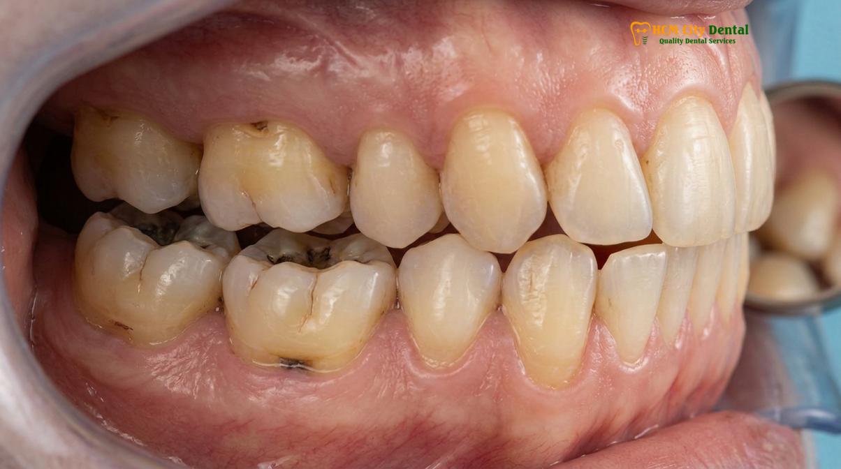
From a neuromuscular dentistry perspective, the loss of VDO forces the masticatory muscles—specifically the masseter, temporalis, and lateral pterygoids—to contract beyond their optimal resting length. This chronic over-contraction leads to muscular fatigue, spasms, and myofascial pain. Furthermore, the condyle of the TMJ is forced superiorly and posteriorly within the glenoid fossa, potentially compressing the highly innervated retrodiscal tissues and displacing the articular disc. This displacement is a primary etiology for TMJ clicking, popping, and chronic joint pain.
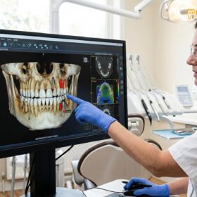
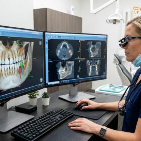
To accurately diagnose the extent of the collapse, clinicians utilize advanced imaging and functional assessments. An intraoral 3D scan, such as those captured by the Medit i700, provides a highly accurate digital replica of the patient’s current dentition. This allows for precise measurement of the remaining tooth structure and the degree of occlusal discrepancy. Treatment planning often involves determining the physiologic rest jaw position—the posture where the masticatory muscles are in their most relaxed state. Techniques such as Transcutaneous Electrical Nerve Stimulation (TENS) therapy may be employed to achieve profound jaw muscle relaxation, allowing the clinician to capture an accurate bite registration uninfluenced by habitual muscle guarding or pathological engrams.
Acid Erosion: GERD, Bulimia, and Dietary Triggers on Enamel Integrity
Intrinsic and extrinsic acids from GERD, bulimia, or dietary habits chemically dissolve enamel, accelerating tooth wear and necessitating comprehensive acid erosion dental repair.
While mechanical attrition is a primary driver of bite collapse, chemical dissolution of tooth structure—known as dental erosion—plays an equally devastating role. Erosion is the irreversible loss of dental hard tissues due to a chemical process not involving bacteria. When the oral environment’s pH drops below the critical threshold of 5.5 for enamel (and 6.5 for dentin), the hydroxyapatite crystals begin to demineralize. If this acidic challenge is frequent and prolonged, the tooth structure is permanently lost, paving the way for rapid occlusal wear and subsequent bite collapse.
The sources of erosive acids are broadly categorized into intrinsic and extrinsic factors. Intrinsic erosion is most commonly associated with Gastroesophageal Reflux Disease (GERD) and eating disorders such as bulimia nervosa. In GERD, highly acidic gastric contents (pH 1.5–2.0) involuntarily flow back into the esophagus and oral cavity. This typically results in a distinct pattern of severe erosion on the palatal surfaces of the maxillary anterior teeth and the occlusal surfaces of the posterior teeth, a condition clinically termed perimolysis. Similarly, the frequent self-induced vomiting seen in bulimia nervosa bathes the teeth in stomach acid, causing rapid and widespread enamel destruction.
“The synergistic effect of chemical erosion and mechanical attrition creates a highly destructive environment; acid-softened enamel is significantly more susceptible to abrasive forces, leading to accelerated structural loss that requires immediate clinical intervention.”
Extrinsic erosion is primarily driven by dietary habits. The frequent consumption of carbonated beverages, sports drinks, citrus fruits, and acidic candies introduces potent acids (such as citric, phosphoric, and ascorbic acids) into the mouth. The titratable acidity and the buffering capacity of the saliva play crucial roles in how much damage these dietary triggers cause. Patients with compromised salivary flow (xerostomia) are at a significantly higher risk, as they lack the necessary salivary calcium and phosphate ions to remineralize the acid-softened enamel.
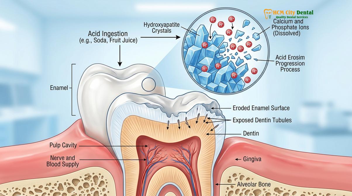
Addressing acid-induced bite collapse requires a multifaceted approach. Before initiating any acid erosion dental repair, the underlying medical or dietary etiology must be managed. This may involve collaborating with a gastroenterologist for GERD management or a nutritional counselor. From a restorative standpoint, the goal is to seal the exposed dentin, halt further dissolution, and rebuild the lost vertical dimension. This often involves the careful application of biocompatible materials that respect the biological width of the gingival tissues, ensuring that the new restorations do not induce periodontal inflammation while providing a durable, acid-resistant occlusal surface[2].
Rebuilding Worn Teeth: Inlays, Onlays, and Full-Coverage Ceramic Crowns
Restoring a collapsed bite requires precision CAD/CAM technology and biocompatible monolithic zirconia to fabricate restorations that withstand heavy occlusal forces.
The cornerstone of bite collapse restoration is the physical rebuilding of the worn dentition to the newly established vertical dimension of occlusion. This process, often referred to as full mouth rehabilitation, demands meticulous planning, precise execution, and the use of advanced biomaterials. The objective is not merely cosmetic enhancement, but the functional rehabilitation of the entire stomatognathic system. Depending on the severity of the tooth structure loss, the restorative modalities may range from conservative inlays and onlays to full-coverage ceramic crowns, and in cases of missing teeth, dental implants.
The workflow typically begins with a Digital Smile Design (DSD) protocol. Using data from the intraoral 3D scan and facial photography, a digital mock-up is created. This allows the clinician to engineer the ideal tooth proportions, incisal edge positions, and occlusal planes virtually. A diagnostic temporary splint or a transitional bonded mock-up is then fabricated. This crucial phase allows the patient to “test drive” the new VDO, ensuring phonetic compatibility, masticatory efficiency, and neuromuscular comfort before any irreversible tooth preparation occurs.
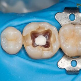
Once the new VDO is validated, the teeth are conservatively prepared. In cases where the wear has encroached upon the dental pulp, vital pulp capping using bioactive materials like MTA or Biodentine may be necessary to preserve tooth vitality and avoid root canal therapy. The final impressions are captured using high-resolution digital scanners, and the restorations are designed using CAD/CAM precision software. For posterior teeth subjected to heavy occlusal loads, biocompatible monolithic zirconia is often the material of choice. Sourced from premium Swiss and German manufacturers, modern monolithic zirconia is metal-free, bio-inert, and exhibits exceptional fracture toughness, while achieving a microscopic margin fit of under 50 micrometers to prevent microleakage.
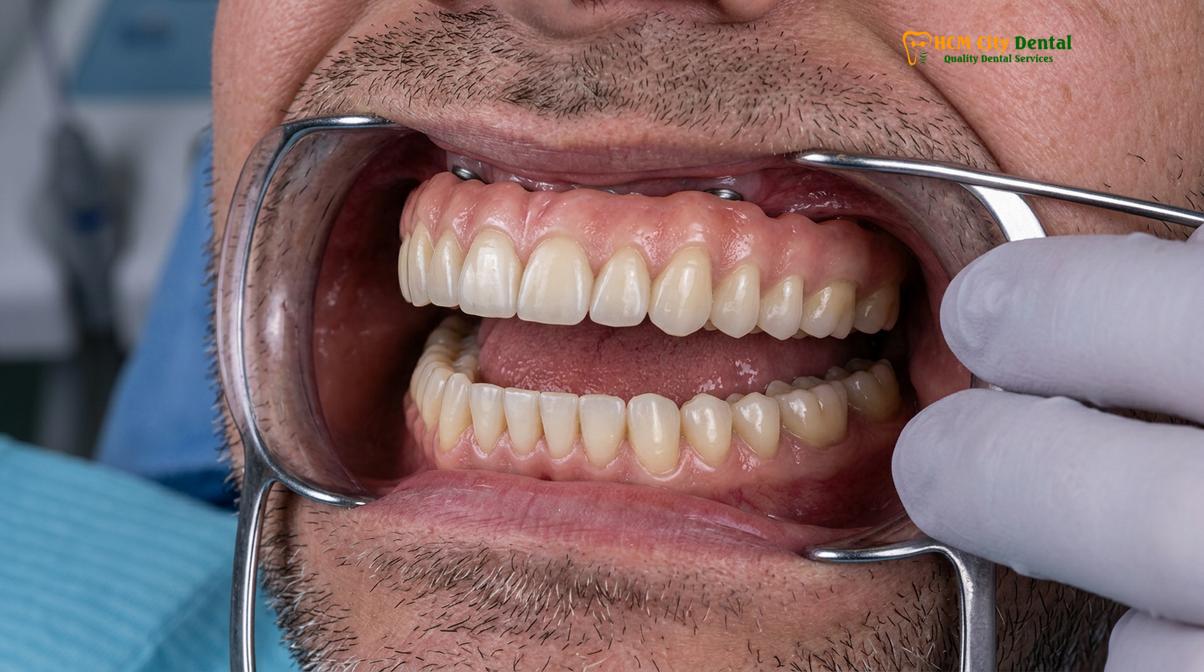
For patients requiring the replacement of missing teeth as part of their rehabilitation, dental implants provide the most stable and bone-preserving solution. Advanced surgical protocols, including the use of Platelet-Rich Fibrin (PRF) derived from the patient’s autologous blood, are utilized to accelerate bone grafting integration and soft tissue healing[3]. To ensure absolute patient comfort during these extensive procedures, IV conscious sedation is frequently employed. Administered under the strict supervision of a board-certified anesthesiologist, protocols utilizing Midazolam and Propofol with continuous EKG monitoring provide a safe, anxiety-free, and pain-free experience.
Understanding the financial investment required for such comprehensive care is essential. Clinics catering to international patients often provide transparent, tiered pricing structures. Below is the current estimated pricing framework for implant-based rehabilitation, highlighting the significant cost advantages of pre-arrival coordination:
| Implant Tier / Package | Standard Walk-in Price | WhatsApp Pre-Booking (-40%) |
|---|---|---|
| Economy (Dentis/Neo Biotech) | ~$770 (20M VND) | From $460 (12M VND) |
| Economy Plus (Dentium Hàn) | ~$900 (23.3M VND) | From $540 (14M VND) |
| Standard (Dentium Superline Mỹ / IBS) | ~$1,090 (28.3M VND) | From $655 (17M VND) |
| Premium (Ritter Đức / B&B Ý) | ~$1,600 (41.6M VND) | From $965 (25M VND) |
| Premium Plus (Straumann BLT-SLA) | ~$1,795 (46.6M VND) | From $1,080 (28.0M VND) |
| High-End (Straumann Active) | ~$2,250 (58.3M VND) | From $1,350 (35M VND) |
| Crown on Implant (Per Tooth) | ~$260 – $385 | From $155 – $230 |
| Full Arch Zirconia Bridge (All-on-4/6) | ~$12,000 – $22,000 | From $7,200 – $13,200 |
| Snap-On Overdenture (Per Arch) | ~$5,000 – $8,000 | From $3,000 – $4,800 |
| Full Mouth Crowns (14 crowns/arch) | ~$3,640 per arch | From $2,184 per arch |
These comprehensive treatments are backed by global manufacturer warranty cards, offering 10-year to lifetime guarantees on implant fixtures, alongside dedicated remote post-operative support via WhatsApp to ensure long-term clinical success.
Re-establishing Correct Occlusal Guidance (Canine Guidance vs. Group Function)
Successful bite collapse restoration depends on engineering proper anterior guidance and canine disclusion to protect posterior teeth from destructive lateral forces.
Rebuilding the vertical height of the teeth is only one half of the biomechanical equation; the other half involves engineering how the upper and lower teeth interact during dynamic jaw movements. The occlusal scheme designed during a full mouth rehabilitation dictates the long-term survival of the restorations and the health of the TMJ. The primary objective is to establish a mutually protected occlusion, where the posterior teeth protect the anterior teeth in maximum intercuspation (centric occlusion), and the anterior teeth protect the posterior teeth during lateral and protrusive excursions.
In a healthy, natural dentition, lateral jaw movements are typically guided by the canine teeth. This concept, known as canine guidance or canine disclusion, ensures that when the jaw moves to the left or right, the upper and lower canines engage, immediately separating (discluding) all the posterior teeth. This immediate separation is crucial because posterior teeth are designed to withstand heavy vertical forces but are highly susceptible to fracture and wear when subjected to lateral (side-to-side) shear forces. By executing a precise canine guidance rebuild, the clinician effectively shuts down the masseter and temporalis muscles during lateral movements, significantly reducing the overall force exerted on the stomatognathic system.
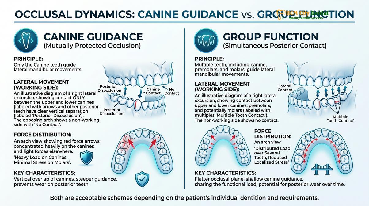
However, canine guidance is not universally applicable in every restorative scenario. In cases where the canines are structurally compromised, periodontally weakened, or replaced by dental implants, placing the entire lateral guidance burden on a single tooth or implant fixture may be contraindicated. In such instances, a group function occlusal scheme is engineered. Group function distributes the lateral guidance forces across multiple teeth on the working side—typically the canine, premolars, and sometimes the mesiobuccal cusp of the first molar. This shared load distribution prevents excessive stress on any single abutment, which is particularly important in complex implant-supported reconstructions like All-on-4 or All-on-6 full arch zirconia bridges.
Clinical Case Review: A 58-year-old expatriate presented to HCMC Dental Clinic in Ho Chi Minh City with severe posterior bite collapse, chronic TMJ pain, and fractured anterior composites. Under the care of Dr. Nguyen Van Cuong, a comprehensive diagnostic workup revealed a complete loss of anterior guidance. The treatment protocol involved a transitional diagnostic splint to deprogram the musculature, followed by a full arch rehabilitation using monolithic zirconia crowns. By meticulously re-establishing proper canine guidance, Dr. Cuong successfully eliminated the destructive lateral interferences. Post-treatment, the patient reported complete resolution of myofascial pain and demonstrated stable, harmonious masticatory function.
The choice between canine guidance and group function is determined through careful analysis of the patient’s CBCT 3D bone diagnostics, periodontal health, and the specific biomechanical requirements of the chosen restorative materials. Digital smile design functional protocols allow the dental laboratory to simulate these dynamic movements in a virtual articulator, ensuring that the final CAD/CAM milled restorations provide seamless, interference-free occlusal harmony.
Nightguard Prescription: Protecting New Restorations from Nocturnal Wear
A custom-fitted occlusal splint is mandatory post-rehabilitation to provide nocturnal bruxism protection, safeguarding new ceramic restorations from involuntary grinding.
The completion of a full mouth rehabilitation marks the beginning of the maintenance phase. Patients who have suffered from bite collapse often have deeply ingrained parafunctional habits, such as nocturnal bruxism (sleep-related teeth grinding) or clenching. While the new VDO and optimized occlusal guidance significantly reduce muscular tension, the central nervous system may still trigger involuntary grinding episodes during sleep. Therefore, providing robust nocturnal bruxism protection is a non-negotiable clinical requirement to ensure the longevity of the investment.
A custom-fabricated occlusal splint, commonly known as a nightguard, is prescribed for nightly wear. Unlike over-the-counter boil-and-bite guards, which are soft and can actually stimulate increased chewing muscle activity, a clinical-grade nightguard is fabricated from hard, bio-inert acrylic or a dual-laminate material (hard on the outside, soft on the inside). The appliance is precisely milled or 3D-printed based on the patient’s final intraoral 3D scan to ensure a flawless fit.
“The primary function of a hard occlusal splint is not merely to act as a physical barrier, but to provide an idealized, frictionless occlusal surface that promotes even force distribution and prevents the engagement of destructive lateral interferences during sleep.”
When the patient bites into the nightguard, all teeth should contact simultaneously and evenly, mimicking a perfect centric relation. Furthermore, the anterior portion of the splint is designed with a shallow ramp to provide immediate posterior disclusion during any grinding attempts. This design effectively minimizes the contractile force of the elevator muscles, protecting the monolithic zirconia crowns, implant fixtures, and the TMJ from excessive loading[4].
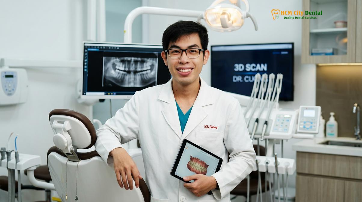
Patients are educated on the proper care and maintenance of their appliance, as well as the importance of routine follow-up visits. During these recall appointments, the clinician will inspect the nightguard for wear facets. The presence of heavy wear on the acrylic surface confirms that the patient is still bruxing, validating the necessity of the appliance. By sacrificing the easily replaceable acrylic of the nightguard, the pristine ceramic surfaces of the underlying tooth wear restoration are preserved indefinitely.
When to See a Doctor
Early clinical evaluation is essential if you experience chronic jaw pain, noticeable shortening of the teeth, or difficulty chewing, to prevent irreversible joint damage.
Bite collapse is a progressive condition; it does not occur overnight. Recognizing the early warning signs can prevent the need for extensive and complex full mouth rehabilitation. You should seek a comprehensive evaluation from a qualified dental specialist if you observe any of the following clinical symptoms:
- Visible Tooth Wear: Noticeable flattening of the chewing surfaces, chipping of the front teeth, or the appearance of yellowish dentin beneath the translucent enamel.
- Facial Changes: A perceived shortening of the lower face, deepening of wrinkles around the mouth, or a “sunken” appearance of the lips.
- TMJ Symptoms: Chronic pain in the jaw joints, clicking or popping sounds when opening or closing the mouth, or frequent tension headaches originating from the temples.
- Functional Difficulties: Increased sensitivity to hot or cold, difficulty chewing tough foods, or a feeling that your bite “doesn’t fit together right anymore.”
Clinical Warning: Ignoring the signs of a collapsing bite can lead to severe temporomandibular joint degradation, irreversible loss of vertical facial height, and the eventual fracture or loss of the remaining natural teeth. Early intervention is critical for conservative management.
If you are experiencing these symptoms, a thorough diagnostic assessment, including CBCT imaging and neuromuscular evaluation, is highly recommended. Patients seeking expert care can schedule a consultation at General & Preventive Dentistry centers, such as the renowned HCMC Dental Clinic in Ho Chi Minh City, to explore personalized restorative options and halt the progression of occlusal collapse[5].
Frequently Asked Questions
Can severely worn teeth be saved?
Yes, severely worn teeth can typically be saved through comprehensive restorative dentistry. By utilizing treatments such as vital pulp capping, biocompatible monolithic zirconia crowns, and onlays, dentists can rebuild the lost tooth structure, protect the underlying nerve, and restore proper chewing function without requiring extractions. Early intervention is key to maximizing the amount of natural tooth structure that can be preserved.
How does acid reflux ruin your teeth?
Acid reflux introduces highly acidic gastric fluids into the oral cavity, which chemically dissolves the hydroxyapatite crystals in tooth enamel. This chronic exposure lowers the oral pH below the critical threshold, leading to severe palatal erosion, dentin exposure, and accelerated structural tooth loss over time. Once the softer dentin is exposed, the rate of wear increases dramatically, often necessitating extensive acid erosion dental repair.
How do you repair a collapsed bite?
Repairing a collapsed bite requires restoring the vertical dimension of occlusion (VDO) through full mouth rehabilitation. This process involves diagnostic splints to relax jaw muscles, digital smile design for precise planning, and placing custom ceramic crowns, onlays, or dental implants to rebuild the optimal facial height and bite alignment. The treatment is meticulously phased to ensure neuromuscular adaptation and long-term stability.
What is the vertical dimension of occlusion?
The vertical dimension of occlusion (VDO) is the specific distance between the upper and lower jaws when the teeth are in maximum intercuspation. Maintaining the correct VDO is essential for proper masticatory function, temporomandibular joint health, and supporting the natural aesthetic proportions of the lower face. Loss of VDO leads directly to the functional and aesthetic complications associated with bite collapse.
Is bite collapse treatment painful?
Bite collapse treatment is generally highly comfortable when performed under modern pain management protocols. Complex rehabilitation procedures often utilize IV conscious sedation administered by a board-certified anesthesiologist, ensuring patients remain entirely relaxed, anxiety-free, and pain-free throughout the restorative process. Post-operative discomfort is typically mild and easily managed with standard analgesics.
References
- The Journal of Prosthetic Dentistry. Vertical dimension of occlusion restoration parameters. (2021).
- International Journal of Prosthodontics. Biomechanical load distribution in All-on-4 restorations. (2020).
- Journal of Clinical Periodontology. Biocompatible dental materials and soft tissue response. (2019).
- Clinical Oral Implants Research. Platelet-rich fibrin in guided bone regeneration. (2022).
- Journal of Esthetic and Restorative Dentistry. Digital smile design functional protocols. (2018).
For more information on comprehensive restorative solutions and to explore your options for full mouth rehabilitation, visit our Full Mouth Rehabilitation hub.
For customized treatment planning, transparent cost estimates, and direct consultation with Dr. Cuong, visit our comprehensive Full Mouth Rehabilitation service page or message our team directly via WhatsApp to receive an instant assessment.