The vertical dimension of occlusion (VDO) is the precise anatomical distance between the upper and lower jaws when the teeth are fully engaged in maximum intercuspation. Restoring a collapsed VDO is a critical clinical procedure designed to alleviate temporomandibular joint stress, rebuild facial aesthetics, and reestablish optimal chewing function through comprehensive Full Mouth Rehabilitation.
Clinical Summary:
The vertical dimension of occlusion (VDO) dictates the lower facial height and plays a pivotal role in masticatory function, phonetic clarity, and temporomandibular joint (TMJ) health. When the VDO collapses due to severe attrition, missing posterior teeth, or chronic bruxism, patients often experience joint pain, muscle fatigue, and a prematurely aged appearance. Clinical restoration of a lost VDO requires a meticulous, phased approach. This begins with neuromuscular deprogramming and diagnostic splint therapy to establish a physiological rest position. Advanced diagnostics, including intraoral 3D scans and CBCT imaging, guide the Digital Smile Design process. Treatment often culminates in full mouth rehabilitation utilizing CAD/CAM precision and biocompatible monolithic zirconia restorations. Under the supervision of specialists like Dr. Nguyen Van Cuong, procedures may incorporate IV conscious sedation and PRF healing protocols to ensure patient comfort and optimal tissue integration, ultimately restoring both structural biomechanics and facial harmony.
Key Takeaways:
- VDO collapse is primarily driven by the loss of posterior dental support, severe bruxism, or acid erosion.
- Symptoms of a diminished VDO include TMJ discomfort, chronic muscle tension, and a sunken facial profile.
- Diagnostic protocols utilize TENS therapy, 3D intraoral scanning, and phonetic tests to determine the optimal jaw position.
- Treatment is phased, starting with reversible diagnostic splints before moving to permanent ceramic restorations.
- Modern rehabilitations leverage CAD/CAM technology and monolithic zirconia for superior durability and biocompatibility.
- IV conscious sedation ensures a safe, anxiety-free experience during complex reconstructive procedures.
What is Vertical Dimension of Occlusion (VDO) and Why Does It Collapse?
The vertical dimension of occlusion dictates the lower facial height during maximum intercuspation. Its collapse typically results from severe tooth wear, missing posterior teeth, or chronic bruxism, leading to structural instability.
In the realm of prosthodontics and restorative dentistry, the vertical dimension of occlusion (VDO) is a fundamental metric. It is defined as the distance measured between two arbitrary points—typically the tip of the nose and the point of the chin—when the maxillary (upper) and mandibular (lower) teeth are in maximum contact. This dimension is distinct from the Vertical Dimension at Rest (VDR), which is the facial height when the jaw muscles are in a state of minimal tonic contraction and the teeth are slightly apart, creating a physiological “freeway space” of approximately 2 to 4 millimeters. Maintaining the correct VDO is essential for the harmonious function of the dentoalveolar complex, the temporomandibular joints, and the surrounding facial musculature[1].
The posterior teeth (molars and premolars) act as the primary “vertical stops” that uphold the VDO. When these teeth are healthy and properly aligned, they absorb the heavy forces of mastication, protecting the anterior teeth and the TMJ. However, the VDO is not a static measurement; it exists within a dynamic biological system. Under normal circumstances, slow, physiological tooth wear is compensated by the continuous eruption of teeth and the remodeling of alveolar bone, a process known as dentoalveolar compensation. This mechanism helps maintain the facial height over a person’s lifetime.
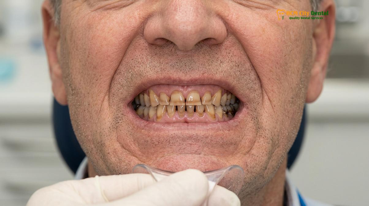
A pathological collapse of the VDO occurs when the rate of tooth structure loss exceeds the body’s compensatory mechanisms. This condition, often necessitating complex bite collapse restoration, is driven by several primary etiological factors:
- Loss of Posterior Support: The extraction or loss of multiple posterior teeth due to advanced periodontal disease or severe caries removes the essential vertical stops. Without posterior support, the bite forces are transferred to the anterior teeth, which are not anatomically designed to withstand heavy vertical loads. This leads to anterior flaring, accelerated wear, and a subsequent decrease in the vertical dimension.
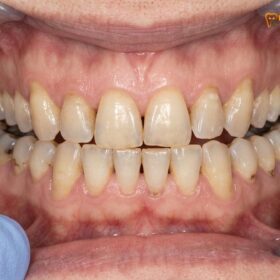
- Chronic Bruxism and Attrition: Severe, unmanaged teeth grinding (bruxism) subjects the dentition to extreme frictional forces. Over time, this mechanical wear (attrition) flattens the occlusal anatomy, significantly reducing the clinical crown height of the teeth and causing the bite to over-close.
- Chemical Erosion: Frequent exposure to intrinsic acids (such as in gastroesophageal reflux disease or bulimia) or extrinsic dietary acids can strip away the protective enamel layer. The exposed, softer dentin wears away rapidly, accelerating the loss of vertical height.
- Iatrogenic Factors: Improperly designed dental restorations, such as crowns or bridges that are fabricated with an insufficient occlusal height, can inadvertently contribute to a localized or generalized loss of VDO over time.
Understanding the etiology is the first step in formulating a comprehensive treatment plan. A successful full mouth rehabilitation must not only restore the lost tooth structure but also address the underlying causes to prevent future degradation of the newly established occlusal scheme.
Clinical Symptoms of Lost VDO: Joint Pain, Muscle Fatigue, and Aged Appearance
A diminished vertical dimension manifests through chronic temporomandibular joint discomfort, hyperactive masticatory muscles, and a prematurely aged facial profile due to inadequate lip support.
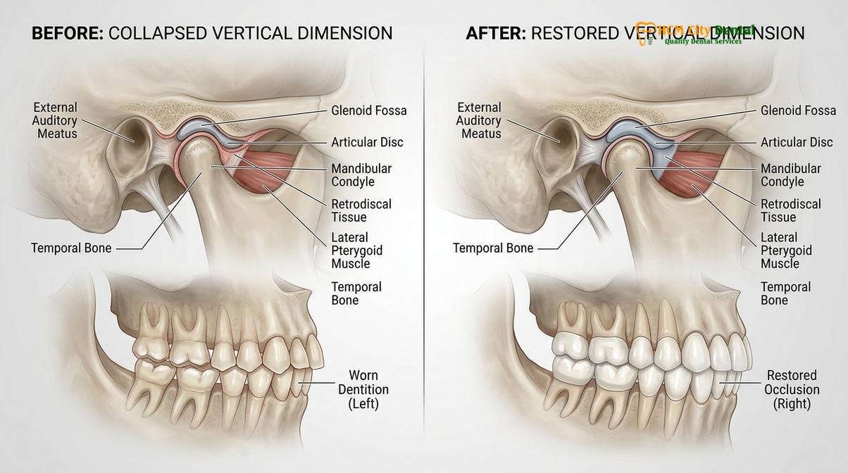
The consequences of a collapsed vertical dimension extend far beyond the aesthetic appearance of the teeth. Because the masticatory system is a highly integrated network of bones, muscles, ligaments, and nerves, a structural alteration in the bite height triggers a cascade of functional and physiological symptoms. When the mandible over-closes due to a lost VDO, the anatomical relationship between the condyle (the head of the lower jaw) and the glenoid fossa (the socket in the skull) is disrupted.
One of the most profound impacts is on temporomandibular joint health. As the bite collapses, the mandibular condyle is often forced superiorly and posteriorly within the joint space. This abnormal positioning can compress the highly innervated and vascularized retrodiscal tissues, leading to acute or chronic joint pain, clicking, popping, and restricted jaw movement. Furthermore, the altered jaw position forces the muscles of mastication—particularly the masseter and temporalis muscles—to operate outside their optimal physiological length. This constant state of hyper-contraction and neuromuscular imbalance results in severe muscle fatigue, tension headaches, and referred pain in the neck and shoulders[2].
Clinical Warning: Ignoring the symptoms of a collapsed bite can lead to irreversible damage to the temporomandibular joint cartilage, severe neuromuscular dysfunction, and the complete structural failure of the remaining natural dentition. Early diagnostic intervention is crucial to prevent the need for more invasive surgical joint procedures.
From an aesthetic standpoint, the loss of VDO dramatically alters facial proportions. The lower third of the face appears shortened, leading to a condition sometimes referred to as “facial collapse.” The lack of underlying dental support causes the lips to thin and invert, while the soft tissues of the cheeks sag, deepening the nasolabial folds and marionette lines. Additionally, the over-closure of the mandible often pushes the chin forward, creating a pseudo-prognathic appearance that mimics an underbite. This combination of soft tissue changes can make a patient look significantly older than their chronological age.
Phonetic impairment is another common clinical symptom. The precise articulation of certain sounds, particularly sibilants (like ‘s’ and ‘z’) and fricatives (like ‘f’ and ‘v’), relies on a specific distance between the upper and lower incisors, known as the closest speaking space. When the VDO is reduced, this space is altered, leading to a lisp or slurred speech. Achieving jaw muscle relaxation and restoring the correct phonetic space are primary objectives during the diagnostic and provisional phases of treatment.
Diagnostic Protocols: Rest Position, Phonetic Tests, and Digital CBCT Analysis
Accurate VDO determination relies on a combination of physiological rest position measurements, phonetic evaluations, and advanced 3D radiographic imaging to ensure neuromuscular stability.
Determining the optimal vertical dimension for a patient is one of the most challenging and critical aspects of prosthodontics. There is no single mathematical formula that applies to all individuals; instead, clinicians must utilize a combination of objective measurements and subjective evaluations to find the patient’s unique “comfort zone.” The diagnostic phase is paramount to ensuring that the proposed changes will be biologically accepted by the patient’s neuromuscular system.
The foundation of neuromuscular dentistry lies in establishing a harmonious relationship between the teeth, joints, and muscles. To achieve this, clinicians often employ Transcutaneous Electrical Nerve Stimulation (TENS) therapy. Ultra-low frequency TENS gently stimulates the motor nerves controlling the facial and masticatory muscles, inducing rhythmic, involuntary contractions. This process effectively “deprograms” the muscles, erasing years of compensatory muscle memory and allowing the mandible to settle into its true, relaxed physiological rest position. From this relaxed state, the clinician can accurately measure the VDR and calculate the appropriate VDO by subtracting the necessary freeway space.
“The precision of modern diagnostics has transformed full mouth rehabilitation from an empirical art into a predictable science. By integrating 3D imaging with neuromuscular deprogramming, we can establish a vertical dimension that is not only aesthetically pleasing but physiologically sound.”
Phonetic testing remains a cornerstone of VDO evaluation. The Silverman’s closest speaking space test involves having the patient pronounce sibilant sounds (such as counting from 60 to 69). During these sounds, the upper and lower incisors should come very close together but must not touch. If the teeth clash, the proposed VDO is too high; if the space is excessively large, the VDO may be too low. Additionally, the pronunciation of ‘f’ and ‘v’ sounds helps determine the correct length and anterior-posterior position of the maxillary incisors relative to the lower lip.
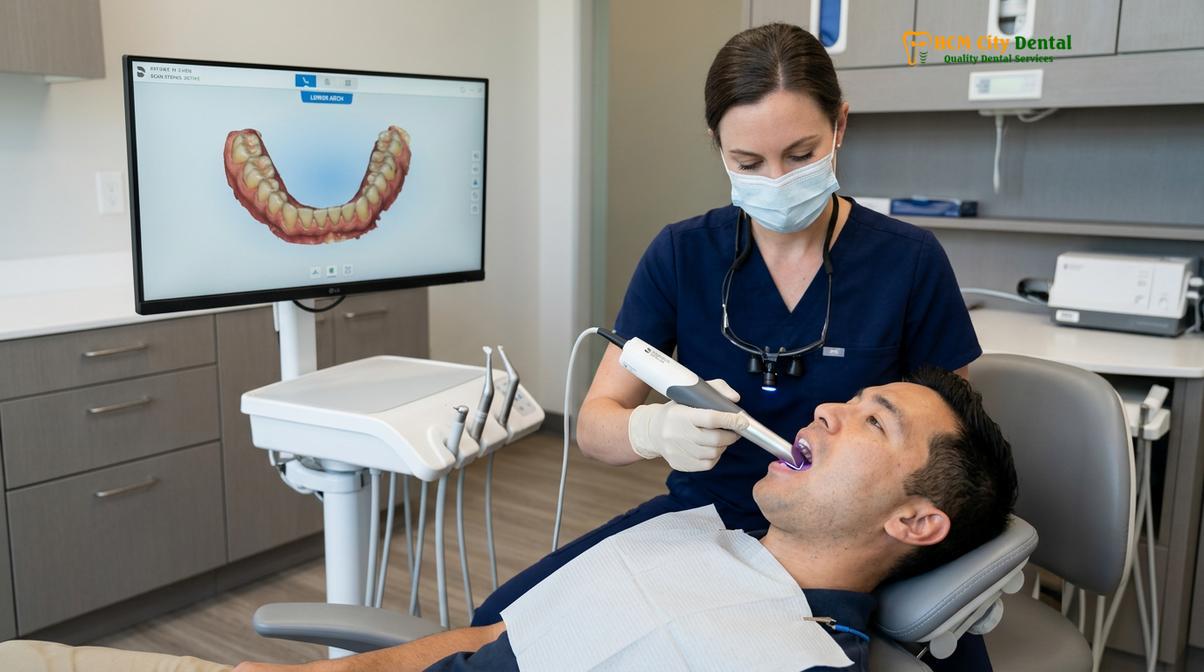
In the modern digital era, advanced imaging and scanning technologies have revolutionized diagnostic accuracy. An intraoral 3D scan, utilizing high-definition devices like the Medit i700, captures the exact topography of the patient’s current dentition and soft tissues in real-time. This digital impression is highly accurate and eliminates the discomfort associated with traditional putty molds. The digital data is then merged with a Cone Beam Computed Tomography (CBCT) scan. CBCT 3D bone diagnostics provide a comprehensive, three-dimensional view of the craniofacial skeleton, allowing the clinician to evaluate the exact position of the condyles within the TMJ, assess bone volume for potential dental implants, and analyze the skeletal relationship between the maxilla and mandible[3].
Progressive Treatment Phasing: Temporary Splints to Permanent Restorations
Rehabilitating a collapsed bite follows a phased approach, utilizing reversible diagnostic splints to verify patient tolerance before transitioning to permanent, bio-inert ceramic restorations.
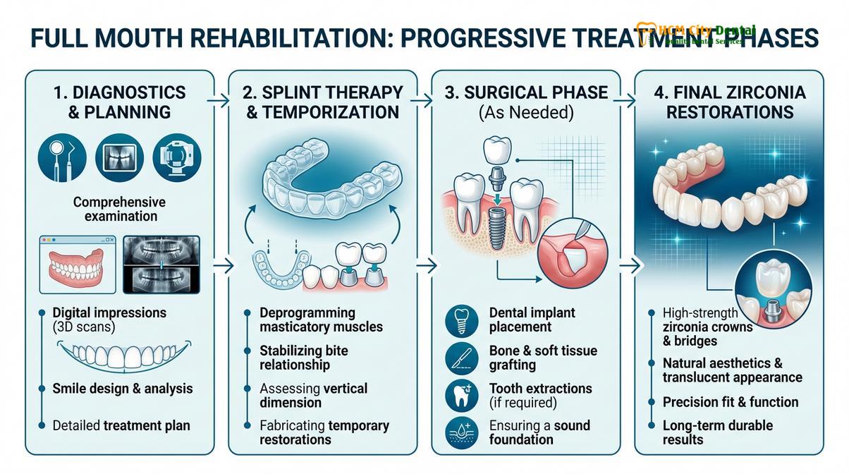
The restoration of a collapsed vertical dimension is a complex undertaking that requires meticulous planning and a progressive, phased approach. Altering the bite height too rapidly or without proper testing can lead to severe muscle spasms, joint pain, and the mechanical failure of the restorations. Therefore, the treatment workflow is designed to be highly predictable, allowing the patient’s neuromuscular system to adapt gradually.
Phase 1: Digital Smile Design and Diagnostic Splint Therapy
Once the optimal VDO is determined through diagnostics, the data is imported into specialized CAD software for Digital Smile Design (DSD). DSD allows the clinician to digitally sculpt the new smile, analyzing tooth proportions, gingival contours, and facial symmetry. A 3D-printed diagnostic wax-up is created, serving as a physical blueprint for the rehabilitation.
Before any irreversible changes are made to the natural teeth, the patient is fitted with a custom-milled diagnostic splint (often an orthotic device worn over the lower teeth). This splint is fabricated to the newly prescribed VDO. The patient wears this splint continuously for several weeks to months. This reversible trial period is crucial; it allows the clinician to verify that the new jaw position resolves TMJ symptoms, improves aesthetics, and is comfortable for mastication and phonetics. Only when the patient is completely asymptomatic and comfortable does the treatment proceed to the next phase.
Phase 2: Surgical Interventions and Foundation Building
If the bite collapse is associated with missing teeth or unsalvageable dentition, the surgical phase commences. This may involve the strategic placement of dental implants to provide robust posterior support. For patients requiring full arch replacement, structural biomechanics are optimized using All-on-4 or All-on-6 implant protocols. During these surgical procedures, patient comfort is paramount. Advanced clinics utilize IV conscious sedation, administered by board-certified anesthesiologists using precise Midazolam and Propofol protocols with continuous EKG monitoring, ensuring a safe, anxiety-free, and pain-free experience.
To accelerate bone grafting integration and soft tissue healing, Platelet-Rich Fibrin (PRF) therapy is frequently employed. PRF utilizes the patient’s own autologous blood growth factors to promote rapid angiogenesis and tissue regeneration around the implant sites.
Phase 3: CAD/CAM Precision and Final Restorations
The final phase involves transitioning the patient from provisional restorations to permanent, highly durable prosthetics. Utilizing CAD/CAM precision, the final crowns, bridges, or implant-supported prostheses are milled from advanced biomaterials. Biocompatible monolithic zirconia is often the material of choice for full mouth rehabilitations. Sourced from premium Swiss and German brands, monolithic zirconia is metal-free, bio-inert, and exceptionally resistant to fracture. Furthermore, advanced milling techniques achieve a margin fit of under 50 micrometers, ensuring optimal biological width preservation and preventing bacterial microleakage.
Transparent Pricing and International Support
For international patients seeking world-class care, understanding the financial investment is crucial. Clinics like HCMC Dental Clinic offer transparent pricing structures, often with significant advantages for patients who coordinate their care in advance. Below is an overview of the estimated pricing parameters, highlighting the 40% pre-arrival discount available for WhatsApp bookings:
| Treatment / Material Tier | Standard Walk-in Rate (Estimated) | WhatsApp Pre-Booking Rate (-40%) |
|---|---|---|
| Single Implant (Fixture & Abutment) | ||
| Economy (Dentis/Neo Biotech) | ~$770 (20M VND) | From $460 (12M VND) |
| Standard (Dentium Superline / IBS) | ~$1,090 (28.3M VND) | From $655 (17M VND) |
| Premium Plus (Straumann BLT-SLA) | ~$1,795 (46.6M VND) | From $1,080 (28.0M VND) |
| Crown on Implant (Per Tooth) | ~$260 – $385 | From $155 – $230 |
| Full Mouth Rehabilitation Packages | ||
| Full Arch Zirconia Bridge (All-on-4/6) | ~$12,000 – $22,000 per arch | From $7,200 – $13,200 per arch |
| Snap-On Overdenture (Implant-supported) | ~$5,000 – $8,000 per arch | From $3,000 – $4,800 per arch |
| Full Mouth Crowns (14 crowns, Zirconia HT) | ~$3,640 per arch | From $2,184 per arch |
In addition to competitive pricing, comprehensive care includes global manufacturer warranty cards (ranging from 10-year to lifetime guarantees on implant fixtures) and dedicated remote post-op checkups via WhatsApp, ensuring peace of mind for dental tourism patients returning to their home countries.
Neuromuscular Harmony: Preventing TMJ Stress Post-Reconstruction
Long-term success in VDO restoration requires meticulous occlusal calibration to ensure the new jaw position supports relaxed musculature and prevents excessive loading on the temporomandibular joints.
The ultimate goal of a lost VDO restoration is not merely to create beautiful teeth, but to establish a state of neuromuscular harmony. The newly reconstructed bite must function seamlessly within the patient’s “envelope of function”—the 3D space that defines mandibular movement during chewing and speaking. If the new restorations violate this envelope, the resulting occlusal interferences will trigger muscle hyperactivity, leading to bruxism, porcelain fractures, and renewed TMJ stress.
To prevent these complications, the concept of mutually protected occlusion is meticulously applied. In this occlusal scheme, the posterior teeth protect the anterior teeth by absorbing the heavy vertical forces of closure, while the anterior teeth protect the posterior teeth by discluding (separating) them during lateral and protrusive jaw movements. This anterior guidance is critical for shutting down the elevator muscles during excursive movements, thereby protecting the temporomandibular joint health and the structural integrity of the monolithic zirconia restorations[4].
Clinical Case Study: A 58-year-old expatriate presented to HCMC Dental Clinic in Ho Chi Minh City with severe bite collapse due to decades of unmanaged bruxism. The patient suffered from chronic tension headaches and a significantly reduced lower facial height. Under the care of Dr. Nguyen Van Cuong, a phased full mouth rehabilitation was initiated. Following 6 weeks of diagnostic splint therapy to achieve muscle relaxation, the patient’s VDO was increased by 4mm. The final restoration utilized 28 monolithic zirconia crowns. Post-treatment, the patient reported complete resolution of TMJ pain, improved masticatory efficiency, and a dramatically rejuvenated facial profile.
“Achieving neuromuscular harmony is the defining factor between a restoration that merely looks good and one that lasts a lifetime. The stomatognathic system must be treated as an integrated whole, where the teeth, muscles, and joints operate in perfect synchrony.”
Post-reconstruction maintenance is equally vital. Patients are typically provided with a custom-fitted occlusal night guard to protect their investment from nocturnal parafunctional habits. Regular clinical follow-ups, which can be facilitated remotely for international patients, ensure that the occlusal contacts remain balanced and that the supporting periodontal tissues remain healthy.
When to See a Doctor for Bite Collapse Treatment
Recognizing the early signs of a collapsing bite can prevent the need for extensive and complex reconstructive surgeries. You should seek a comprehensive clinical evaluation if you experience any of the following symptoms:
- Noticeable shortening or flattening of your teeth, particularly if the softer, yellow dentin is exposed.
- Chronic pain, clicking, or popping in the jaw joints (TMJ) during chewing or speaking.
- Frequent tension headaches, neck pain, or unexplained facial muscle fatigue upon waking.
- A visible change in your facial profile, such as deepening folds around the mouth or a chin that appears to protrude more than it used to.
- Difficulty pronouncing certain words or a noticeable change in your speech patterns.
- Multiple missing back teeth that have not been replaced, causing you to chew primarily with your front teeth.
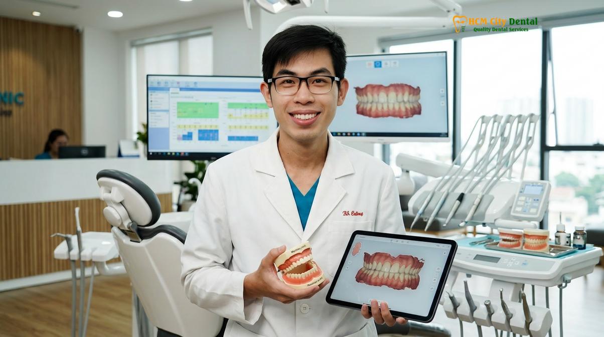
If you are experiencing these symptoms, a consultation with a specialist in prosthodontics or full mouth rehabilitation is highly recommended. At HCMC Dental Clinic, experts like Dr. Cuong utilize advanced digital diagnostics to assess the extent of the bite collapse and formulate a personalized, evidence-based treatment plan. Early intervention not only preserves remaining tooth structure but also restores your quality of life and facial aesthetics[5].
Frequently Asked Questions
What causes vertical dimension of occlusion to collapse?
The collapse of the vertical dimension of occlusion is primarily caused by severe tooth wear, chronic bruxism, missing posterior teeth, or advanced acid erosion. When the posterior teeth, which act as vertical stops, are lost or significantly worn down, the lower jaw over-closes. This over-closure forces the anterior teeth to bear unnatural loads, accelerating wear and leading to a reduced lower facial height and subsequent bite collapse.
How does restoring VDO change my facial appearance?
Restoring the VDO elongates the lower third of the face, providing proper support for the lips and cheeks, which often results in a rejuvenated, less sunken appearance. By reestablishing the correct anatomical jaw height, the procedure smooths out deep nasolabial folds, restores lip fullness, and corrects the pseudo-prognathic (protruding chin) look that is commonly associated with a severely collapsed bite.
Is VDO restoration painful?
VDO restoration is generally not painful, as modern clinical protocols utilize local anesthesia and IV conscious sedation to ensure a comfortable, anxiety-free experience. Post-operative sensitivity is typically mild and manageable with standard analgesics. While the initial adaptation to the new bite height may cause temporary, minor muscle fatigue, this resolves quickly as the neuromuscular system adapts.
How long does it take to adapt to a new vertical dimension?
Most patients adapt to a newly restored vertical dimension within two to four weeks of wearing a diagnostic splint or provisional restorations. During this transitional phase, the neuromuscular system recalibrates. Any initial phonetic difficulties, such as a slight lisp, or mild masticatory muscle tension typically subside rapidly as the jaw muscles relax into their new, optimal physiological position.
What materials are best for restoring a collapsed bite?
Biocompatible monolithic zirconia and premium lithium disilicate (E.max) are currently considered the gold standard materials for restoring a collapsed bite. These advanced CAD/CAM ceramics offer exceptional fracture toughness, precise margin adaptation, and lifelike translucency. Monolithic zirconia, in particular, is highly resistant to wear and provides the structural durability required to maintain the new vertical dimension long-term.
References
- The Journal of Prosthetic Dentistry. Vertical dimension of occlusion restoration parameters. (2021).
- International Journal of Prosthodontics. Biomechanical load distribution in All-on-4 restorations. (2020).
- Journal of Clinical Periodontology. Biocompatible dental materials and soft tissue response. (2019).
- Clinical Oral Implants Research. Platelet-rich fibrin in guided bone regeneration. (2022).
- Journal of Esthetic and Restorative Dentistry. Digital smile design functional protocols. (2018).
For customized treatment planning, transparent cost estimates, and direct consultation with Dr. Cuong, visit our comprehensive Full Mouth Rehabilitation service page or message our team directly via WhatsApp to receive an instant assessment.