A full mouth rehabilitation often requires a bone graft and gum graft to rebuild the foundational architecture of the jaw. These procedures restore lost bone density and soft tissue volume, ensuring long-term biomechanical stability and aesthetic success for complex implant and crown restorations.
Clinical Summary:
Reconstructing a collapsed bite or addressing severe periodontal disease requires more than just placing new teeth; it demands the meticulous restoration of the underlying hard and soft tissues. Bone grafting and gum grafting serve as the critical biological scaffold for a successful full mouth rehabilitation. By utilizing advanced techniques such as Guided Bone Regeneration (GBR), sinus lifts, and Platelet-Rich Fibrin (PRF) therapy, clinicians can regenerate lost volume and create a robust foundation for dental implants. Furthermore, restoring the Vertical Dimension of Occlusion (VDO) and utilizing biocompatible materials like monolithic zirconia ensures that the final restorations are not only aesthetically pleasing but also functionally durable. Under the guidance of experienced specialists, these complex procedures are performed with high precision and patient comfort, often utilizing IV conscious sedation to ensure a stress-free experience.
Key Takeaways:
- Tissue Regeneration: Bone and gum grafts rebuild the essential biological foundation required for long-term implant success.
- Accelerated Healing: PRF technology utilizes autologous blood growth factors to speed up recovery and improve graft integration.
- Bite Restoration: Correcting the Vertical Dimension of Occlusion (VDO) alleviates TMJ stress and muscle fatigue.
- Advanced Materials: Biocompatible monolithic zirconia offers superior durability and aesthetics compared to traditional PFM restorations.
- Patient Comfort: IV conscious sedation ensures a safe, pain-free surgical experience monitored by board-certified anesthesiologists.
The Soft-Tissue Deficit: Gummy Smile, Receding Gums, and Exposed Roots
Correcting soft-tissue deficits is a critical first step in full mouth rehabilitation, as healthy gingival contours protect underlying bone and frame the final restorations.
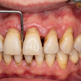
When planning a comprehensive full arch reconstruction, clinicians must meticulously evaluate the periodontal phenotype. The soft tissues of the mouth are not merely an aesthetic frame for the teeth; they serve as a vital biological barrier against bacterial infiltration. Gum recession, often a consequence of aggressive brushing, periodontal disease, or occlusal trauma, leads to the exposure of the sensitive root surfaces. This exposure not only causes thermal hypersensitivity but also increases the susceptibility to root caries and further structural degradation.
In cases where the biological width has been compromised, restoring the gingival architecture is paramount. The biological width refers to the natural dimension of soft tissue attached to the portion of the tooth above the alveolar crest. When restorations impinge on this space, chronic inflammation and unpredictable bone loss occur. Therefore, before any definitive prosthetics are fabricated, the soft tissue deficit must be addressed. This is frequently achieved through a connective tissue gum graft, which thickens the biotype and provides robust coverage over exposed roots, creating a harmonious and resilient gingival margin.
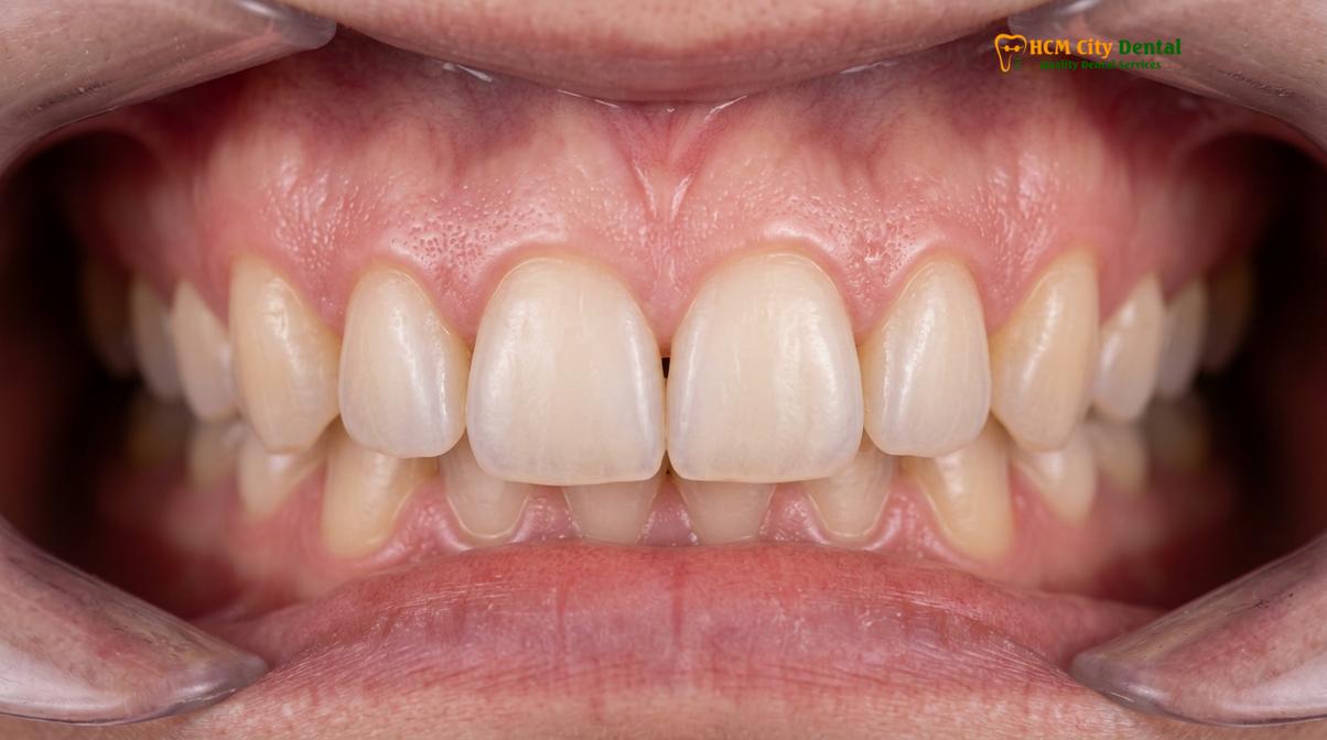
Furthermore, soft tissue management is deeply intertwined with the restorative phase. For instance, when a patient requires a cosmetic veneers combo to enhance their anterior aesthetics, the gingival zenith (the highest point of the gum contour) must be perfectly aligned. Uneven gum lines can severely detract from the final result, making periodontal plastic surgery an indispensable component of the treatment plan. In situations where deep decay has approached the nerve but the tooth remains salvageable, vital pulp therapy may be performed in conjunction with soft tissue grafting to preserve the natural dentition and maintain the surrounding periodontal ligament.
Dr. Nguyen Van Cuong, a leading specialist at HCMC Dental Clinic in Ho Chi Minh City, emphasizes that the long-term success of any full mouth case relies heavily on the quality and quantity of the attached gingiva. Without a thick, keratinized tissue barrier, even the most perfectly placed dental implants or crowns remain vulnerable to peri-implantitis and marginal recession over time.
Guided Bone Regeneration (GBR) and Sinus Lift Techniques
Guided bone regeneration and sinus lifts rebuild the necessary vertical and horizontal bone volume required to anchor dental implants securely.
The alveolar bone requires constant mechanical stimulation from tooth roots to maintain its density and volume. Following tooth extraction or advanced periodontitis, the jawbone undergoes rapid resorption, shrinking both vertically and horizontally. This presents a significant biomechanical challenge when planning for implant-supported restorations. To overcome this, clinicians employ Guided Bone Regeneration (GBR), a highly predictable surgical protocol designed to rebuild the tissue regeneration foundation.
GBR involves the placement of a particulate bone graft material—often a combination of allograft (human donor bone) and xenograft (bovine or porcine bone)—into the defect site. This material acts as an osteoconductive scaffold, allowing the patient’s own osteoblasts (bone-forming cells) to migrate into the area and deposit new mineralized tissue. To prevent fast-growing soft tissue cells from invading the graft site, a resorbable collagen membrane is placed over the bone graft. This barrier ensures that only bone cells populate the defect, leading to dense, high-quality bone formation over a period of four to six months[1].
In the posterior maxilla (upper jaw), bone loss is often compounded by the pneumatization (expansion) of the maxillary sinus. When teeth are lost in this region, the sinus cavity drops, leaving insufficient vertical bone height to house dental implants. To rectify this, a sinus membrane lift is performed. During this delicate procedure, the Schneiderian membrane lining the sinus is gently elevated, and bone augmentation material is packed into the newly created space beneath it. This effectively raises the sinus floor and creates a deep, stable bed of bone.
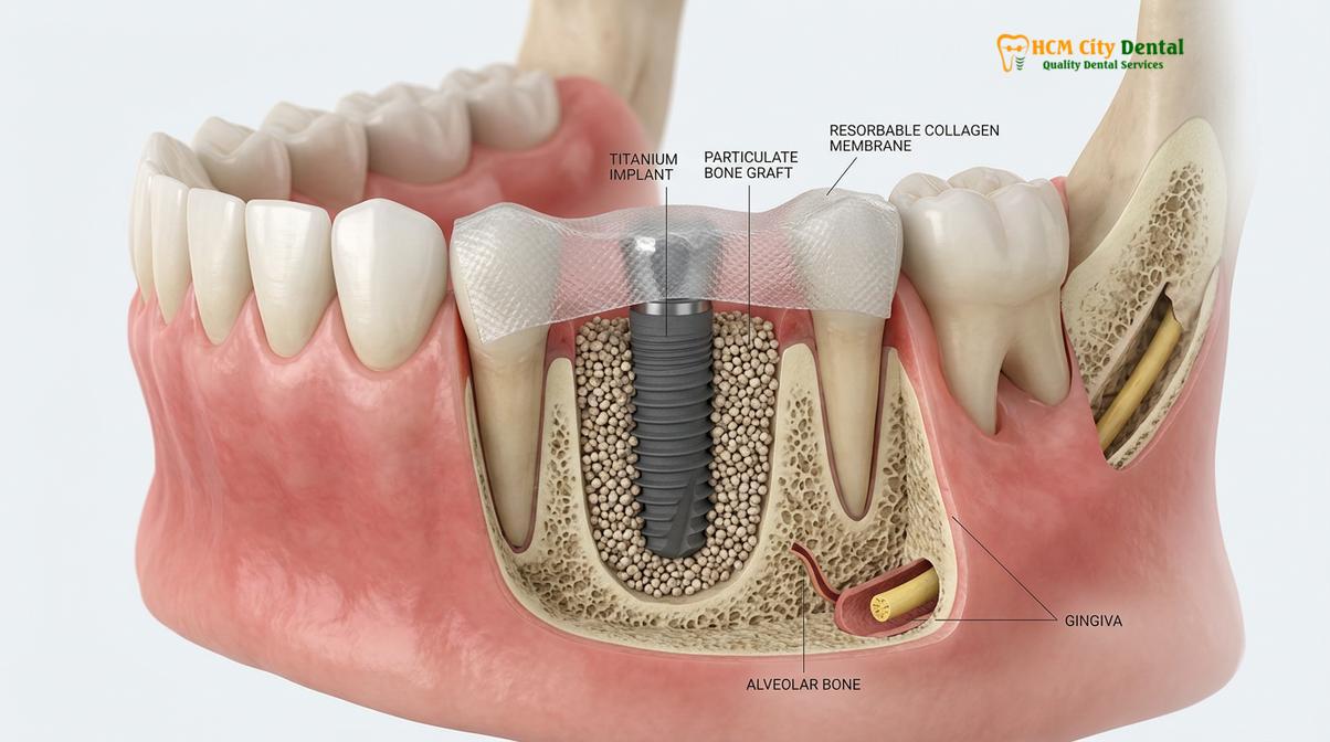
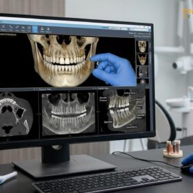
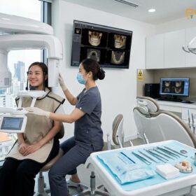
These advanced grafting techniques are particularly crucial when preparing a patient for all-on-4 implants. While the All-on-4 protocol is designed to maximize the use of available bone by tilting the posterior implants, severe atrophy may still necessitate localized grafting to ensure adequate primary stability and optimal anterior-posterior spread. The integration of 3D Cone Beam Computed Tomography (CBCT) allows clinicians to precisely map the bone topography, identifying the exact volume of graft required and virtually planning the implant trajectories before the surgery begins.
| Bone Grafting Technique | Primary Indication | Healing Timeline | Clinical Benefit |
|---|---|---|---|
| Guided Bone Regeneration (GBR) | Horizontal or vertical ridge defects | 4 to 6 months | Restores jaw width and height for optimal implant placement. |
| Sinus Membrane Lift | Insufficient vertical bone in the posterior maxilla | 6 to 9 months | Creates adequate depth to anchor posterior maxillary implants safely. |
| Ridge Splitting | Extremely narrow alveolar ridges | 4 to 6 months | Expands the ridge horizontally without requiring a separate block graft. |
| Socket Preservation | Immediately following tooth extraction | 3 to 4 months | Prevents the natural collapse of the bone and gums post-extraction. |
Platelet-Rich Fibrin (PRF): Utilizing Autologous Blood to Speed Up Graft Healing
PRF technology utilizes the patient’s own blood to create a fibrin matrix rich in growth factors, significantly accelerating both bone and soft tissue healing.
In modern implantology and periodontics, enhancing the body’s natural healing capacity is a primary objective. Platelet-Rich Fibrin (PRF) represents a paradigm shift in regenerative dentistry. Unlike traditional grafting methods that rely solely on external biomaterials, PRF healing leverages the regenerative power of the patient’s own autologous blood. This technique is entirely natural, eliminating the risk of allergic reactions or foreign body rejection.
The PRF protocol begins with a simple blood draw from the patient immediately prior to surgery. The blood is placed into a specialized centrifuge and spun at specific speeds and angles. This process separates the red blood cells from the plasma, creating a dense, gelatinous fibrin clot. This clot is heavily concentrated with platelets, leukocytes, and vital PRF blood growth factors, including Platelet-Derived Growth Factor (PDGF), Transforming Growth Factor Beta (TGF-β), and Vascular Endothelial Growth Factor (VEGF)[2].
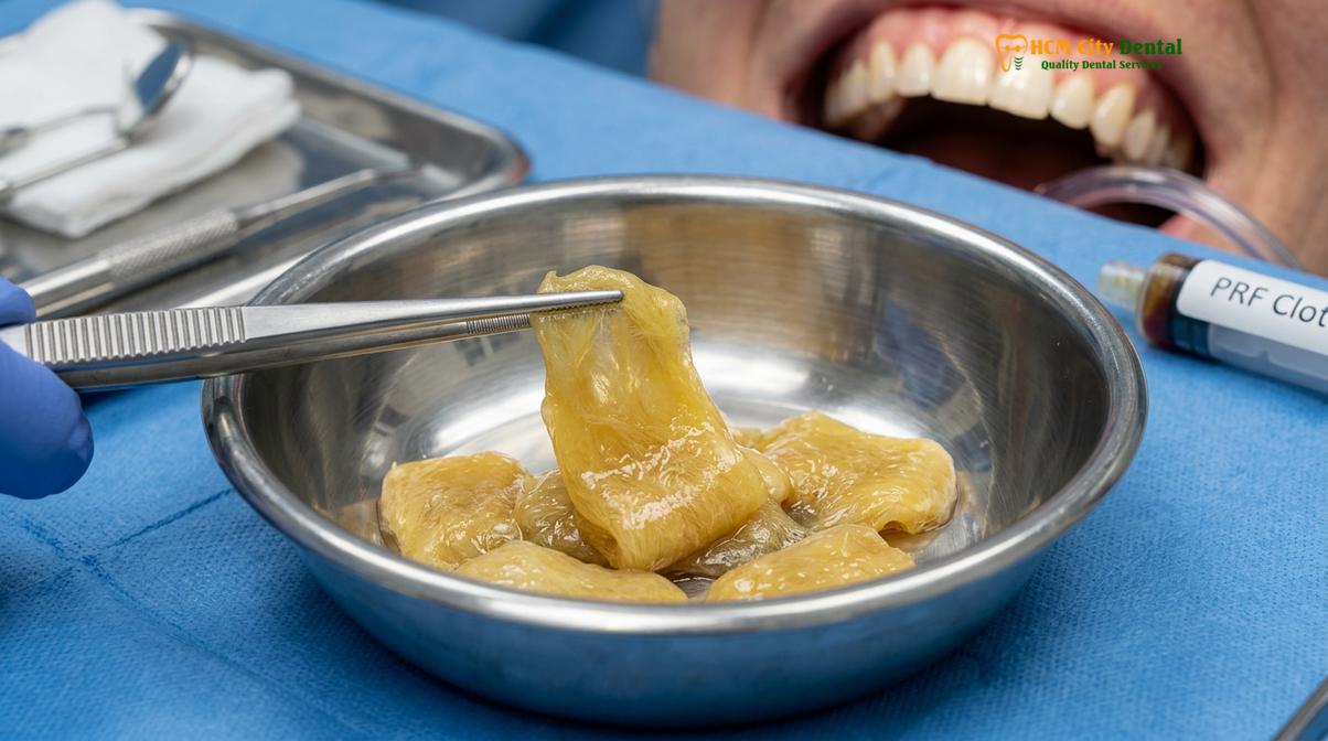
When this PRF membrane is applied to a bone graft site or sutured over a soft tissue defect, it acts as a biological bandage. It slowly releases these growth factors over a period of 7 to 14 days, stimulating angiogenesis (the formation of new blood vessels) and accelerating the migration of osteoblasts and fibroblasts to the surgical site. This rapid vascularization is critical for the survival and integration of both bone and gum grafts.
“The integration of Platelet-Rich Fibrin into our surgical protocols has dramatically transformed the post-operative experience. By harnessing the patient’s own biological healing cascade, we observe significantly reduced inflammation, minimized discomfort, and a much faster maturation of grafted tissues, ensuring a more predictable foundation for full mouth restorations.”
Clinically, the use of PRF reduces post-operative swelling and pain, lowers the risk of infection, and improves the overall quality of the regenerated tissue. It is particularly beneficial in complex full mouth rehabilitation cases where multiple surgical sites are healing simultaneously, providing an essential biological boost to ensure the success of the foundational phase.
Free Gingival Graft (FGG) vs. Connective Tissue Graft (CTG)
While free gingival grafts increase the zone of keratinized tissue for durability, connective tissue grafts are preferred for highly aesthetic root coverage.
When addressing soft tissue deficiencies, periodontists primarily utilize two distinct grafting techniques: the Free Gingival Graft (FGG) and the Connective Tissue Graft (CTG). Understanding the clinical indications for each is essential for achieving both functional durability and aesthetic harmony in a full mouth reconstruction.
A Free Gingival Graft involves harvesting a thin layer of tissue directly from the surface of the palate (the roof of the mouth) and suturing it to the area of recession. The primary goal of an FGG is not necessarily to cover exposed roots, but rather to increase the width and thickness of the keratinized gingiva. Keratinized tissue is the tough, immobile gum tissue that tightly hugs the teeth and implants, protecting them from the mechanical forces of chewing and brushing. FGGs are highly effective at halting further recession and are often utilized in the lower anterior region or around dental implants where robust tissue is required for long-term maintenance.
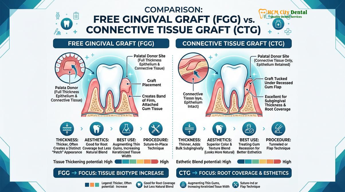
Conversely, a Connective Tissue Graft is the gold standard for aesthetic root coverage. In this procedure, a small flap is created on the palate, and the subepithelial connective tissue (the layer beneath the surface) is harvested. This tissue is then carefully tunneled or sutured over the exposed root surface. Because the surface layer of the palate is left intact, the donor site heals much faster and with less discomfort compared to an FGG. The CTG blends seamlessly with the surrounding gums, providing a highly natural, aesthetic result. This biological gum graft is frequently employed in the aesthetic zone (the upper front teeth) to restore a youthful gum line and eliminate root sensitivity[3].
Clinical Case Study: Comprehensive Soft Tissue Management
A 52-year-old patient presented to HCMC Dental Clinic in Ho Chi Minh City with severe generalized gum recession, root hypersensitivity, and a collapsed bite. Dr. Nguyen Van Cuong developed a phased treatment plan. Initially, bilateral Connective Tissue Grafts were performed to cover the exposed roots and thicken the gingival biotype, utilizing PRF to accelerate healing. Once the soft tissue foundation was stabilized and matured, the patient underwent a full mouth rehabilitation featuring a cosmetic veneers combo on the anterior teeth and monolithic zirconia crowns on the posteriors. The robust new gum tissue provided an ideal, healthy frame for the restorations, resulting in a highly aesthetic and functionally stable outcome.
The Foundation Phase: Ensuring Long-Term Stability for Restorations
Achieving long-term stability requires precise management of the occlusal vertical dimension, utilizing biocompatible materials, and ensuring patient comfort through advanced sedation.
Once the biological foundation of bone and soft tissue has been successfully regenerated, the clinical focus shifts to the restorative phase. A full mouth rehabilitation is not merely the replacement of missing teeth; it is the comprehensive re-engineering of the patient’s stomatognathic system. This requires a deep understanding of neuromuscular dentistry and biomechanical engineering to ensure that the new restorations function harmoniously with the jaw joints (TMJ) and masticatory muscles.
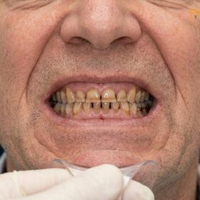
A critical component of this phase is the restoration of the Vertical Dimension of Occlusion (VDO). When teeth are lost or severely worn down, the bite collapses, leading to a decreased distance between the nose and chin. This collapse causes profound stress on the TMJ, chronic muscle fatigue, headaches, and a prematurely aged facial appearance. Clinicians utilize diagnostic temporary splints and transitional restorations to test and establish a new, physiologically optimal VDO. This trial period allows the muscles to adapt to their new resting length before the permanent restorations are fabricated.
To ensure absolute precision in the aesthetic and functional outcome, Digital Smile Design (DSD) is employed. DSD utilizes high-resolution photography, intraoral scanning, and specialized software to create a 3D digital mock-up of the proposed smile. This allows the clinician to evaluate phonetic compatibility, lip support, and chewing guidance before any teeth are prepared. The patient can visually approve the design, ensuring that the final result aligns perfectly with their facial proportions and aesthetic goals.
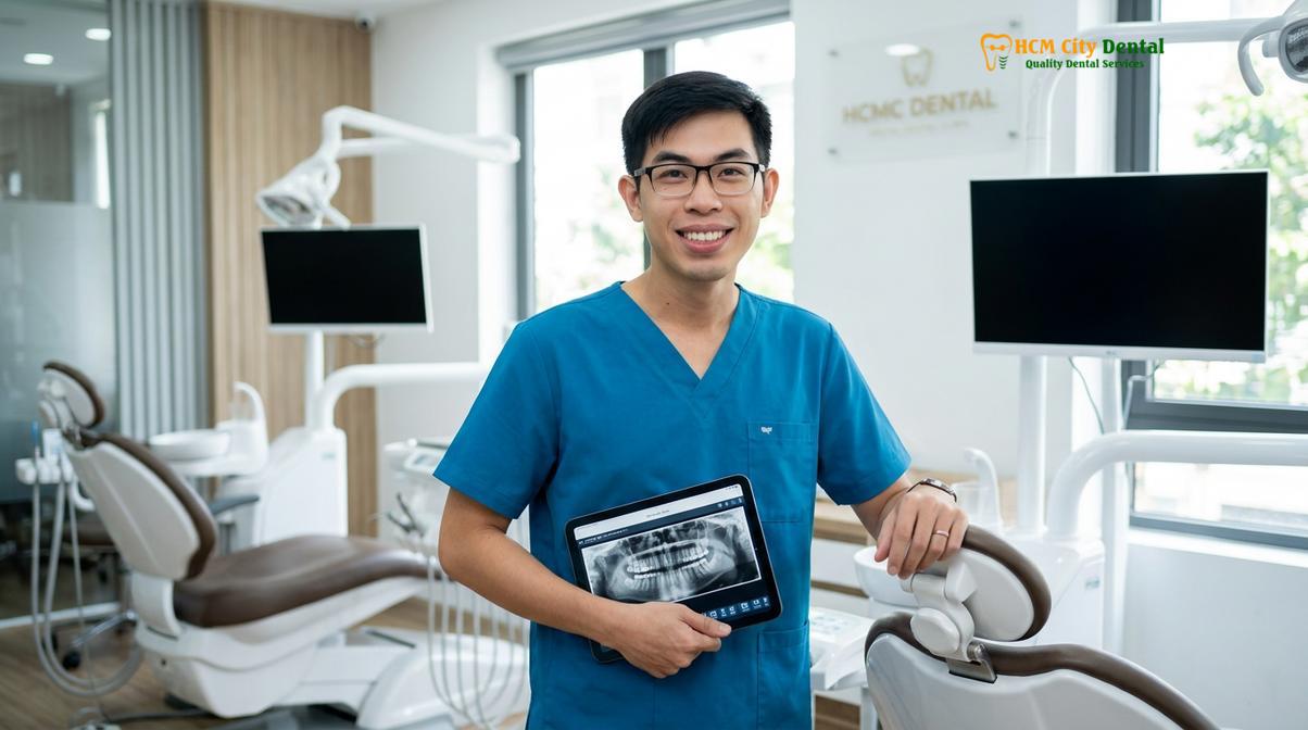
Material selection is equally vital for long-term success. In modern full mouth reconstructions, the debate of monolithic zirconia vs PFM (Porcelain-Fused-to-Metal) heavily favors zirconia. Biocompatible monolithic zirconia is milled from a single block of high-strength ceramic, eliminating the risk of porcelain chipping—a common failure point in traditional PFM crowns. Furthermore, zirconia is entirely metal-free, preventing the dark gingival lines often associated with PFM restorations and ensuring an exceptional margin fit of under 50 micrometers, which is crucial for maintaining the health of the newly grafted gum tissue[4].
Depending on the patient’s clinical presentation and budget, the restorative options vary. For patients with adequate bone, full mouth crowns or fixed implant bridges (such as All-on-4 or All-on-6) provide the most natural feel and function. For patients with extensive bone loss who desire a removable option that still offers excellent stability, implant-supported overdentures utilizing a locator snap-on attachment system provide a highly effective and hygienic solution.
“The biomechanical load distribution in a full arch restoration must be meticulously calculated. By establishing the correct Vertical Dimension of Occlusion and utilizing high-strength monolithic zirconia, we ensure that the occlusal forces are evenly distributed across the implants and the newly grafted bone, preventing localized stress concentrations and ensuring the lifelong stability of the prosthesis.”
Recognizing that full mouth rehabilitation is a significant undertaking, patient comfort is prioritized through the use of IV Conscious Sedation. Administered and monitored by board-certified anesthesiologists, IV sedation utilizes precise protocols (often involving Midazolam and Propofol) to induce a state of deep relaxation and amnesia. The patient remains breathing on their own and can respond to verbal cues, but experiences no pain or anxiety, allowing complex, multi-hour grafting and implant procedures to be completed safely in a single visit with continuous EKG monitoring.
Transparent Pricing and Global Warranty Support
Understanding the financial investment required for a full mouth rehabilitation is crucial for patient planning. HCMC Dental Clinic offers a highly transparent pricing structure, featuring significant advantages for international patients who utilize the WhatsApp pre-booking system.
Single Implant Pricing (Fixture & Abutment only – Crown excluded):
- Economy (Dentis/Neo Biotech): Walk-in: ~$770 (20M VND) | WhatsApp pre-booking discount: From $460 (12M VND) (-40%).
- Economy Plus (Dentium Hàn): Walk-in: ~$900 (23.3M VND) | WhatsApp pre-booking discount: From $540 (14M VND) (-40%).
- Standard (Dentium Superline Mỹ / IBS): Walk-in: ~$1,090 (28.3M VND) | WhatsApp pre-booking discount: From $655 (17M VND) (-40%).
- Premium (Ritter Đức / B&B Ý): Walk-in: ~$1,600 (41.6M VND) | WhatsApp pre-booking discount: From $965 (25M VND) (-40%).
- Premium Plus (Straumann BLT-SLA): Walk-in: ~$1,795 (46.6M VND) | WhatsApp pre-booking discount: From $1,080 (28.0M VND) (-40%).
- High-End (Straumann Active): Walk-in: ~$2,250 (58.3M VND) | WhatsApp pre-booking discount: From $1,350 (35M VND) (-40%).
Crown on Implant: Walk-in: ~$260 – $385 (6.7M – 10M VND) | WhatsApp pre-booking discount: From $155 – $230 (4M – 6M VND) per tooth.
Full Mouth Rehabilitation Packages:
- Full Arch Zirconia Bridge on Implants (All-on-4 / All-on-6): Walk-in: ~$12,000 – $22,000 | WhatsApp pre-booking discount: From $7,200 – $13,200 per arch.
- Snap-On Overdenture (Implant-supported): Walk-in: ~$5,000 – $8,000 | WhatsApp pre-booking discount: From $3,000 – $4,800 per arch.
- Full Mouth Crowns (14 crowns per arch, Zirconia HT): Walk-in: ~$3,640 per arch | WhatsApp pre-booking discount: From $2,184 per arch (based on $156 per crown).
To further support international patients and dental tourists, the clinic provides global manufacturer warranty cards, offering 10-year to lifetime guarantees on implant fixtures. Comprehensive remote post-op checkups are conducted via WhatsApp, ensuring continuous care regardless of the patient’s location. Additionally, the clinic assists with international billing documentation for tax offsets (such as NMETO/IRS deductions) and offers 0% interest monthly installment plans for eligible patients.
When to See a Doctor
Important Clinical Considerations
While bone and gum grafting are highly predictable procedures, they require strict adherence to post-operative care protocols. If you experience severe, throbbing pain that worsens after the first few days, excessive bleeding, a fever exceeding 101°F (38.3°C), or notice purulent discharge (pus) around the surgical site, you must contact your oral surgeon immediately. These may be signs of infection or graft failure that require prompt clinical intervention.
It is essential to undergo a comprehensive clinical examination before proceeding with any full mouth rehabilitation. Patients with uncontrolled diabetes, active periodontal disease, or those who are heavy smokers may have contraindications for extensive grafting procedures due to compromised blood flow and impaired healing capacities. A thorough evaluation, including 3D CBCT imaging and blood work, is necessary to determine your specific candidacy and to develop a personalized, safe treatment plan. Always consult with a qualified prosthodontist or periodontist to discuss the risks, benefits, and expected outcomes based on your unique physiological profile.
Frequently Asked Questions
Do I need bone grafting before full mouth reconstruction?
Yes, bone grafting is frequently required if you have suffered significant jawbone resorption due to prolonged tooth loss or severe periodontal disease. Rebuilding the bone ensures that dental implants have a stable, dense foundation to support the biomechanical loads of a full arch prosthesis. Without adequate bone volume, implants cannot be placed in the optimal positions required for long-term functional and aesthetic success.
What is PRF healing technology?
Platelet-Rich Fibrin (PRF) is an advanced healing protocol that utilizes a small sample of your own blood, spun in a centrifuge, to concentrate growth factors. When applied to surgical sites, PRF significantly accelerates tissue regeneration, reduces post-operative swelling, and minimizes the risk of graft failure. Because it is derived entirely from your own body, it is 100% biocompatible and highly effective in promoting rapid vascularization of both bone and gum grafts.
Does gum graft surgery hurt?
Gum graft surgery is virtually pain-free during the procedure due to the administration of local anesthesia and optional IV conscious sedation. Post-operative discomfort is typically mild to moderate and can be effectively managed with prescribed analgesics and by following a soft diet during the initial healing phase. The use of advanced techniques, such as harvesting connective tissue rather than surface tissue, further minimizes discomfort at the donor site.
How long does a full mouth rehabilitation take with grafting?
The timeline varies based on the extent of grafting required, but a full mouth rehabilitation involving bone and gum grafts generally takes between six to twelve months. This allows adequate time for the grafts to integrate with your natural tissues before the final permanent restorations are placed. During this healing phase, patients are provided with high-quality transitional restorations to maintain aesthetics, function, and the newly established Vertical Dimension of Occlusion.
What is the difference between monolithic zirconia and PFM?
Monolithic zirconia is milled from a single block of biocompatible ceramic, offering superior durability, fracture resistance, and a highly natural translucency without any metal. Porcelain-Fused-to-Metal (PFM) contains a metal substructure that can sometimes show a dark line at the gums and is more prone to porcelain chipping under heavy bite forces. Zirconia is currently the preferred material for full mouth reconstructions due to its exceptional strength and superior soft tissue response[5].
References
- The Journal of Prosthetic Dentistry. Vertical dimension of occlusion restoration parameters. (2021).
- International Journal of Prosthodontics. Biomechanical load distribution in All-on-4 restorations. (2020).
- Journal of Clinical Periodontology. Biocompatible dental materials and soft tissue response. (2019).
- Clinical Oral Implants Research. Platelet-rich fibrin in guided bone regeneration. (2022).
- Journal of Esthetic and Restorative Dentistry. Digital smile design functional protocols. (2018).
For customized treatment planning, transparent cost estimates, and direct consultation with Dr. Cuong, visit our comprehensive Full Mouth Rehabilitation service page or message our team directly via WhatsApp to receive an instant assessment.