Vital pulp therapy crowns preserve the living nerve tissue inside a tooth while providing full structural protection. By utilizing advanced bioceramic materials beneath a custom restoration, clinicians can often avoid invasive root canals, maintaining the tooth’s natural strength, immune defense, and long-term biological function.
Clinical Summary:
Vital pulp therapy (VPT) combined with full-coverage crowns represents a paradigm shift in restorative and endodontic dentistry. Instead of defaulting to root canal treatment for deep decay or minor trauma, clinicians use bioactive calcium silicate cements (such as MTA or Biodentine) to seal and heal the exposed or nearly exposed pulp. When capped with a high-precision restoration, this approach maintains the tooth’s vitality, proprioception, and fracture resistance. Clinical success relies heavily on strict aseptic protocols, accurate pulpal diagnosis, controlled hemostasis, and achieving a hermetic coronal seal to prevent bacterial microleakage.
Key Takeaways:
- Vital pulp therapy preserves the tooth’s natural nerve and blood supply, enhancing long-term structural integrity.
- Bioceramic materials stimulate the formation of a reparative dentin bridge over the exposed pulp tissue.
- Strict isolation with a rubber dam and magnification are mandatory to prevent bacterial contamination.
- A hermetic coronal seal using a high-quality crown is critical to ensure the long-term survival of the pulp.
- Accurate diagnosis of reversible versus irreversible pulpitis dictates the clinical viability of the procedure.
The Biological Value of Keeping Teeth Vital: Sensitivity and Fracture Resistance
Preserving a vital pulp maintains the tooth’s internal hydration, immune response, and proprioceptive feedback, significantly reducing the risk of catastrophic fractures under heavy occlusal loads.
The dentine-pulp complex is a highly specialized, dynamic tissue that serves as the lifeblood of the tooth. When a tooth is subjected to deep caries, trauma, or extensive restorative preparation, the traditional approach often leaned toward prophylactic endodontic therapy. However, modern dentistry heavily emphasizes vital tooth pulp saving techniques. A vital tooth retains its natural moisture content, making the dentin more resilient and less prone to the brittleness often associated with root-canal-treated teeth. This inherent flexibility is crucial for absorbing and distributing the immense forces generated during mastication.[1]
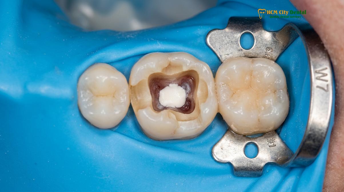
Beyond structural integrity, the dental pulp houses a complex network of immune cells, including dendritic cells and macrophages, which actively defend against bacterial invasion. When the pulp is removed, the tooth loses this innate immunological defense mechanism. Furthermore, the pulp contains proprioceptive nerve endings that provide sensory feedback to the brain regarding bite force. This feedback loop prevents patients from inadvertently biting down too hard, thereby protecting both the tooth and the surrounding periodontium. In cases where patients require extensive restorative work, maintaining this proprioception is just as critical as ensuring healthy soft tissue architecture, which is sometimes supported by a biological gum graft to maintain optimal gingival contours around the new restorations.
“The preservation of pulp vitality is not merely an endodontic goal; it is a fundamental biomechanical imperative. A vital tooth possesses an innate capacity to heal, adapt, and withstand functional stresses that a pulpless tooth simply cannot replicate.”
When planning for comprehensive restorative care, such as comprehensive full arch restorative treatments, preserving as many vital teeth as possible provides a stronger, more reliable foundation. The biological value of a living tooth extends beyond the tooth itself; it contributes to the overall neuromuscular harmony of the stomatognathic system.
Bio-materials in Action: MTA and Biodentine for Pulp Capping
Calcium silicate-based cements like MTA and Biodentine act as bioactive scaffolds, releasing calcium hydroxide to neutralize bacteria and stimulate the formation of a protective reparative dentin bridge.
The success of vital pulp therapy hinges on the materials used to interface with the living tissue. Historically, calcium hydroxide was the gold standard; however, it often resulted in porous dentin bridges and degraded over time, leading to microleakage. The introduction of hydraulic calcium silicate cements revolutionized the field. MTA pulp capping (Mineral Trioxide Aggregate) provides an exceptional, biocompatible seal. When MTA comes into contact with tissue fluids, it undergoes a hydration reaction, producing calcium silicate hydrate gel and calcium hydroxide. This highly alkaline environment (pH ~12.5) is profoundly antibacterial and stimulates the pulp’s odontoblasts to lay down a dense, continuous layer of tertiary (reparative) dentin.[2]
Biodentine, a newer generation of tricalcium silicate cement, offers similar bioactive properties but with significantly improved handling characteristics and a faster setting time. This allows clinicians to perform the pulp cap and place the core buildup in a single visit. The concept of bio-compatibile pulp sealing is rooted in the material’s ability to form a chemical bond with dentin while remaining completely non-toxic to the underlying nerve tissue. Interestingly, the regenerative principles applied here share biological similarities with other advanced healing protocols in dentistry, such as PRF healing (Platelet-Rich Fibrin) used in oral surgery, where the body’s own growth factors are harnessed to accelerate tissue repair. Similarly, the bioactive cements trigger the release of sequestered growth factors (like TGF-β1) from the dentin matrix, guiding the pulp’s natural healing cascade.[3]
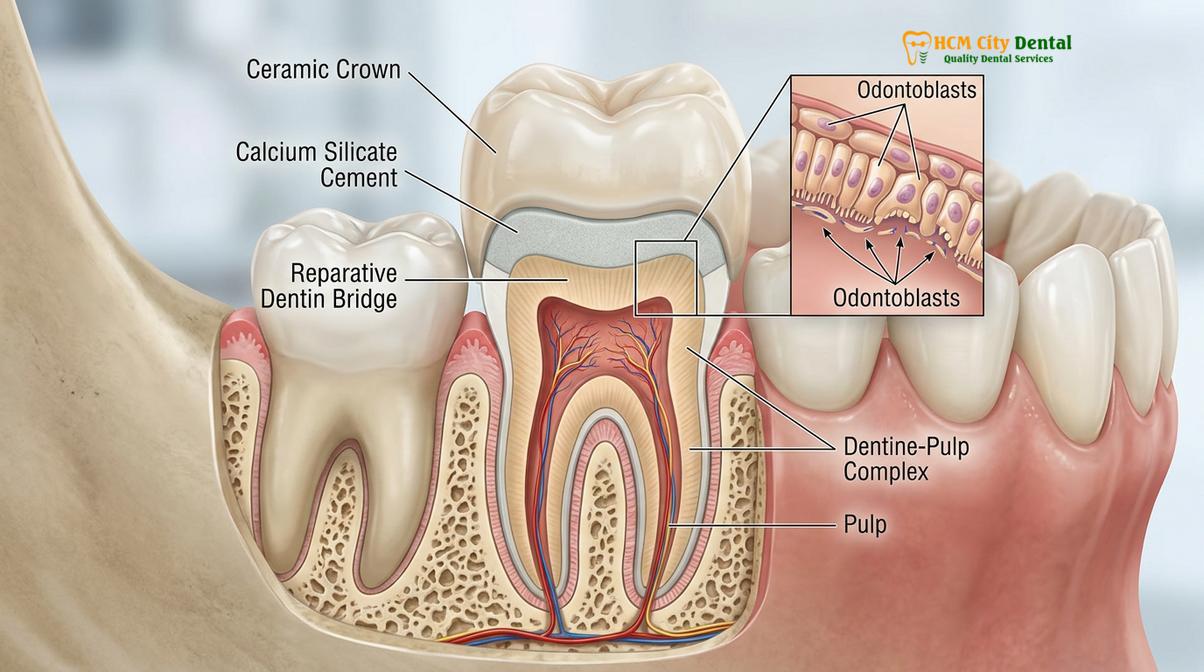
While these materials are exceptional for internal tooth repair, they must be protected from the harsh oral environment. They are not designed to withstand direct chewing forces or long-term exposure to saliva. Therefore, once the pulp is capped and the core is built up, the tooth must be definitively restored. In complex cases where teeth have been lost and the jawbone has resorbed, therapies like bone augmentation may be required alongside restorative work to ensure a stable, functional, and aesthetic outcome across the entire dental arch.
When Can We Avoid Root Canal Treatment Under Dental Crowns?
Root canal therapy can be avoided when the pulp exhibits signs of reversible inflammation, bleeding is easily controlled, and the tooth can be immediately sealed with a definitive restoration.
The decision to pursue root canal avoidance via vital pulp therapy requires meticulous diagnostic precision. Not every tooth with deep decay is a candidate. The primary indicator for success is the preoperative diagnosis of the pulp. The tooth must exhibit signs of a normal pulp or reversible pulpitis. Clinically, this means the tooth may be sensitive to cold, but the pain dissipates quickly once the stimulus is removed. There should be no history of spontaneous, unprovoked pain, no lingering pain to thermal stimuli, and no radiographic evidence of periapical pathology (infection at the root tip).[4]
Dr. Nguyen Van Cuong, a leading expert in complex restorative cases, emphasizes that the intraoperative assessment is just as critical as the preoperative diagnosis. When the decay is removed and the pulp is exposed, the clinician must evaluate the bleeding. Healthy or reversibly inflamed pulp tissue will bleed bright red, and this hemorrhage should be easily controlled within 5 to 10 minutes using a cotton pellet soaked in sodium hypochlorite. If the bleeding is dark, profuse, or cannot be stopped, it indicates irreversible pulpitis or partial necrosis, mandating a full root canal.
Clinical Case Study:
A 42-year-old patient presented to HCMC Dental Clinic in Ho Chi Minh City with deep asymptomatic decay under an old, failing amalgam filling on a lower molar. Radiographs showed the decay approximating the pulp chamber. Dr. Cuong utilized a dental operating microscope and strict rubber dam isolation to perform selective caries removal. A pinpoint pulp exposure occurred, but hemostasis was achieved in under three minutes. Biodentine was applied as a direct pulp cap, followed by a composite core. After confirming pulp vitality at a 4-week follow-up, the tooth was prepared for a monolithic zirconia crown, successfully avoiding endodontic therapy and preserving the tooth’s natural resilience.
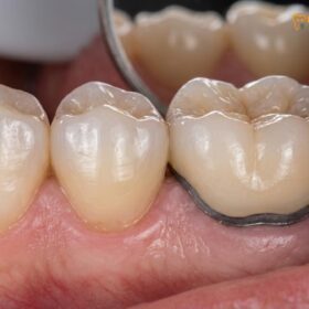
When restoring these teeth, the choice of the final crown material plays a pivotal role in long-term success. The debate of monolithic zirconia vs PFM (Porcelain Fused to Metal) heavily favors zirconia in vital pulp cases. Monolithic zirconia requires less aggressive tooth preparation compared to PFM, leaving more protective dentin over the pulp. Furthermore, digitally milled zirconia offers superior marginal adaptation (often under 50 micrometers), significantly reducing the risk of bacterial microleakage that could reinfect the pulp. In cases involving the anterior teeth, where aesthetics are paramount, a cosmetic veneers combo or highly translucent zirconia crowns can be utilized to achieve a flawless smile while still adhering to minimally invasive, pulp-preserving principles.
Clinical Protocol: Direct and Indirect Pulp Capping Procedures
The clinical workflow demands strict aseptic isolation, precise caries excavation, controlled hemostasis with sodium hypochlorite, and the immediate application of a bioactive sealer beneath the final crown.
Executing vital pulp therapy requires a rigorous, step-by-step protocol. Any deviation, particularly regarding infection control, dramatically increases the risk of failure. For patients undergoing extensive procedures, IV conscious sedation is often utilized. Administered under the supervision of a board-certified anesthesiologist using protocols involving Midazolam and Propofol, this ensures the patient remains completely relaxed, pain-free, and still, which is crucial when working microscopically near the delicate nerve tissue.
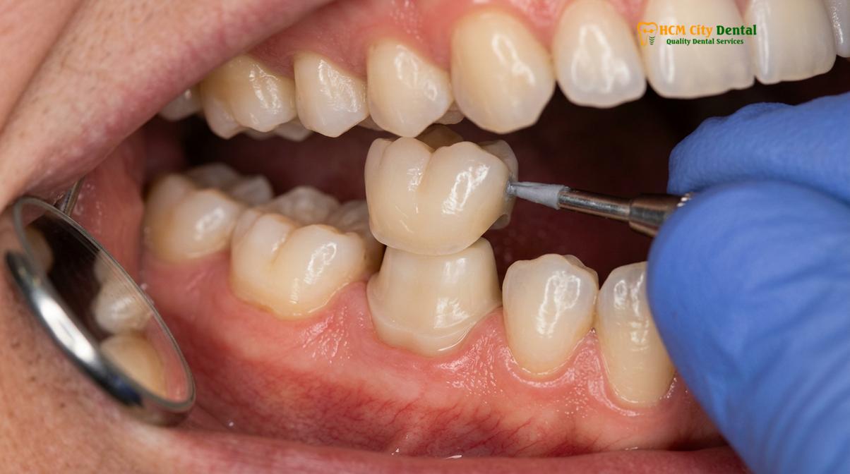
The procedure begins with the mandatory placement of a rubber dam to isolate the tooth from salivary bacteria. Caries excavation is performed from the periphery toward the pulp. The goal is to establish a clean, hard dentin seal at the margins (the peripheral seal zone). In an indirect pulp cap, the deepest layer of affected (but not infected) dentin is left in place to avoid exposing the pulp, and a bioceramic liner is placed over it. In a direct pulp cap, where the pulp is visibly exposed, the area is gently bathed in 1.5% to 2.5% sodium hypochlorite to disinfect the exposure site and control bleeding. Once hemostasis is achieved, the MTA or Biodentine is carefully flowed over the exposure, creating a bacteria-tight seal.
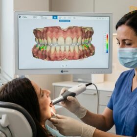
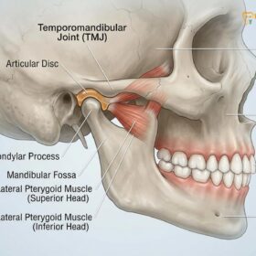
Following the pulp therapy, the tooth must be restored. In comprehensive rehabilitation cases, clinicians utilize Digital Smile Design (DSD) to plan the final restorations. This involves creating 3D digital mock-ups to evaluate phonetic compatibility and chewing guidance before any teeth are prepared. Additionally, establishing the correct Vertical Dimension of Occlusion (VDO) is vital. If a patient suffers from a collapsed bite, diagnostic temporary splints are used to find the physiologic rest jaw position, relieving muscle fatigue and TMJ stress before transitioning to permanent bio-inert restorations.
Understanding the financial investment for these advanced restorative procedures is important for patients. Below is an overview of the pricing structure for various treatments, highlighting the significant advantages of pre-booking.
| Treatment / Material Category | Standard Walk-in Rate (Estimated) | WhatsApp Pre-Booking Rate (-40%) |
|---|---|---|
| Crown on Implant (Per Tooth) | ~$260 – $385 | From $155 – $230 |
| Full Mouth Crowns (14 crowns/arch, Zirconia HT) | ~$3,640 per arch | From $2,184 per arch |
| Single Implant: Economy (Dentis/Neo Biotech) | ~$770 | From $460 |
| Single Implant: Standard (Dentium Superline/IBS) | ~$1,090 | From $655 |
| Single Implant: Premium Plus (Straumann BLT-SLA) | ~$1,795 | From $1,080 |
| Full Arch Zirconia Bridge (All-on-4 / All-on-6) | ~$12,000 – $22,000 per arch | From $7,200 – $13,200 per arch |
Long-Term Monitoring: Success Rates and Diagnostic Testing
Post-operative monitoring utilizes thermal sensibility testing and 3D CBCT imaging to verify pulp vitality, absence of apical pathology, and the continuous formation of reparative dentin.
The placement of the final crown does not mark the end of the vital pulp therapy journey; rigorous long-term monitoring is essential. Success is defined clinically by the absence of pain or swelling, and radiographically by the lack of periapical radiolucencies and the visible formation of a dentin bridge beneath the capping material. Follow-up appointments typically occur at 3, 6, and 12 months. During these visits, thermal sensibility testing (using cold spray) is carefully applied to the cervical area of the crowned tooth to confirm that the nerve remains responsive and vital.[5]
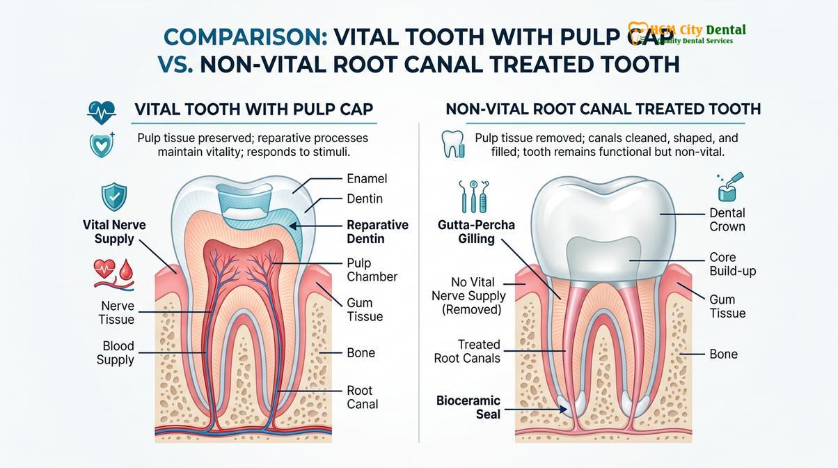
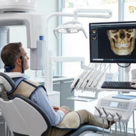
Dr. Nguyen Van Cuong advocates for the use of high-resolution CBCT (Cone Beam Computed Tomography) 3D bone diagnostics when evaluating complex cases or if a patient reports ambiguous symptoms. CBCT provides a three-dimensional view of the root apex, allowing clinicians to detect early signs of endodontic failure long before they become visible on traditional 2D X-rays. For international patients visiting HCMC Dental Clinic, remote post-op checkups via WhatsApp, supported by global manufacturer warranty cards, provide peace of mind and continuity of care.
It is a clinical reality that not all vital pulp therapies succeed indefinitely. If a tooth eventually succumbs to necrosis or suffers a catastrophic root fracture years later, extraction may become necessary. In such scenarios, modern implantology offers robust solutions. For single teeth, a biocompatible titanium or zirconia implant can be placed. For patients facing the loss of multiple teeth or an entire arch, all-on-4 implants provide a fixed, highly aesthetic, and functional replacement. Alternatively, for patients seeking a removable but stable option, implant-supported overdentures utilizing a locator snap-on attachment system offer excellent retention and improved chewing efficiency compared to traditional dentures.
When to See a Doctor (Important Clinical Considerations)
While vital pulp therapy is highly successful, patients must be vigilant during the healing phase. Mild, transient sensitivity to cold or biting pressure is normal for the first few weeks as the pulp recovers and lays down new dentin. However, certain symptoms indicate that the pulp is failing to heal and is transitioning into irreversible pulpitis or necrosis.
Seek immediate dental evaluation if you experience:
- Spontaneous, unprovoked throbbing pain that wakes you up at night.
- Severe, lingering pain after consuming hot or cold foods (lasting longer than 10-15 seconds).
- Swelling in the gums around the crowned tooth or in the face/jaw.
- A persistent pimple-like bump (fistula) on the gums that may drain pus or taste salty.
- Extreme tenderness when tapping on the tooth or chewing soft foods.
Clinical Disclaimer: Vital pulp therapy is not universally applicable. Teeth with a history of spontaneous pain, necrotic pulps, or existing periapical infections are strictly contraindicated for this procedure and require full endodontic treatment or extraction. Always consult with a qualified dental professional for an individualized diagnostic assessment.
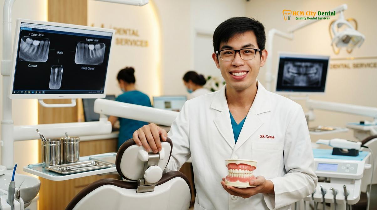
If you have been told you need a root canal but wish to explore minimally invasive, pulp-preserving alternatives, a comprehensive clinical and radiographic examination is the first step. Advanced diagnostics and bioceramic technologies have expanded our ability to save natural tooth structure safely and predictably.
Frequently Asked Questions
Is it always necessary to get a root canal before a crown?
No, a root canal is not always necessary before a crown. If the tooth’s nerve is healthy or only reversibly inflamed, vital pulp therapy can preserve the living tissue beneath the crown, maintaining the tooth’s natural strength and immune defenses. Root canals are only mandatory when the pulp is irreversibly damaged, infected, or necrotic.
What is the success rate of vital pulp therapy?
Vital pulp therapy boasts a high success rate, often exceeding 85% to 90% in properly selected cases. Success depends heavily on achieving strict hemorrhage control, using biocompatible calcium silicate cements, and ensuring a hermetic seal with a high-quality final restoration to prevent bacterial microleakage.
How do you keep a tooth alive under a dental crown?
A tooth is kept alive under a crown by carefully removing decay, disinfecting the area, and applying a bioactive material like MTA or Biodentine over the pulp. This stimulates the formation of a protective dentin bridge before the final crown is cemented, shielding the nerve from thermal and bacterial insults.
Can a tooth with irreversible pulpitis be saved with pulp capping?
Traditionally, irreversible pulpitis required a root canal, but modern partial pulpotomy techniques using bioceramics can sometimes save the remaining healthy radicular pulp. However, if bleeding cannot be controlled within minutes during the procedure, full endodontic therapy is mandatory to prevent systemic infection.
Does vital pulp therapy cause pain after the procedure?
Mild, transient sensitivity to cold or biting is normal for a few days as the pulp heals and forms secondary dentin. However, severe, spontaneous, or throbbing pain indicates treatment failure and necessitates immediate clinical re-evaluation, as it likely signals that the nerve is dying.
References
- Journal of Endodontics. AAE Position Statement on Vital Pulp Therapy. (2021).
- International Endodontic Journal. European Society of Endodontology position statement: Management of deep caries and the exposed pulp. (2019).
- Journal of Dentistry. Vital pulp therapy: histopathology and histobacteriology-based guidelines. (2019).
- Clinical Oral Investigations. Long-term outcomes of direct pulp capping using calcium silicate cements. (2020).
- The Journal of Prosthetic Dentistry. Marginal adaptation and microleakage of monolithic zirconia crowns. (2022).
For customized treatment planning, transparent cost estimates, and direct consultation with Dr. Cuong, visit our comprehensive Full Mouth Rehabilitation service page or message our team directly via WhatsApp to receive an instant assessment.