When comparing monolithic zirconia vs PFM (porcelain fused to metal) for dental restorations, monolithic zirconia offers superior flexural strength and eliminates the risk of porcelain chipping. Unlike PFM crowns, which often expose a dark metal margin over time, zirconia provides highly biocompatible, lifelike aesthetics for long-term restorative success.
Clinical Summary:
Monolithic zirconia has largely superseded PFM in modern restorative dentistry due to its single-phase structural integrity and exceptional tissue biocompatibility. PFM restorations rely on a bilayer design that is susceptible to delamination and often causes metal ceramic grey margins as gums recede. For patients undergoing extensive dental reconstructions, zirconia delivers predictable durability, optimal crown weight comfort, and seamless integration with digital smile design workflows. Advanced clinical protocols further enhance the patient experience during the transition to metal-free restorations.
Key Takeaways:
- Monolithic zirconia eliminates the porcelain-to-metal interface, drastically reducing the risk of chipping or fracture under heavy occlusal loads.
- PFM crowns frequently develop a dark grey line at the gingival margin due to metal oxidation and natural tissue recession.
- Zirconia exhibits superior tissue biocompatibility, promoting healthier gum attachment and significantly less plaque accumulation.
- Advanced digital workflows allow precise vertical dimension of occlusion (VDO) restoration using lightweight, highly aesthetic ceramic materials.
- International patients can access premium monolithic zirconia restorations at significant savings through specialized pre-booking programs.
Structural Integrity: Why Monolithic Zirconia Prevents Porcelain Chipping
Monolithic zirconia is milled from a single solid block, providing immense flexural strength that prevents the delamination and chipping commonly seen in bilayered PFM restorations.
The structural biomechanics of dental restorations dictate their longevity, especially in patients with heavy bite forces or parafunctional habits like bruxism. Historically, porcelain-fused-to-metal (PFM) crowns were the standard for combining the strength of a cast metal substructure with the tooth-colored aesthetics of feldspathic porcelain. However, this bilayered approach introduces a critical vulnerability: the interface between the metal core and the veneering porcelain. Differences in the coefficient of thermal expansion between these two distinct materials can create residual stresses during the manufacturing process, leading to a high susceptibility to cohesive fractures and porcelain chipping under cyclic masticatory loading[1].
In stark contrast, monolithic zirconia is engineered as a single, homogenous material. Milled via advanced CAD/CAM technology from a solid block of yttria-stabilized zirconium oxide and subsequently sintered at extreme temperatures, it achieves a flexural strength frequently exceeding 1,000 to 1,200 MPa. Because there is no separate veneering layer to delaminate, the risk of catastrophic porcelain fracture is virtually eliminated. This single-phase structural integrity makes a monolithic zirconia arch the preferred choice for complex, high-load scenarios, such as full arch reconstructions, where biomechanical stability is paramount.
“The transition from bilayered metal-ceramic systems to monolithic ceramics represents a paradigm shift in prosthodontics. By eliminating the weakest link—the porcelain-metal interface—clinicians can offer restorations that withstand the rigorous demands of the posterior dentition while maintaining exceptional aesthetic fidelity.”
Furthermore, the monolithic nature of zirconia allows for more conservative tooth preparation. Because PFM crowns require sufficient thickness to mask the underlying dark metal and provide adequate bulk for the porcelain layer, more healthy tooth structure must be removed. Zirconia, possessing inherent strength even at reduced thicknesses, allows clinicians to preserve more natural dentin and enamel. The ability to achieve a precise margin fit further ensures that the restoration seals the underlying tooth structure against bacterial microleakage, promoting long-term structural integrity.
The Dark Gumline Problem: How PFM Causes Grey Margins Over Time
PFM crowns contain a metal substructure that oxidizes and becomes visible as a dark grey line when the surrounding gum tissue naturally recedes over time.
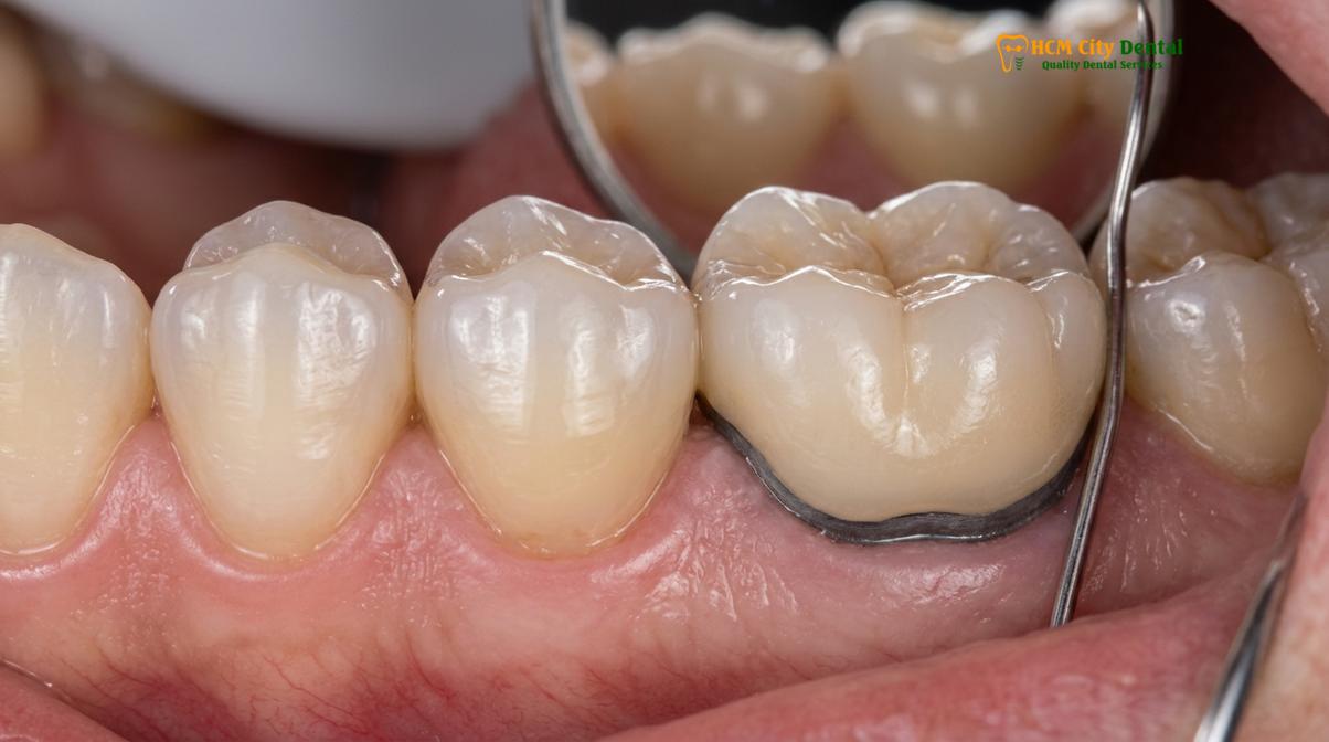
One of the most prevalent aesthetic complaints associated with legacy dental restorations is the appearance of a dark, unsightly line at the junction where the crown meets the gum tissue. This phenomenon is intrinsically linked to the material composition of PFM crowns. The metal alloy substructure, which provides the foundation for the porcelain veneer, is completely opaque. To prevent this dark metal from showing through the porcelain, dental technicians must apply an opaque masking layer. However, at the very edge of the crown—the margin—the porcelain is often too thin to completely conceal the metal, or the metal collar is intentionally left exposed for marginal adaptation.
As patients age, it is common for the gingival tissues to undergo slight physiological recession. When the gums recede even a fraction of a millimeter, the hidden metal margin of a PFM crown becomes exposed to the oral environment, resulting in highly visible metal ceramic grey margins. Additionally, certain non-precious metal alloys used in older PFM crowns can undergo oxidation and corrosion over time. This process can cause metallic ions to leach into the surrounding gingival tissues, a condition sometimes referred to as metallosis or the “umbrella effect,” which permanently tattoos the gums with a bluish-grey hue[2].
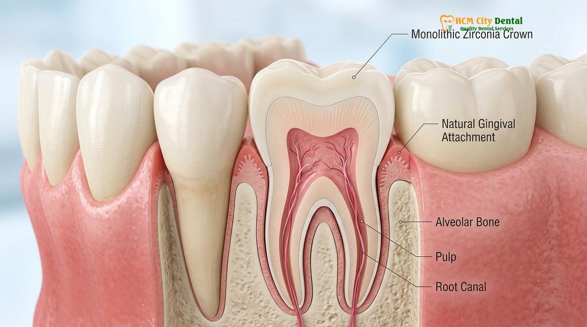
Monolithic zirconia completely circumvents this aesthetic dilemma. Being entirely metal-free, zirconia restorations possess a natural translucency that mimics the optical properties of natural dentin and enamel. Even if gingival recession occurs years after the placement of a full arch case, there is no dark substructure to expose. The margin remains tooth-colored and seamlessly blends with the root surface. In cases where previous PFM crowns have caused severe tissue discoloration or recession, a biological gum graft may be recommended alongside the transition to zirconia to restore a healthy, natural-looking gingival architecture.
Bio-compatibility: Preventing Gum Inflammation and Recession
Zirconia is highly bio-inert, meaning it resists plaque accumulation and promotes healthy gingival attachment without triggering the allergic or inflammatory responses sometimes associated with metal alloys.
The interaction between restorative materials and the surrounding periodontal tissues is a critical determinant of long-term clinical success. The concept of biological width—the natural dimension of soft tissue attached to the tooth above the alveolar bone—must be respected during any restorative procedure. When materials irritate this delicate tissue, chronic inflammation, bleeding on probing, and eventual bone loss can occur. PFM crowns, particularly those fabricated with base metal alloys containing nickel or beryllium, carry a documented risk of eliciting localized allergic reactions or hypersensitivity in susceptible individuals.
Monolithic zirconia is celebrated for its exceptional tissue biocompatibility. As a bio-inert ceramic, it does not corrode, oxidize, or release ions into the oral environment. Clinical studies have consistently demonstrated that human gingival fibroblasts adhere favorably to polished zirconia surfaces, promoting a robust hemidesmosomal attachment that seals the biological width against bacterial infiltration[3]. This favorable soft tissue response is crucial when managing complex cases involving bone augmentation and subsequent implant placement, where maintaining a healthy mucosal barrier is essential for preventing peri-implantitis.
Furthermore, the surface free energy of highly polished or glazed monolithic zirconia is significantly lower than that of veneering porcelain or natural enamel. This physical property translates to a reduced affinity for plaque accumulation. Patients with full arch zirconia bridges often report that their restorations feel exceptionally smooth and are easier to keep clean. When combined with advanced healing protocols like PRF (Platelet-Rich Fibrin), which utilizes the patient’s own autologous blood growth factors to accelerate soft tissue recovery[4], the integration of zirconia restorations yields highly predictable and biologically harmonious outcomes that align with modern prosthodontic standards[6].
Weight and Comfort: The Lightweight Advantage of Ceramic Materials
Modern zirconia restorations offer optimal crown weight comfort, reducing the biomechanical load on the temporomandibular joint and supporting a more natural proprioceptive feel during mastication.
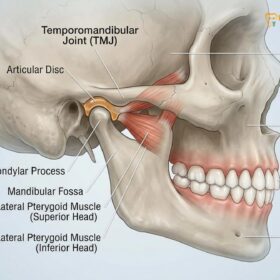
The physical weight of a dental prosthesis plays a subtle yet significant role in patient comfort and neuromuscular adaptation, particularly in extensive full arch reconstructions. Traditional PFM bridges, especially those utilizing high-density precious or semi-precious metal alloys, can be noticeably heavy. When an entire arch is restored with metal-based prostheses, the increased mass can alter the proprioceptive feedback mechanism of the periodontal ligaments or the osseoperception in dental implants. This added weight can contribute to masticatory muscle fatigue and place unnecessary stress on the temporomandibular joint (TMJ).
Monolithic zirconia offers a distinct advantage in terms of crown weight comfort. Despite its immense compressive and flexural strength, zirconium oxide is significantly less dense than most dental casting alloys. This lightweight characteristic allows clinicians to design robust, long-span bridges that feel remarkably natural to the patient. The reduction in prosthetic mass facilitates a smoother transition during the restorative phase, allowing the neuromuscular system to adapt more readily to the newly established occlusal scheme.
This lightweight advantage is particularly beneficial when correcting a collapsed bite. In cases of severe occlusal wear or missing teeth, the Vertical Dimension of Occlusion (VDO) must be carefully re-established to restore facial harmony, alleviate TMJ compression, and optimize chewing efficiency. Using diagnostic temporary splints, the clinician determines the physiologic rest jaw position. Once the ideal VDO is confirmed, transitioning to permanent, lightweight monolithic zirconia restorations ensures that the masticatory muscles are not overburdened, promoting long-term neuromuscular stability and patient comfort[5].
Advanced Clinical Workflows for Full Mouth Rehabilitation
Comprehensive rehabilitation utilizes digital smile design and meticulous planning to seamlessly integrate complex treatments like implant-supported prostheses and full mouth crowns.
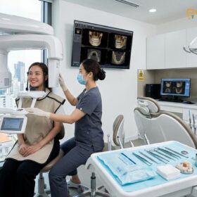
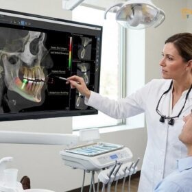
The successful execution of a full arch restoration requires a meticulous, multidisciplinary approach that blends advanced diagnostics, surgical precision, and state-of-the-art prosthodontics. The workflow typically begins with comprehensive 3D CBCT bone diagnostics. This high-resolution imaging allows the clinical team to evaluate bone volume, identify anatomical landmarks, and plan precise trajectories for dental implants. If bone resorption is severe, targeted bone augmentation procedures are performed to create a stable foundation for the future prostheses.
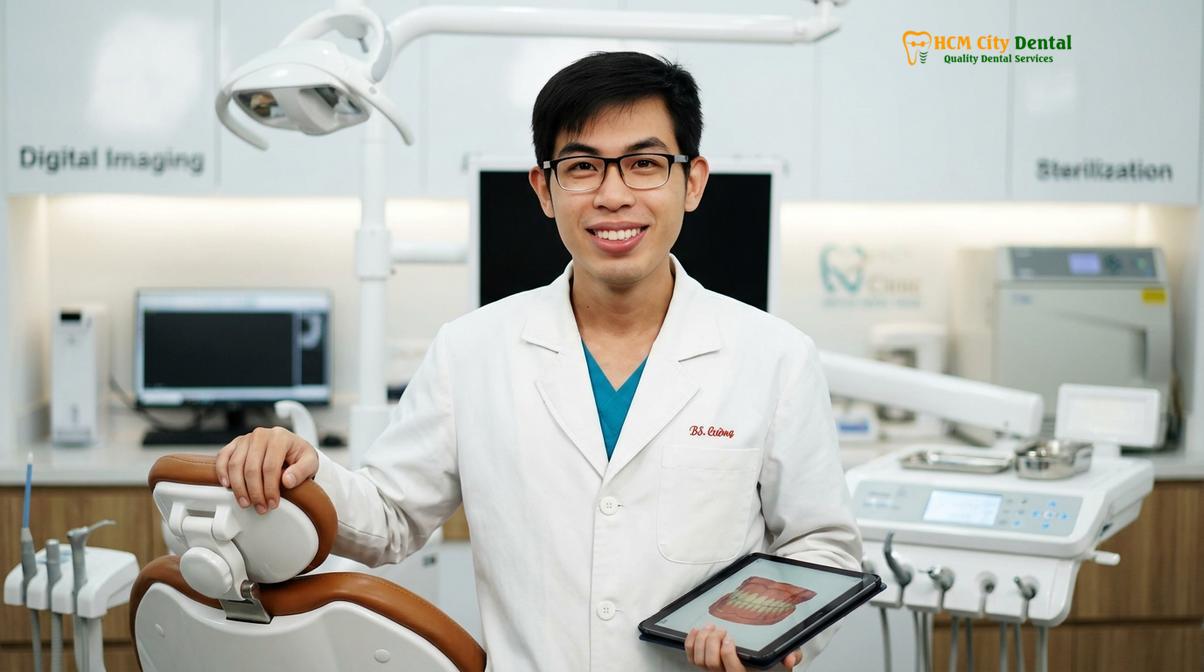
Dr. Nguyen Van Cuong, a leading expert in complex rehabilitations, emphasizes the importance of meticulous planning. He notes that evaluating phonetic compatibility and lip support before any teeth are prepared is essential for predictable outcomes. Under his guidance, patients receive tailored care that prioritizes both structural longevity and facial harmony.
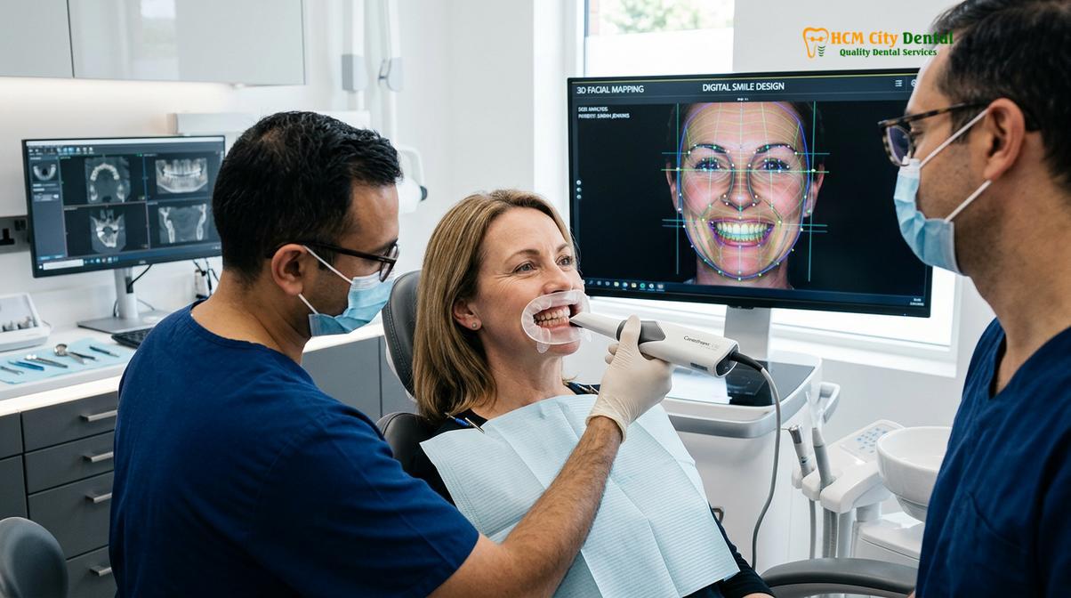
A cornerstone of modern restorative planning is Digital Smile Design (DSD). This innovative protocol involves capturing dynamic video and high-resolution photography of the patient’s face, lips, and teeth in motion. Using specialized software, the clinician creates a 3D digital mock-up of the proposed restorations. By creating a digital wax-up before any teeth are prepared, the clinical team can evaluate phonetic compatibility, lip support, and anterior guidance. The patient can visually approve their new smile, ensuring that the final monolithic zirconia restorations perfectly complement their unique facial aesthetics.
Clinical Case Overview: A 58-year-old patient presented to HCMC Dental Clinic in Ho Chi Minh City with severe bite collapse, generalized attrition, and failing PFM bridges exhibiting dark grey margins. The treatment plan involved the removal of unsalvageable teeth, bilateral bone augmentation utilizing PRF healing protocols, and the placement of premium implants in the upper arch. Following a period of osseointegration, the patient was restored with a custom-milled monolithic zirconia arch. The result was a complete restoration of the Vertical Dimension of Occlusion, elimination of TMJ discomfort, and a highly aesthetic, metal-free smile.
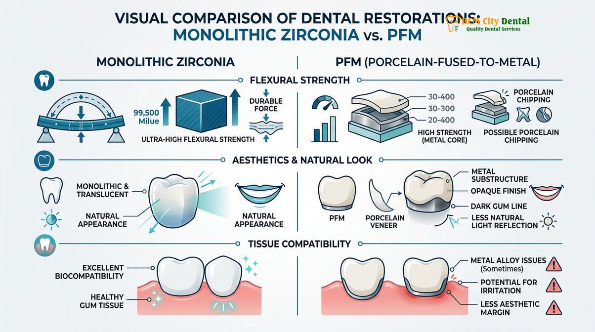
| Clinical Parameter | Monolithic Zirconia | Porcelain-Fused-to-Metal (PFM) |
|---|---|---|
| Flexural Strength | Very High (>1,000 MPa) | Moderate (Porcelain layer prone to chipping) |
| Aesthetics & Margins | Highly translucent, no dark margins | Opaque core, risk of metal ceramic grey margins |
| Tissue Biocompatibility | Excellent (Bio-inert, low plaque affinity) | Variable (Risk of metal allergies/inflammation) |
| Structural Design | Single-phase monolithic block | Bilayered (Metal substructure + Porcelain veneer) |
| Crown Weight Comfort | Lightweight, natural proprioception | Heavier, depending on the metal alloy used |
When to Consult a Prosthodontic Specialist
Recognizing the signs of failing legacy restorations is crucial for preventing further damage to the underlying tooth structure and surrounding periodontal tissues.
Patients who currently have older PFM restorations should monitor their oral health closely for signs of material fatigue or biological incompatibility. If you notice a persistent dark line near the gumline, experience chronic localized gum bleeding despite good oral hygiene, or observe visible chipping of the porcelain layer, it is highly recommended to seek a professional evaluation. These symptoms often indicate that the restoration’s marginal integrity has been compromised or that the underlying metal is causing an adverse tissue reaction.
Important Clinical Note: Ignoring a persistent dark line or chronic gum bleeding around old PFM crowns can lead to severe localized periodontitis. Prompt evaluation is necessary to prevent irreversible bone loss around the affected teeth or implants.
“Timely intervention is critical when legacy restorations begin to fail. Replacing compromised PFM crowns before severe periodontal breakdown occurs can preserve vital bone and soft tissue architecture.”
During a consultation, a specialist will utilize digital radiography and periodontal probing to assess the health of the abutment teeth or implants beneath the existing crowns. If a transition to monolithic zirconia is deemed appropriate, a customized treatment plan will be developed to safely remove the old prostheses, address any underlying decay or inflammation, and restore the arch with modern, biocompatible ceramics.
Cost Analysis: Walk-in Prices vs. WhatsApp Discount Value
Understanding the financial parameters of full arch restorations helps patients secure premium monolithic zirconia treatments at the best possible value.
For walk-in patients, the cost of a full monolithic zirconia arch reflects the premium nature of the CAD/CAM milling process and the high-grade ceramic materials utilized. Standard clinic rates account for the extensive clinical time, digital smile design, and laboratory fabrication required to produce a flawless, metal-free restoration. While this investment yields exceptional long-term durability and aesthetics, the initial out-of-pocket expense can be significant for those paying standard walk-in fees.
However, many international dental centers offer structured pathways to make these advanced treatments more accessible. By utilizing WhatsApp pre-booking programs, patients can often secure comprehensive treatment packages at a significant discount compared to standard walk-in rates. This streamlined communication channel allows the clinical team to review preliminary X-rays and provide a transparent cost estimate before the patient even travels. Ultimately, investing in monolithic zirconia through these optimized booking channels provides exceptional clinical value, minimizing the need for future repairs while delivering a highly aesthetic, durable smile.
If you are considering upgrading your smile from legacy PFM crowns to modern ceramics, contact HCMC Dental Clinic in Ho Chi Minh City to explore your options for full mouth rehabilitation and discover how our advanced digital workflows can restore your confidence and oral health.
References
- The Journal of Prosthetic Dentistry. Flexural strength and fracture resistance of monolithic zirconia versus porcelain-fused-to-metal restorations. (2021).
- Journal of Clinical Periodontology. Gingival response and biological width maintenance around biocompatible dental materials. (2019).
- International Journal of Prosthodontics. Biomechanical load distribution and proprioception in full-arch implant prostheses. (2020).
- Clinical Oral Implants Research. Efficacy of platelet-rich fibrin in guided bone regeneration and soft tissue healing. (2022).
- Journal of Esthetic and Restorative Dentistry. Digital smile design and vertical dimension of occlusion restoration protocols. (2018).
- Vietnam Odonto-Stomatology Association (VOSA). National guidelines on restorative materials and biocompatibility in prosthodontics. (2023).
For customized treatment planning, transparent cost estimates, and direct consultation with Dr. Cuong, visit our comprehensive Full Mouth Rehabilitation service page or message our team directly via WhatsApp to receive an instant assessment.