Crown lengthening before a crown is a minor surgical procedure that reshapes gum tissue and underlying bone to expose more healthy tooth structure. This ensures the final dental restoration has a solid foundation, preventing chronic inflammation and ensuring long-term structural success.
Clinical Summary:
When a tooth is severely decayed or fractured below the gumline, placing a crown directly can violate the biologic width and lead to premature failure. Crown lengthening surgically lowers the gingival margin and crestal bone, providing adequate supracrestal tooth structure. This establishes a proper ferrule effect, allowing the prosthodontist to secure the restoration effectively while maintaining optimal periodontal health. The procedure requires a dedicated healing phase before digital impressions and final cementation can occur.
Key Takeaways:
- Exposes healthy tooth structure to provide a secure, stable foundation for dental crowns.
- Prevents biological width violations, which can cause chronic gum inflammation and bone loss.
- Creates a mandatory 1.5 to 2.0 mm ferrule effect for the structural durability of the restored tooth.
- Requires a tissue maturation and healing period of 4 to 8 weeks before final digital impressions.
- Integrates seamlessly with advanced CAD/CAM milling and digital intraoral scanning workflows.
What is Functional Crown Lengthening?
Functional crown lengthening is a targeted periodontal surgery designed to expose sound tooth structure hidden beneath the gumline, enabling the secure and biologically sound placement of a dental restoration.
In restorative dentistry, the foundation upon which a prosthesis is built dictates its longevity. When a tooth suffers from extensive caries, severe attrition, or a fracture that extends subgingivally (below the gumline), there is often insufficient clinical crown height remaining to support a restoration. Attempting to place a crown on such a compromised foundation frequently results in poor retention, recurrent decay, and periodontal complications. To resolve this, a periodontist performs a surgical crown extension.
Unlike a simple gingivectomy, which only removes soft tissue, functional crown lengthening typically involves the careful recontouring of both the gingiva and the underlying alveolar bone. The goal is to apically reposition the supporting structures, thereby exposing a greater amount of healthy enamel and dentin. This newly exposed structure allows the restorative dentist to properly prepare the tooth, ensuring that the margins of the final restoration sit on sound, clean tooth structure rather than compromised tissue or restorative filling material.
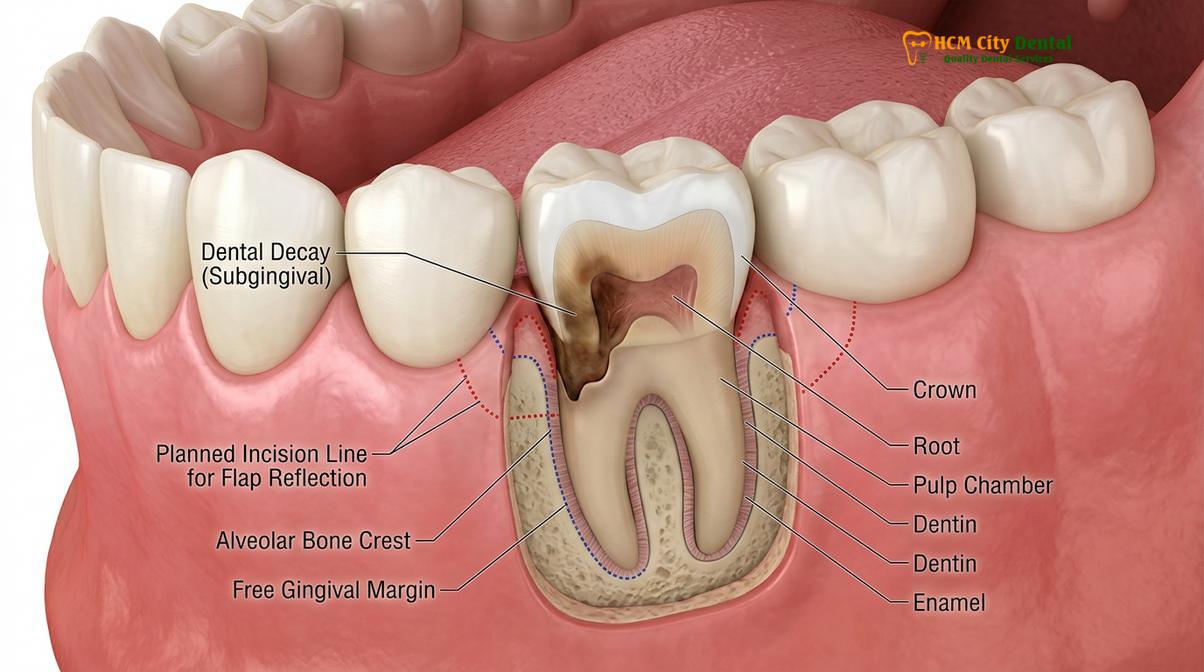
The diagnostic phase preceding this procedure is meticulous. Clinicians utilize high-resolution periapical radiographs and periodontal probing to assess the crown-to-root ratio, the anatomy of the adjacent roots, and the thickness of the buccal alveolar bone. If the removal of bone to expose one tooth would severely compromise the support of adjacent teeth, alternative treatments such as orthodontic forced eruption or extraction followed by a dental implant may be considered. However, when indicated, functional crown lengthening is a highly predictable procedure that rescues teeth that might otherwise be deemed non-restorable.
Establishing the Ferrule Effect for Durability
The ferrule effect is a 360-degree collar of healthy tooth structure above the gumline that the crown grips, which is essential for preventing tooth fracture and crown dislodgment.
One of the most critical biomechanical concepts in prosthodontics is the ferrule effect dental principle. When a tooth is prepared for a crown, especially one that has undergone a root canal and requires a post and core buildup, the crown must encircle a band of solid, healthy dentin. This band acts much like the metal rings around a wooden barrel, binding the structure together and distributing occlusal (chewing) forces evenly down the root.
Clinical guidelines dictate that a minimum of 1.5 to 2.0 mm of vertical tooth structure, with at least 1.0 mm of dentin thickness, must be present above the restorative margin to achieve an adequate ferrule. Without this collar, the functional loads placed on the tooth during mastication are transferred directly to the post or the core material, drastically increasing the risk of catastrophic root fracture or the decementation of the crown.[1]
“The presence of an adequate ferrule is the single most important factor in the long-term survival of endodontically treated teeth restored with post, core, and crown systems. It significantly enhances fracture resistance and structural integrity.”
When decay or trauma destroys this vital supragingival structure, crown lengthening becomes the necessary intervention to recreate it. By lowering the bone level, the surgeon artificially lengthens the clinical crown, providing the restorative dentist with the necessary real estate to establish this protective collar. This meticulous preparation ensures that whether the final restoration is a traditional PFM crown or a modern monolithic zirconia crown, it will possess the mechanical retention required to withstand decades of use.
Correcting Biologic Width Violations
The biologic width is the natural protective attachment of gum tissue to the tooth; invading this space with a crown margin causes chronic inflammation and unpredictable bone loss.
The periodontium has a strict anatomical dimension known as the biologic width, recently re-termed supracrestal tissue attachment. This space, averaging approximately 2.04 mm in height, consists of the junctional epithelium and the supracrestal connective tissue attachment. It serves as a vital biological seal, protecting the underlying alveolar bone from the bacteria-rich environment of the oral cavity.[2]
A biological width violation occurs when a dental restoration, such as a deep filling or a crown margin, is placed too far subgingivally, encroaching upon this sacred space. The body’s immune system recognizes the restorative material as a foreign invader and initiates a chronic inflammatory response. Clinically, this presents as localized gingival erythema (redness), persistent bleeding on probing, localized pain, and eventually, unpredictable osteoclastic activity leading to bone loss and periodontal pocket formation.
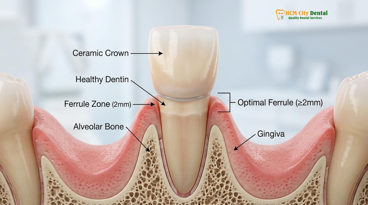
To correct or prevent this violation, crown lengthening is employed to surgically move the bone crest further down the root. The general rule in restorative dentistry is to establish at least 3.0 mm of space between the restorative margin and the alveolar bone crest. This allows 2.0 mm for the re-establishment of the biologic width and 1.0 mm for the gingival sulcus. By respecting these microscopic dimensions, clinicians ensure that the soft tissues surrounding the new dental crowns remain pink, firm, and free of inflammation.
The Surgical Procedure: Expectations and Healing
The procedure involves local anesthesia, careful reflection of the gum tissue, precise bone recontouring, and suturing, followed by a mandatory healing phase before final crown fabrication.
Understanding the surgical workflow helps alleviate patient anxiety and highlights the precision required for successful outcomes. The procedure begins with the administration of profound local anesthesia. Once the area is completely numb, the periodontist makes precise incisions, often utilizing a sulcular or submarginal approach depending on the amount of keratinized tissue present. A full-thickness mucoperiosteal flap is then carefully elevated to expose the underlying tooth root and alveolar bone.
The most critical phase of the surgery is the osseous recontouring. Using specialized rotary instruments and hand chisels, the surgeon performs an ostectomy (removal of supporting bone) and osteoplasty (reshaping of non-supporting bone). The bone is meticulously sculpted to follow the natural scalloped architecture of the cementoenamel junction (CEJ), ensuring that the newly established biologic width will be uniform around the entire circumference of the tooth. Once the desired architecture is achieved, the surgical site is thoroughly irrigated, and the flap is apically repositioned and secured with fine sutures.[3]
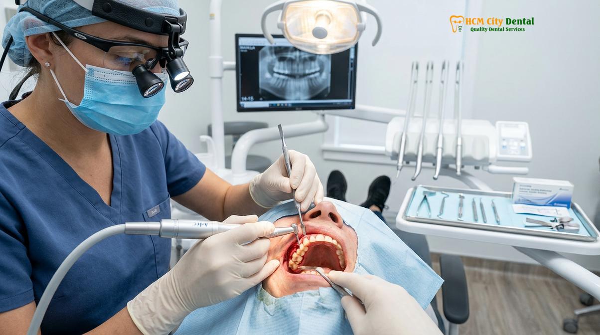
Following the surgery, a temporary crown is often placed or adjusted to protect the tooth, but its margins are kept strictly away from the healing tissues. The recovery phase is paramount. Patients are advised to maintain a soft diet, avoid mechanical brushing at the surgical site, and use prescribed antimicrobial rinses (such as chlorhexidine) to prevent infection.
Clinical Warning: Rushing the restorative phase after crown lengthening can compromise the final result. The soft tissues require a minimum of 4 to 8 weeks to mature and stabilize. In the esthetic zone, waiting up to 3 to 6 months may be necessary to prevent gingival recession after the permanent crown is cemented.
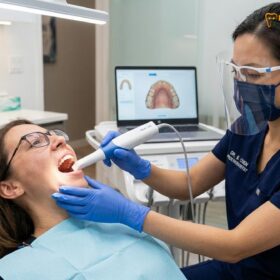
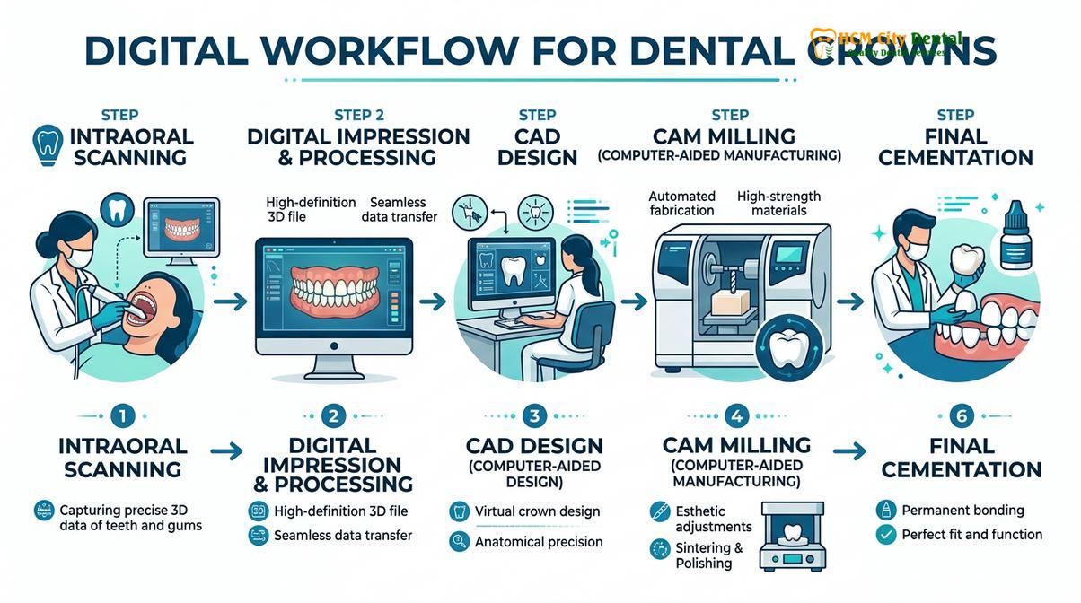
Dr. Nguyen Van Cuong emphasizes the importance of this maturation period. “Patience during the healing phase is non-negotiable,” notes Dr. Cuong. “We must wait for the gingival zenith to completely stabilize before we utilize our digital scanners. Capturing the margin too early risks a discrepancy between the crown and the final gumline.” Once healing is complete, advanced tools like the iTero scanner or Medit i700 are used to capture highly accurate, comfortable digital impressions, eliminating the need for messy traditional silicone materials.
Esthetic Integration: Aligning Gumlines for a Balanced Smile
Beyond functional necessity, crown lengthening harmonizes uneven gingival contours and corrects gummy smiles, ensuring that the final ceramic restorations look naturally proportioned.
While functional crown lengthening focuses on structural salvage, esthetic crown lengthening is an art form dedicated to visual harmony. Many patients present with altered passive eruption—a condition where the gum tissue fails to recede normally during tooth development, resulting in short, square-looking teeth and excessive gingival display. When preparing these teeth for cosmetic restorations, a gummy smile crown prep approach is required to establish the correct width-to-length ratio (the golden proportion) of the teeth.
In the esthetic zone (the front teeth), symmetry is paramount. The periodontist carefully maps out the ideal gingival zenith (the highest point of the gumline) for each tooth. By surgically lifting the gumline, the true anatomical crown is revealed. This procedure is frequently performed in conjunction with full smile makeovers involving multiple dental crowns or veneers.[4]
“Esthetic crown lengthening transforms the canvas of the smile. By establishing symmetrical gingival architecture, we provide the ideal framework for high-translucency ceramics to mimic natural dentition flawlessly.”
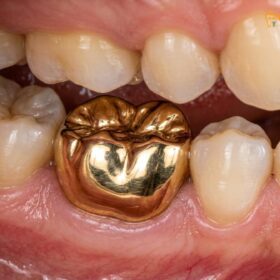
Material selection plays a crucial role in these highly visible areas. Following the healing of the esthetic crown lengthening, clinicians often choose premium materials such as E.max (Ivoclar, Liechtenstein) lithium disilicate for its outstanding lifelike aesthetics and translucency on front teeth. For areas requiring higher bite force resistance while maintaining esthetics, Zirconia HT (Cercon HT, Germany) is utilized. The combination of precise soft tissue management and elite material science yields results that are virtually indistinguishable from natural teeth.
Clinical Case Example: A patient visited HCMC Dental Clinic in Ho Chi Minh City presenting with a severe gummy smile and fractured anterior teeth. The treatment plan initiated with esthetic crown lengthening across the upper six anterior teeth to establish proper proportions. After a 12-week tissue maturation phase, the teeth were prepared, and digital impressions were taken using the Medit i700. The patient was restored with six custom-shaded E.max crowns, resulting in a perfectly balanced, radiant smile with optimal periodontal health.
Advanced Technology, Materials, and Treatment Workflows
Modern restorative workflows combine digital intraoral scanning, premium certified materials, and direct laboratory partnerships to deliver precise, highly aesthetic crowns efficiently.
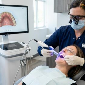
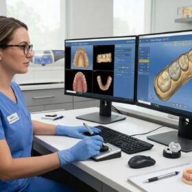
The landscape of restorative dentistry has been revolutionized by digital technology. At the forefront of this shift is the integration of CAD/CAM milling and advanced intraoral scanning. Devices like the iTero scanner and the Medit i700 provide 3D precision that traditional impression materials simply cannot match. These scanners capture the subgingival margins exposed during crown lengthening with micron-level accuracy, ensuring that the final restoration fits seamlessly, thereby preventing microleakage and secondary decay.[5]
For international patients engaging in dental tourism, efficiency without compromising quality is essential. Through a Direct Lab Partnership in Ho Chi Minh City, the collaboration between the prosthodontist and the master ceramist is immediate and highly controlled. This synergy allows for an Express 3-5 Day Turnaround, enabling patients to undergo complex treatments—including multiple crowns or a dental bridge—within a single week. Every restoration utilizes authentic, Material Certification-backed ceramics from Germany and Liechtenstein, ensuring global standards of biocompatibility and strength.
Dr. Nguyen Van Cuong frequently highlights the peace of mind provided by modern clinical protocols. “Beyond the precision of the fit and the beauty of the shade matching, we ensure our patients are supported long after they leave the clinic,” Dr. Cuong explains. This commitment is backed by a comprehensive global warranty policy, which includes remote follow-up support via WhatsApp, utilizing structured photo and video assessment protocols to monitor the health of the restorations and surrounding gums.
To make these premium treatments accessible, transparent pricing and flexible financials, including 0% interest monthly credit card installment plans, are standard. Below is the current pricing structure, highlighting the significant advantages of pre-arrival coordination.
| Restoration Type / Material | Standard Walk-in Fee | WhatsApp Pre-Booking (-40%) |
|---|---|---|
| PFM Crown (Porcelain Fused to Metal) | ~$140 (3.5M VND) | From $84 (2.1M VND) |
| Zirconia Crown (Cercon, Germany) | ~$200 (5.0M VND) | From $120 (3.0M VND) |
| Zirconia HT Crown (Cercon HT, Germany) | ~$260 (6.5M VND) | From $156 (3.9M VND) |
| E.max Crown (Ivoclar, Liechtenstein) | ~$280 (7.0M VND) | From $168 (4.2M VND) |
| 3-Unit Bridge (Zirconia HT) | ~$480 (12.0M VND) | From $288 (7.2M VND) |
| 4-Unit Bridge (Zirconia HT) | ~$640 (16.0M VND) | From $384 (9.6M VND) |
| Inlay / Onlay (Sứ Lab) | ~$180 (4.5M VND) | From $108 (2.7M VND) |
When to See a Doctor
While dental restorations are designed to be highly durable, the underlying biological structures require ongoing monitoring. You should seek a professional clinical examination if you experience persistent bleeding, swelling, or redness around an existing crown, as this may indicate a biological width violation or recurrent subgingival decay. Additionally, if a tooth breaks off near the gumline, prompt evaluation is necessary to determine if crown lengthening can save the root before extraction becomes the only viable option. Always consult with a qualified prosthodontist or periodontist to receive an individualized diagnostic assessment, as early intervention significantly increases the success rate of restorative treatments.
For those seeking expert evaluation, exploring premium dental crowns and bridges at HCMC Dental Clinic ensures access to state-of-the-art diagnostics and comprehensive restorative care.
Frequently Asked Questions
Why do I need crown lengthening before a crown?
Crown lengthening is required when there is insufficient healthy tooth structure above the gumline to support a restoration. By surgically lowering the gum and bone level, the procedure exposes enough enamel and dentin to create a secure foundation, preventing the new crown from failing or causing chronic gum inflammation.
Is crown lengthening painful?
The procedure itself is performed under profound local anesthesia, ensuring you feel no pain during the surgery. Post-operative discomfort is typically mild to moderate and can be effectively managed with prescribed analgesics and cold compresses during the first few days of the healing phase.
How long after crown lengthening can I get my permanent crown?
You must typically wait 4 to 8 weeks after the surgery before the final impressions for your permanent crown can be taken. In the esthetic zone (front teeth), your prosthodontist may recommend waiting up to 3 to 6 months to ensure the gingival margins have completely stabilized.
Does crown lengthening affect the adjacent teeth?
Yes, the procedure often involves minor recontouring of the gum and bone around neighboring teeth to ensure a smooth, natural, and harmonious gingival architecture. A skilled periodontist carefully blends the surgical site with the adjacent tissues to prevent abrupt changes in the gumline.
Can I wear a temporary crown during the healing phase?
Yes, a temporary crown is usually placed to protect the underlying tooth structure, maintain your bite, and provide esthetic continuity while the gums heal. However, the margin of the temporary crown must be carefully managed so it does not irritate the healing surgical site.
References
- Journal of Periodontology. The relationship between biologic width and restorative margins. (2019).
- International Journal of Prosthodontics. Biomechanical principles of the ferrule effect in endodontically treated teeth. (2020).
- American Academy of Periodontology. Clinical guidelines for functional and esthetic crown lengthening. (2021).
- Journal of Esthetic and Restorative Dentistry. Soft tissue management and material selection in the esthetic zone. (2022).
- Journal of Dentistry. Accuracy of digital intraoral scanners in capturing subgingival margins. (2023).