Dental calculus directly contributes to gum recession by harboring pathogenic bacteria that trigger chronic gingival inflammation. As the immune system responds to this calcified biofilm, the resulting inflammatory cascade destroys the delicate connective tissue fibers, causing the gum margin to pull away from the tooth root.
Clinical Summary:
The relationship between dental calculus and gum recession is a fundamental concept in periodontology. When soft bacterial plaque is not adequately removed, it mineralizes into calculus (tartar), creating a porous, rough surface that facilitates further bacterial colonization. This subgingival biofilm triggers a chronic immune response, leading to the destruction of the periodontal ligament and alveolar bone. As the underlying bone resorbs, the gingival tissue follows, resulting in clinical gum recession and exposed root surfaces. Modern periodontal therapy relies on advanced mechanical debridement to arrest this disease process. Utilizing a dual-scaler approach—combining piezoelectric and magnetostrictive technologies—alongside the Swiss Guided Biofilm Therapy (GBT) protocol, clinicians can safely eradicate subgingival calculus without damaging the delicate root cementum. While scaling cannot regenerate lost tissue, it is the critical first step in stabilizing periodontal health and preventing eventual tooth loss.
Key Takeaways:
- Dental calculus is mineralized plaque that cannot be removed by brushing; it requires professional ultrasonic scaling.
- Subgingival calculus triggers chronic inflammation, leading to the destruction of connective tissue and subsequent gum recession.
- Heavy tartar can act as an artificial splint, making teeth feel temporarily loose once the calculus is professionally removed.
- Lost gingival tissue does not grow back naturally; treatment focuses on halting progression and maintaining remaining attachment.
- Guided Biofilm Therapy (GBT) uses warm water and low-abrasive erythritol powder to remove biofilm painlessly and preserve enamel.
Pathological Process
The transition from soft plaque to mineralized calculus initiates a chronic inflammatory response that systematically degrades the gingival attachment apparatus.
To understand how receding gums tartar pathologies develop, one must first examine the biochemical lifecycle of the oral microbiome. The oral cavity is a dynamic environment where a thin, acellular protein film known as the acquired pellicle forms on the tooth surface within minutes of brushing. This pellicle acts as a biological adhesive, allowing primary colonizing bacteria to attach and multiply. Within 24 to 72 hours, if this soft biofilm (plaque) is not mechanically disrupted through effective oral hygiene, it begins to undergo a process of calcification.
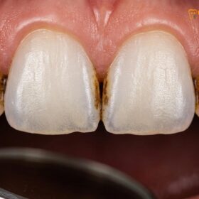
The mineralization of plaque into dental calculus is driven by the precipitation of mineral salts. Supragingival calculus (above the gumline) derives its minerals primarily from saliva, resulting in a composition rich in calcium phosphate, hydroxyapatite, and octacalcium phosphate. Subgingival calculus (below the gumline), however, derives its minerals from the gingival crevicular fluid—an inflammatory exudate. This subgingival tartar is typically darker, often brown or black, due to the incorporation of blood pigments and the presence of anaerobic, black-pigmented bacteria such as Porphyromonas gingivalis [1].
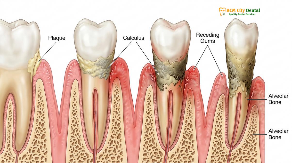
Calculus itself is not the direct cause of tissue destruction; rather, it is the highly porous, rough surface of the calculus that acts as a permanent reservoir for living, pathogenic biofilm. This unyielding bacterial presence induces plaque-induced gingivitis, the earliest stage of periodontal disease. The body’s immune system detects the bacterial endotoxins (lipopolysaccharides) and mounts a defense, sending neutrophils and macrophages to the site of infection. However, because the immune cells cannot phagocytize the calcified mass, the inflammatory response becomes chronic.
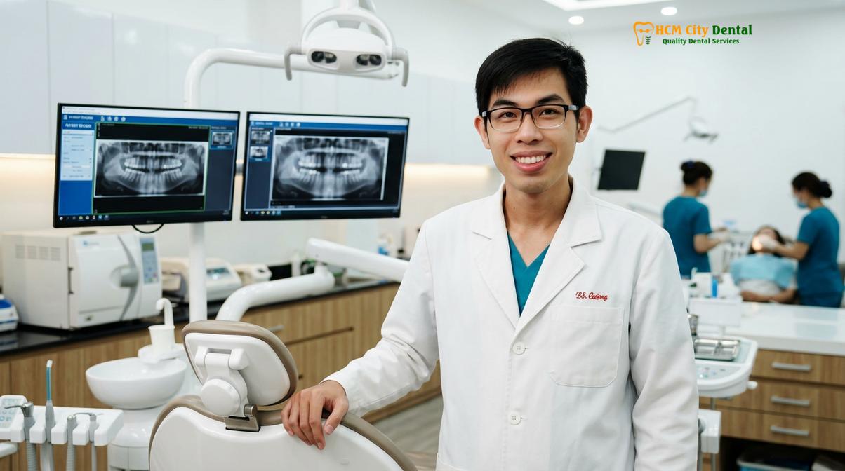
Dr. Nguyen Van Cuong emphasizes that the eradication of this subgingival biofilm is the cornerstone of periodontal stabilization. When calculus is allowed to persist within the gingival sulcus, the continuous release of inflammatory cytokines (such as Interleukin-1β and Tumor Necrosis Factor-alpha) begins to break down the collagen fibers of the periodontal ligament. As these fibers are destroyed, the gingival margin loses its tight seal against the tooth, creating a periodontal pocket. The deeper the pocket, the more anaerobic the environment becomes, favoring highly virulent bacteria that accelerate tissue destruction and drive the gum margin further down the root surface.
Alveolar Bone Loss
Unresolved gingival inflammation inevitably progresses to the underlying alveolar bone, where bacterial endotoxins stimulate osteoclastic activity and irreversible bone resorption.
The progression from superficial gingival inflammation to deep structural damage marks the transition into periodontitis. The alveolar bone is the specialized ridge of bone that houses the tooth sockets. It is highly responsive to both mechanical forces and biochemical signals. In a healthy state, there is a continuous, balanced cycle of bone formation by osteoblasts and bone resorption by osteoclasts. However, the chronic presence of subgingival calculus disrupts this delicate homeostasis.
As the periodontal pocket deepens due to the initial loss of connective tissue attachment, the bacterial flora shifts dramatically. The environment becomes dominated by the “Red Complex” bacteria—Porphyromonas gingivalis, Treponema denticola, and Tannerella forsythia. These highly pathogenic microorganisms produce potent enzymes, such as collagenases and proteases, which directly degrade the structural proteins of the periodontium. Furthermore, they release toxins that penetrate the gingival tissues and reach the alveolar bone [2].
The immune system’s attempt to isolate this severe infection inadvertently causes the most significant damage. Fibroblasts and macrophages in the inflamed tissue secrete matrix metalloproteinases (MMPs) and prostaglandins (specifically PGE2). Prostaglandin E2 is a potent stimulator of osteoclasts—the cells responsible for breaking down bone tissue. The body essentially resorbs its own bone to move it away from the advancing front of bacterial infection, a process clinically identified as bone loss periodontitis.
“The destruction of alveolar bone in periodontitis is a defensive retreat by the host immune system. By resorbing the bone, the body attempts to create a spatial buffer between the vital bone marrow and the advancing bacterial biofilm harbored on the calculus.”
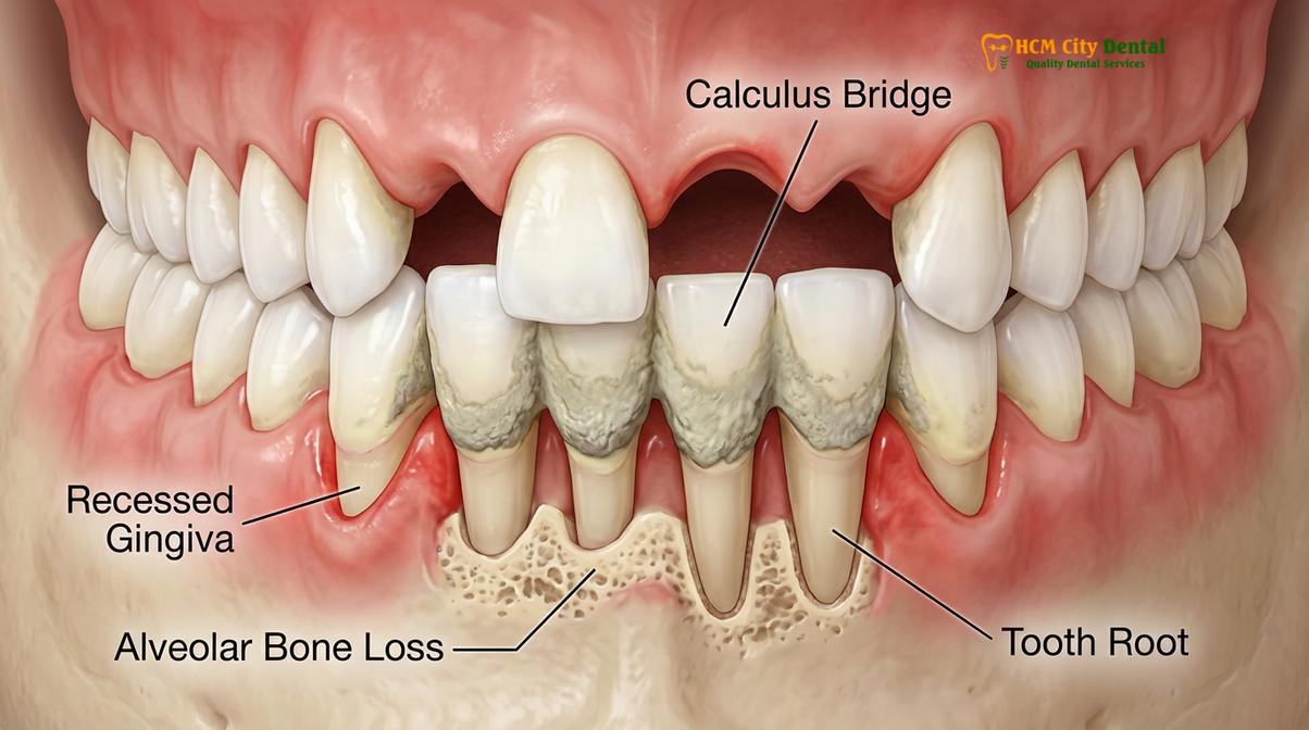
As the alveolar crest resorbs, the overlying gingival tissue often follows the receding bone level, leading to visible gum recession. In some cases, the gums may remain swollen and fibrotic, masking the underlying bone loss and creating deep “pseudo-pockets.” Regardless of the visual presentation, the loss of alveolar bone compromises the foundational support of the tooth. The exposed root surfaces, now devoid of their protective bone and gingival covering, are highly susceptible to root caries, severe dentinal hypersensitivity, and further mechanical abrasion from aggressive brushing.
Tartar “Holding” Loose Teeth
Heavy calculus deposits can artificially splint mobile teeth together, masking severe periodontal destruction until the tartar is professionally removed.
One of the most paradoxical and distressing experiences for a patient undergoing periodontal therapy is the sensation of increased tooth mobility immediately following a deep cleaning. This phenomenon is deeply tied to the biomechanics of advanced periodontitis and the physical properties of massive calculus deposits.
In cases of severe, long-standing neglect, supragingival and subgingival calculus can accumulate to such an extent that it bridges across multiple teeth. This is most commonly observed on the lingual (tongue-side) surfaces of the lower anterior teeth, an area bathed in mineral-rich saliva from the submandibular ducts. As the calculus calcifies into a solid, concrete-like mass, it physically connects the teeth. Simultaneously, the underlying periodontal disease is silently destroying the alveolar bone and periodontal ligament that normally secure the teeth in the jaw.
Clinical Warning: Patients with heavy calculus bridges often delay treatment because their teeth feel secure. However, this artificial stability masks ongoing, severe bone destruction. Delaying professional removal increases the risk of spontaneous tooth exfoliation (falling out) as the disease progresses unchecked.
Because the teeth are splinted together by the tartar, the patient does not perceive the true extent of their bone loss. The calculus acts as a rigid cast. When a clinician performs a thorough scaling and root planing procedure, this calculus bridge is shattered and removed. Suddenly, the teeth are no longer artificially supported by the tartar. The true clinical picture is revealed: teeth with significantly reduced bone support that now exhibit noticeable mobility.
This situation often leads to the misconception that the loose teeth scaling procedure itself caused the damage. In reality, the scaling merely uncovered the damage that the calculus had been causing for years. The removal of the calculus is absolutely critical. If left in place, the chronic infection would eventually destroy the remaining bone, leading to the complete loss of the dentition. Following the removal of the calculus, the gums will undergo a healing phase. As the inflammation subsides, the gingival tissues will tighten around the roots, and in many cases, the perceived mobility will decrease over the following weeks. In cases of severe mobility, the dentist may need to apply a temporary or permanent dental splint (using composite resin and wire) to stabilize the teeth while the periodontium heals [3].
Can Gums Grow Back?
Once gingival tissue and underlying bone are lost to periodontal disease, the gums cannot regenerate naturally, though surgical interventions can restore the gumline.
A primary concern for patients diagnosed with periodontal disease is whether their receded gums will grow back once the calculus is removed and the infection is cured. From a strict biological standpoint, the answer is no. The gingival tissue and the alveolar bone do not possess the intrinsic ability to regenerate vertically once they have been destroyed by chronic periodontitis.
When the calculus is removed, the immediate clinical result is a reduction in inflammation. The swollen, edematous gums will shrink as the excess fluid and inflammatory infiltrate dissipate. This healing process can sometimes make the recession appear slightly worse initially, as the “puffy” gums deflate to reveal the true level of attachment loss. However, this shrinkage is a sign of healing. The remaining gingival tissue will form a tight, healthy seal against the clean root surface, known as a long junctional epithelium. This halts the progression of the disease and prevents further recession.
For patients seeking to restore the lost tissue for aesthetic reasons or to treat severe root sensitivity, periodontal plastic surgery is required. Procedures such as connective tissue grafts, free gingival grafts, or the pinhole surgical technique involve taking soft tissue (often from the patient’s palate) and suturing it over the exposed roots. These procedures can successfully cover exposed roots, thicken the gingival biotype, and halt further recession, provided the underlying periodontal disease has been completely stabilized.
“The goal of non-surgical periodontal therapy is not regeneration, but stabilization. By removing the etiologic factors—plaque and calculus—we arrest the disease process, allowing the body to repair the inflamed tissues and maintain the current level of attachment.”
In cases where surgery is not indicated or desired, management focuses on protecting the exposed root surfaces. The root cementum and underlying dentin are much softer than enamel and are highly susceptible to decay and abrasion. Clinical protocols often involve enamel remineralization therapies, including the application of high-concentration fluoride varnish or amorphous calcium phosphate (ACP) pastes. These treatments occlude the open dentinal tubules, significantly reducing hypersensitivity and strengthening the root surface against bacterial acids.
Advanced Clinical Removal Protocols
Modern periodontal therapy utilizes advanced ultrasonic technology and guided biofilm therapy to safely eradicate calculus without damaging the delicate root cementum.
The removal of tenacious calculus requires precision, appropriate technology, and a deep understanding of root anatomy. At HCMC Dental Clinic in Ho Chi Minh City, the approach to professional dental cleaning transcends traditional hand scraping. The clinic employs a sophisticated, multi-modal approach designed to maximize calculus removal while minimizing trauma to the soft tissues and preserving the integrity of the tooth structure.
The Dual Scaler Advantage
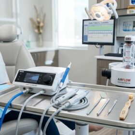
Not all calculus is created equal, and therefore, a one-size-fits-all approach to scaling is clinically inadequate. HCMC Dental Clinic utilizes a “Dual Scaler Advantage,” equipping clinicians with both Piezoelectric and Magnetostrictive ultrasonic technologies to customize the treatment based on the patient’s specific presentation:
- Piezoelectric Scalers (e.g., Acteon/Satelec): These devices utilize ceramic discs that expand and contract when electrical current is applied, producing a linear, back-and-forth motion at the tip. Operating at frequencies up to 32 kHz, piezoelectric scalers generate very little heat and require less water for cooling. Because only the lateral sides of the tip are active, they offer precise control and are significantly gentler. This makes them the preferred choice for patients with sensitive teeth, exposed roots, children, and those undergoing orthodontic treatment.
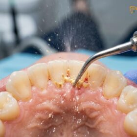
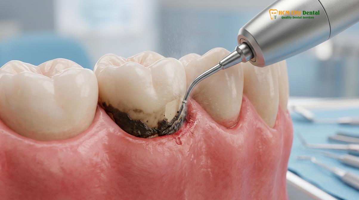
- Magnetostrictive Scalers (e.g., Dentsply Cavitron): These units rely on a stack of metal strips that change dimension within a magnetic field, creating an elliptical, figure-eight motion at the tip. This means all surfaces of the tip (front, back, and sides) are active. This omnidirectional energy is highly effective for breaking down massive, heavy calculus bridges, performing full mouth debridement, and navigating deep subgingival pockets where heavy tartar is firmly embedded.
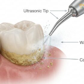
Both technologies rely on the cavitation effect—the rapid formation and collapse of microscopic water bubbles generated by the high-frequency vibration. When these bubbles collapse, they release shockwaves that tear apart bacterial cell walls and flush debris out of the periodontal pocket via acoustic microstreaming [4].
Guided Biofilm Therapy (GBT)
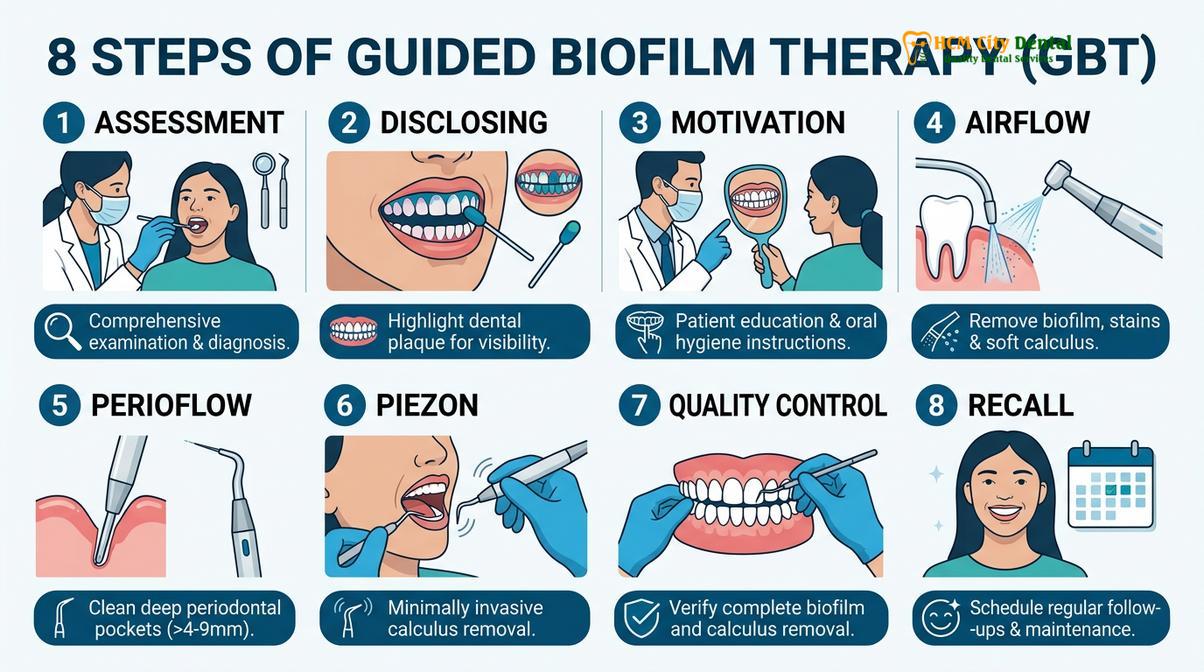
For the ultimate in patient comfort and clinical efficacy, the clinic features the premium Swiss EMS Guided Biofilm Therapy (GBT) protocol. Dr. Nguyen Van Cuong emphasizes that GBT represents a paradigm shift in preventive dentistry. Instead of scaling first and polishing last, GBT targets the biofilm first, making the subsequent calculus removal faster, easier, and less invasive.
The GBT protocol utilizes the Airflow Prophylaxis system, which combines warm water and a highly specialized, low-abrasive erythritol powder. With a microscopic grain size of just 14 to 25μm, erythritol is minimally abrasive. It safely and painlessly blasts away biofilm, early plaque, and extrinsic stains without scratching natural enamel, titanium implants, porcelain crowns, or delicate veneers. By removing the biofilm first, the clinician can clearly see the remaining mineralized calculus, allowing for targeted, conservative use of the ultrasonic scaler only where absolutely necessary.
Clinical Case Study: A 45-year-old patient presented to HCMC Dental Clinic in Ho Chi Minh City with generalized grade II mobility, severe halitosis, and heavy subgingival calculus. The patient was terrified of dental pain. Dr. Cuong utilized the Dual Scaler approach, starting with the Magnetostrictive scaler to break the heavy calculus bridges, followed by the Piezoelectric scaler for gentle subgingival debridement. The session concluded with the GBT Airflow protocol using erythritol powder to eradicate residual biofilm. Within four weeks, the gingival inflammation resolved, the tissues tightened, and the perceived tooth mobility significantly decreased, saving the dentition from extraction.
Clinical Pricing Structure
Transparency in treatment planning is essential for patient compliance. The cost of calculus removal varies based on the severity of the buildup and the specific technologies utilized. According to the latest clinic fee schedule, the pricing parameters are structured as follows:
| Treatment Protocol | Clinical Indication | Estimated Cost (VND) | Estimated Cost (USD) |
|---|---|---|---|
| Standard Ultrasonic Scaling & Polishing | Routine maintenance, mild to moderate supragingival calculus. | 500,000 – 800,000 (Walk-in: 800k – 1.3m) |
~$20 – $32 |
| Airflow Prophylaxis / GBT | Premium biofilm eradication, heavy staining, implant maintenance. | 1,500,000 – 2,000,000 (Walk-in: 2.5m – 3.3m) |
~$60 – $80 |
| Scaling & Root Planing (SRP) | Deep subgingival calculus, active periodontitis (priced per quadrant). | 1,000,000 – 1,500,000 (Walk-in: 1.6m – 2.5m) |
~$40 – $60 |
| Full Mouth Debridement | Massive calculus bridges, severe neglect, pre-surgical clearing. | 2,000,000 – 3,000,000 (Walk-in: 3.3m – 5m) |
~$80 – $120 |
Note: Patients utilizing the dedicated WhatsApp booking channel can access a -40% discount on the standard walk-in rates, ensuring high-quality periodontal care remains accessible.
Early Prevention
Preventing calculus-induced recession requires a combination of meticulous daily mechanical plaque control and strictly adhered professional periodontal maintenance schedules.
The most effective strategy against gum recession is preventing the formation of calculus entirely. Because plaque begins to mineralize within 24 to 72 hours, daily mechanical disruption of the biofilm is non-negotiable. Patients must adopt the Modified Bass brushing technique, angling the toothbrush bristles at 45 degrees toward the gumline to sweep plaque out of the shallow gingival sulcus. Furthermore, because a toothbrush cannot reach the interproximal spaces (between the teeth), daily use of dental floss or interdental brushes is critical. These areas are the most common starting points for undetected subgingival calculus.
Chemical adjuncts can support mechanical cleaning. Toothpastes containing pyrophosphates or zinc citrate act as tartar-control agents by binding to calcium in the saliva, inhibiting the crystal growth necessary for plaque mineralization. However, these agents only prevent new calculus from forming; they cannot dissolve calculus that has already hardened [5].
For patients who have already experienced periodontal disease and subsequent recession, standard six-month dental visits are insufficient. These patients require a strict periodontal maintenance schedule, typically every three to four months. This interval is clinically determined based on the repopulation rate of pathogenic bacteria within the periodontal pockets. By intervening before the biofilm has the opportunity to mature and calcify, clinicians can maintain the health of the reduced periodontium indefinitely.
When to See a Doctor
Immediate clinical evaluation is required if you experience signs of acute periodontal infection or rapid structural changes in your dentition.
While chronic periodontitis is often painless and progresses silently, certain symptoms warrant urgent evaluation by a dental professional. You should schedule a clinical examination if you experience:
- Spontaneous bleeding: Gums that bleed unprovoked, or heavily during normal brushing and eating.
- Acute swelling or purulence: The presence of pus expressing from the gumline, often accompanied by a localized, painful swelling (periodontal abscess).
- Sudden tooth mobility: Teeth that suddenly feel loose, shift position, or create a change in how your upper and lower teeth bite together.
- Severe root sensitivity: Intense, lingering pain when consuming hot, cold, or sweet foods, indicating significant root exposure and potential nerve irritation.
Early diagnostic intervention, including periodontal probing and radiographic assessment, is crucial to halting tissue destruction and preserving your natural teeth.
Frequently Asked Questions
Can scaling reverse gum recession?
No, scaling cannot reverse true gum recession, but it halts further progression. Once the gingival tissue and underlying bone are lost, they do not grow back naturally. However, removing the calculus eliminates the chronic inflammation, allowing the remaining gums to reattach firmly to the root surface and preventing further tissue loss.
Is it normal for teeth to feel loose after tartar removal?
Yes, it is common for teeth to feel temporarily loose after heavy tartar removal because the calculus was acting as an artificial splint. When massive calculus bridges are removed, the underlying bone loss becomes apparent. The teeth will often stabilize over a few weeks as the gums heal and tighten around the roots.
Does plaque destroy jawbone?
Yes, when plaque hardens into subgingival calculus, it triggers an immune response that activates bone-destroying cells, leading to jawbone loss. The bacteria within the calculus release toxins that cause chronic inflammation, prompting the body’s immune system to break down the alveolar bone in an attempt to isolate the infection.
How often should I get a dental cleaning if I have receding gums?
Patients with receding gums and a history of periodontitis should undergo professional periodontal maintenance every three to four months. This accelerated schedule is necessary because pathogenic bacteria repopulate periodontal pockets within 90 to 120 days, and frequent intervention prevents the biofilm from calcifying into destructive subgingival calculus.
Is ultrasonic scaling painful on exposed roots?
Ultrasonic scaling can cause mild sensitivity on exposed roots, but modern piezoelectric scalers and local anesthetics ensure a comfortable experience. Exposed dentin lacks the protective enamel layer, making it sensitive to temperature and vibration. Clinicians can adjust the power settings and use warm water to minimize any discomfort during the procedure.
References
- Journal of Clinical Periodontology. Pathogenesis of plaque-induced periodontal diseases. (2021).
- Periodontology 2000. The role of dental calculus and its removal in periodontal therapy. (2020).
- International Journal of Dental Hygiene. Clinical efficacy of Guided Biofilm Therapy. (2022).
- Journal of Periodontology. Biomechanics of tooth mobility and occlusal trauma. (2019).
- Clinical Oral Investigations. Erythritol air-polishing powder safety on root cementum. (2023).