Recognizing gingivectomy infection signs early is critical for preserving gum health and ensuring optimal aesthetic results. Key indicators include persistent throbbing pain, worsening swelling after 48 hours, yellow or white pus discharge, and severe bad breath. Prompt clinical evaluation ensures proper healing and prevents damage to the underlying alveolar bone.
Clinical Summary:
While a gingivectomy is a highly predictable and safe procedure, post-operative infections can occur if oral bacteria infiltrate the surgical site. Normal healing involves a temporary white film of granulation tissue, but true infections present with purulent discharge, escalating pain, and systemic fever. Utilizing advanced diode lasers significantly reduces these risks by instantly cauterizing tissue, while strict adherence to post-operative hygiene protocols ensures a seamless, complication-free recovery.
Key Takeaways:
- Normal healing includes a white or yellowish film (fibrin), which is a biological dressing and should not be confused with pus.
- Severe, throbbing pain that worsens after 3 to 4 days is a primary indicator of a dental post op infection.
- Smoking drastically increases infection risk by restricting blood flow and oxygen to healing periodontal tissues.
- Using an 810nm diode laser minimizes bleeding and sterilizes the surgical margin, accelerating recovery to just 48 hours.
- Immediate clinical intervention is required if you experience a fever, spreading swelling, or notice foul-tasting discharge.
Understanding the Risk of Infection in the Oral Cavity
The oral cavity harbors billions of bacteria, making surgical sites susceptible to infection if the biological width is violated or hygiene is poor. Advanced laser technology mitigates this risk by sterilizing the incision instantly.
The human mouth is a complex microbiome containing hundreds of different bacterial species. While most of these microorganisms exist in a symbiotic balance, a surgical procedure like a gingivectomy creates a temporary vulnerability. When the protective epithelial barrier is removed to reshape the gums, the underlying connective tissues are exposed. If pathogenic bacteria—such as Porphyromonas gingivalis or Streptococcus mutans—colonize the surgical wound, a gum infection after surgery can rapidly develop. Understanding the anatomical and microbiological factors at play is the first step in preventing post-operative complications.
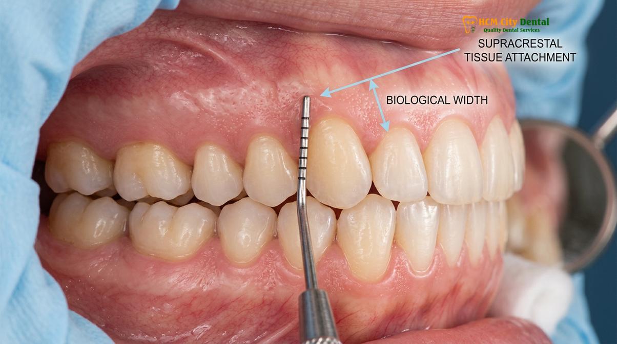
A critical concept in periodontal surgery is the preservation of the biological width, modernly referred to as the supracrestal tissue attachment. This refers to the natural dimension of space occupied by the junctional epithelium and connective tissue attachment above the alveolar bone, typically measuring around 2 millimeters[1]. If a gingivectomy removes too much tissue and encroaches upon this biological width, the body responds with chronic inflammation, unpredictable bone resorption, and a significantly higher susceptibility to localized infections. Therefore, precise preoperative sounding of the bone level is mandatory to ensure the surgical margin remains at a safe distance from the osseous crest.
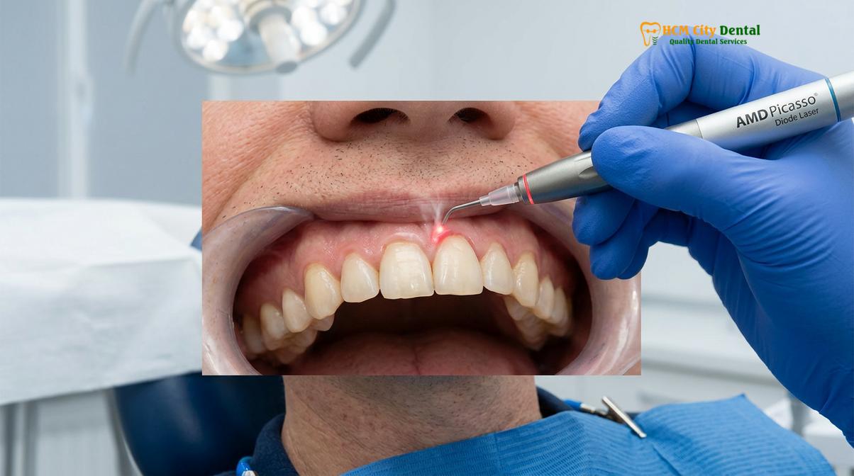
To minimize the risk of bacterial infiltration, modern clinical workflows have largely transitioned from traditional steel scalpels to advanced photothermal devices. The use of AMD Picasso Lasers (Diode soft tissue laser, 810nm) represents the gold standard in aesthetic gum contouring. The 810nm wavelength is highly absorbed by melanin and hemoglobin, allowing for bloodless, sutureless cuts. As the laser energy vaporizes the excess tissue, it simultaneously creates a zone of thermal coagulation. This instant cauterization seals blood vessels and lymphatic endings, effectively creating a sterile barrier that blocks bacterial entry[2]. Consequently, patients experience a minimally invasive procedure with reduced postoperative pain, minimal swelling, and a rapid 48-hour initial recovery phase.
Dr. Nguyen Van Cuong emphasizes that the success of a gingivectomy is not solely dependent on the cutting instrument, but on comprehensive preoperative preparation. “Before any soft tissue modification, we must ensure the oral environment is as pristine as possible. Performing a gingivectomy in the presence of active periodontal disease is a recipe for severe infection,” notes Dr. Cuong. Therefore, a thorough dental scaling and root planing session is often required prior to surgery to eliminate calculus and reduce the overall bacterial load in the oral cavity.
Warning Signs: White Pus, Throbbing Pain, and Persistent Bad Breath
Distinguishing between normal healing tissue and an active infection is crucial. While a white fibrin patch is normal, thick yellow pus, escalating throbbing pain, and severe halitosis are definitive signs of a clinical infection.
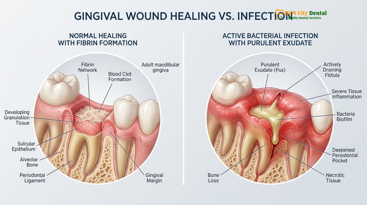
One of the most common sources of anxiety for patients recovering from gum surgery is misinterpreting the visual changes in their mouth. The oral mucosa heals differently than skin on the outside of the body. Because the mouth is a wet environment, a scab cannot form. Instead, the body produces a layer of fibrin and granulation tissue to protect the raw surgical site. This often appears as a white patch gums healing process. This whitish-yellow film is a completely normal biological dressing and is a sign that the body is repairing the epithelial layer.
However, it is vital to differentiate this healthy fibrin layer from purulent exudate (pus). Pus is a thick, opaque, yellowish-green fluid composed of dead white blood cells, tissue debris, and bacteria. If the white material on your gums is accompanied by a foul taste, easily wipes away to reveal angry, bleeding tissue, or is actively oozing from the gumline, it is a strong indicator of a dental post op infection. Furthermore, an infection will typically cause the surrounding tissues to become highly erythematous (deep red), swollen, and warm to the touch.
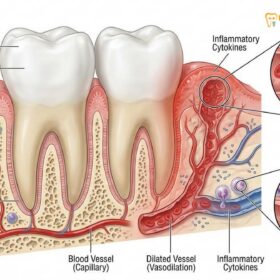
Pain is another critical diagnostic differentiator. It is entirely normal to experience mild to moderate soreness or a dull ache for the first 24 to 48 hours following a gingivectomy. This discomfort is usually well-managed with over-the-counter analgesics like ibuprofen or acetaminophen. However, if the pain transitions into a severe, pulsating, or throbbing sensation that radiates to the jaw or ear, and specifically if this pain worsens after the third day, it signals acute inflammation often driven by bacterial infection. The throbbing nature of the pain is caused by vasodilation and the accumulation of inflammatory fluids compressing local nerve endings.
| Clinical Indicator | Normal Healing Profile | Signs of Active Infection |
|---|---|---|
| Visual Appearance | Thin, stable white/yellow fibrin patch; pink surrounding gums. | Thick, oozing yellow/green pus; deep red, angry surrounding tissue. |
| Pain Levels | Mild soreness peaking at 24-48 hours, then steadily decreasing. | Severe, throbbing pain that escalates or returns after 3-4 days. |
| Swelling | Slight, localized puffiness that resolves within a few days. | Spreading, tense swelling that may affect the cheek or jawline. |
| Odor / Taste | Normal breath or slight metallic taste from minor bleeding. | Persistent, severe bad breath (halitosis) and a foul, bitter taste. |
| Systemic Signs | Normal body temperature, normal energy levels. | Fever, chills, lethargy, or swollen lymph nodes in the neck. |
Persistent bad breath, or halitosis, that does not improve with prescribed oral hygiene measures is another red flag. While a slight change in breath is expected due to altered brushing habits immediately post-op, a strong, putrid odor is caused by volatile sulfur compounds released by anaerobic bacteria multiplying in the infected site. If you experience this combination of symptoms, immediate clinical intervention is necessary to prevent the infection from spreading to the deeper periodontal ligaments or alveolar bone.
Why Tobacco Smoking Destroys Gum Healing Cells and Triggers Infection
Smoking introduces toxins that cause severe vasoconstriction, starving the gums of oxygen and immune cells. This hypoxic environment directly facilitates bacterial overgrowth and significantly delays surgical wound closure.
The detrimental effects of combustible tobacco on periodontal health are well-documented, but the risks are exponentially magnified following oral surgery. When a patient smokes after a gingivectomy, they are directly sabotaging the body’s cellular repair mechanisms. Nicotine is a potent vasoconstrictor; it causes the peripheral blood vessels in the gingival tissues to clamp down and narrow. This severe reduction in blood flow means that the surgical site is deprived of essential oxygen, nutrients, and the white blood cells necessary to fight off opportunistic oral bacteria.
Furthermore, the carbon monoxide present in cigarette smoke binds to hemoglobin in the blood much more readily than oxygen does, creating a state of localized hypoxia (oxygen starvation) in the healing tissues. Fibroblasts, the specialized cells responsible for producing collagen and rebuilding the gum tissue, cannot function in a hypoxic environment. As a result, the wound closure is delayed, leaving the internal connective tissues exposed to the bacteria-rich oral environment for a prolonged period. This delayed healing is a primary catalyst for post-operative infections.
“The introduction of combustible tobacco toxins into a fresh surgical wound not only paralyzes the local immune response but fundamentally alters the microvascular architecture, making the incidence of necrotic tissue and severe infection highly predictable in non-compliant patients.”
In cases where the gingivectomy is combined with functional or esthetic crown lengthening—which may involve minor bone contouring (osteoplasty or osteotomy)—the risks of smoking are even more severe. Exposed or newly contoured alveolar bone relies entirely on the overlying soft tissue for its blood supply. If smoking compromises the gingival blood flow, the underlying bone can become necrotic, leading to severe pain, bone loss, and the failure of the entire aesthetic procedure. Patients are strictly advised to cease smoking for at least 48 hours prior to surgery and a minimum of 7 to 14 days post-operatively to ensure safe and predictable healing.
How to Clean the Surgical Area Safely to Keep Plaque Away
Meticulous, gentle oral hygiene is the primary defense against infection. Utilizing prescribed antimicrobial rinses and modifying brushing techniques ensures the surgical margin remains clean without disrupting the delicate healing tissues.
Maintaining a pristine oral environment is the most effective way to prevent gingivitis after cut and avoid post-operative infections. However, the approach to oral hygiene must be carefully modified during the initial healing phase. Aggressive brushing or the use of harsh chemical mouthwashes can tear the delicate fibrin clot, induce bleeding, and irritate the regenerating epithelial cells. A balanced, clinically guided approach is essential.
For the first 24 hours following the procedure, patients are generally advised to avoid rinsing vigorously or spitting forcefully, as the negative pressure can dislodge the initial blood clot. After the first day, the cornerstone of post-operative care is chemical plaque control. Dentists typically prescribe an antimicrobial mouthwash containing 0.12% chlorhexidine gluconate. Chlorhexidine is highly substantive, meaning it binds to the oral tissues and slowly releases its antibacterial properties over several hours, effectively suppressing the bacterial load without the need for mechanical scrubbing[3].
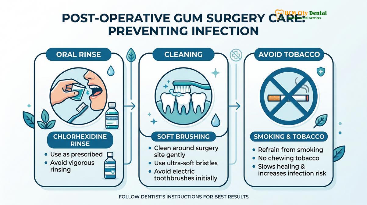
When it comes to mechanical cleaning, patients must use an ultra-soft surgical toothbrush. The teeth in the non-surgical areas should be brushed and flossed normally to maintain overall oral health. In the surgical area, the bristles should only touch the clinical crown of the tooth, carefully avoiding the healing gum margin. Water flossers and electric toothbrushes should be strictly avoided in the surgical zone for at least one to two weeks, as the high-pressure water or intense vibrations can easily damage the fragile supracrestal tissue attachment as it re-establishes itself.
A dental tourism patient visiting HCMC Dental Clinic in Ho Chi Minh City underwent a comprehensive full-arch laser gum contouring procedure to correct an asymmetrical gummy smile. Because the patient was traveling, strict adherence to post-operative hygiene was critical. Dr. Nguyen Van Cuong provided a detailed digital smile design (DSD) recovery protocol, emphasizing the use of chlorhexidine rinses and soft-diet modifications. By following the precise cleaning instructions and avoiding mechanical trauma to the newly established gingival zenith points, the patient achieved a flawless, infection-free recovery within 7 days, allowing them to enjoy the remainder of their trip safely.
Advanced Clinical Workflows and Pricing at HCMC Dental Clinic
Comprehensive aesthetic gum contouring requires precise architectural planning and transparent cost structures. From minimally invasive laser treatments to complex orthognathic collaborations, modern clinics offer tailored solutions.
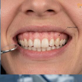
Achieving a harmonious, symmetrical smile goes beyond simple tissue removal; it requires a deep understanding of dental aesthetics, including the calibration of gingival zenith points (the highest point of the gum scallop, displaced slightly distal to the long axis of the tooth) and the principles of digital smile design (DSD). At leading institutions, the approach to Gum Contouring & Gummy Smile correction is highly individualized, utilizing a spectrum of advanced techniques to address both soft tissue and skeletal discrepancies.
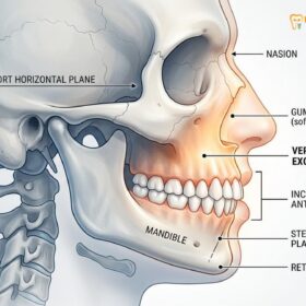
For patients with mild to moderate gingival overgrowth, the AMD Picasso Lasers provide an elegant, minimally invasive solution. However, for severe skeletal cases—diagnosed as Vertical Maxillary Excess (VME)—soft tissue lasers alone are insufficient. In these complex scenarios, HCMC Dental Clinic works in collaboration with Surgical Jaw Partners at the National Hospital of Odonto-Stomatology in Ho Chi Minh City (Bệnh viện Răng Hàm Mặt Trung Ương TP.HCM). This partnership allows for advanced orthognathic interventions, such as the Le Fort I osteotomy, which physically repositions the maxilla superiorly to reduce gingival display, ensuring the highest level of clinical safety and authority[4].
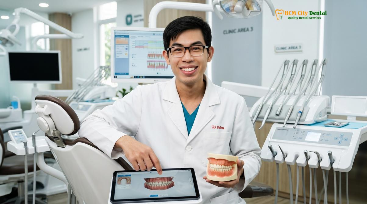
Understanding the financial investment is also a crucial part of the patient journey. According to the latest clinic fee schedule, the pricing structure is designed to be transparent and accessible, particularly for international patients seeking high-quality care. The current estimated costs are as follows:
- Laser Gingivectomy (per tooth): 1,500,000 VND (~$60 USD) with advance booking (Walk-in: 2,500,000 VND).
- Full-Arch Laser Gum Contouring (up to 10 teeth): 12,000,000 VND (~$480 USD) with advance booking (Walk-in: 20,000,000 VND).
- Functional/Esthetic Crown Lengthening (per tooth, including bone contouring): 3,000,000 VND (~$120 USD) with advance booking (Walk-in: 5,000,000 VND).
- Surgical Lip Repositioning / Lip Lowering: 15,000,000 VND (~$600 USD) with advance booking (Walk-in: 25,000,000 VND). This procedure restricts the upward pull of the lip muscles[5].
- Botox Gummy Smile Correction (both sides): 4,500,000 VND (~$180 USD) with advance booking (Walk-in: 7,500,000 VND). This is a non-surgical option to temporarily relax a hyperactive levator muscle.
Patients utilizing the dedicated communication channels can often access a -40% WhatsApp booking discount, making world-class aesthetic periodontics highly attainable. Whether the treatment plan involves a simple diode laser contouring or a complex multidisciplinary approach, the focus remains on preserving biological health while maximizing aesthetic outcomes.
“Esthetic crown lengthening and gingival contouring are not merely cosmetic endeavors; they are precise architectural modifications that must respect the biological width to ensure long-term periodontal stability and prevent chronic inflammatory responses.”
When to See a Doctor
While minor swelling and mild discomfort are expected parts of the healing journey, certain symptoms require immediate professional evaluation. You should contact your dental provider without delay if you experience any of the following red flags:
- A persistent fever exceeding 100.4°F (38°C) or experiencing sudden chills.
- Swelling that continues to increase dramatically after the first 48 to 72 hours, especially if it spreads to the neck, cheek, or under the jaw.
- Uncontrolled bleeding that does not stop after applying firm, continuous pressure with a clean gauze pad for 20 minutes.
- The presence of thick, foul-tasting yellow or green pus oozing from the surgical site.
- Severe, throbbing pain that is not relieved by prescribed or over-the-counter pain medications.
Early intervention is the key to managing post-operative complications effectively. Your dentist can perform a thorough clinical examination, safely irrigate the area, and prescribe targeted antibiotics if a bacterial infection is confirmed, ensuring your recovery gets back on track.
Frequently Asked Questions
What does a healing gum look like vs. an infected gum?
A healing gum typically features a thin, whitish-yellow film of fibrin that is painless and stable. In contrast, an infected gum presents with thick, foul-smelling yellow pus, severe redness, escalating throbbing pain, and swelling that worsens after 48 hours. The fibrin layer is a normal biological dressing, while pus indicates active bacterial destruction.
Are antibiotics necessary after a laser gingivectomy?
Routine antibiotics are rarely necessary after a laser gingivectomy because the diode laser sterilizes the surgical margin. However, they may be prescribed if the patient has underlying systemic conditions, a compromised immune system, or if clear signs of a bacterial infection develop during the recovery phase.
What should I do if I suspect an infection?
If you suspect an infection, contact your dental provider immediately for a clinical evaluation. Do not attempt to scrape the area, apply unapproved topical ointments, or self-medicate with leftover antibiotics, as this can disrupt healing, mask symptoms, and worsen the bacterial imbalance.
How long does the white patch on gums last during healing?
The white patch of granulation tissue and fibrin typically lasts for 5 to 7 days. As the underlying epithelial cells regenerate and mature, this temporary biological dressing will naturally slough off, revealing healthy, newly contoured pink gum tissue underneath.
Can I use salt water rinses to prevent infection?
Yes, gentle warm salt water rinses starting 24 hours after surgery can help soothe the tissues and maintain a clean environment. However, it should complement, not replace, prescribed antimicrobial rinses like chlorhexidine during the initial healing phase. Ensure the rinsing action is gentle to avoid dislodging the blood clot.
References
- Journal of Clinical Periodontology. Esthetic crown lengthening outcomes and biological width. (2021).
- International Journal of Periodontics & Restorative Dentistry. Diode laser vs scalpel gingivectomy healing. (2020).
- Journal of Aesthetic and Restorative Dentistry. Management of post-operative periodontal infections. (2019).
- Journal of Oral and Maxillofacial Surgery. Le Fort I osteotomy stability in gummy smile correction. (2022).
- Clinical Oral Investigations. Surgical lip repositioning longevity and complication rates. (2018).