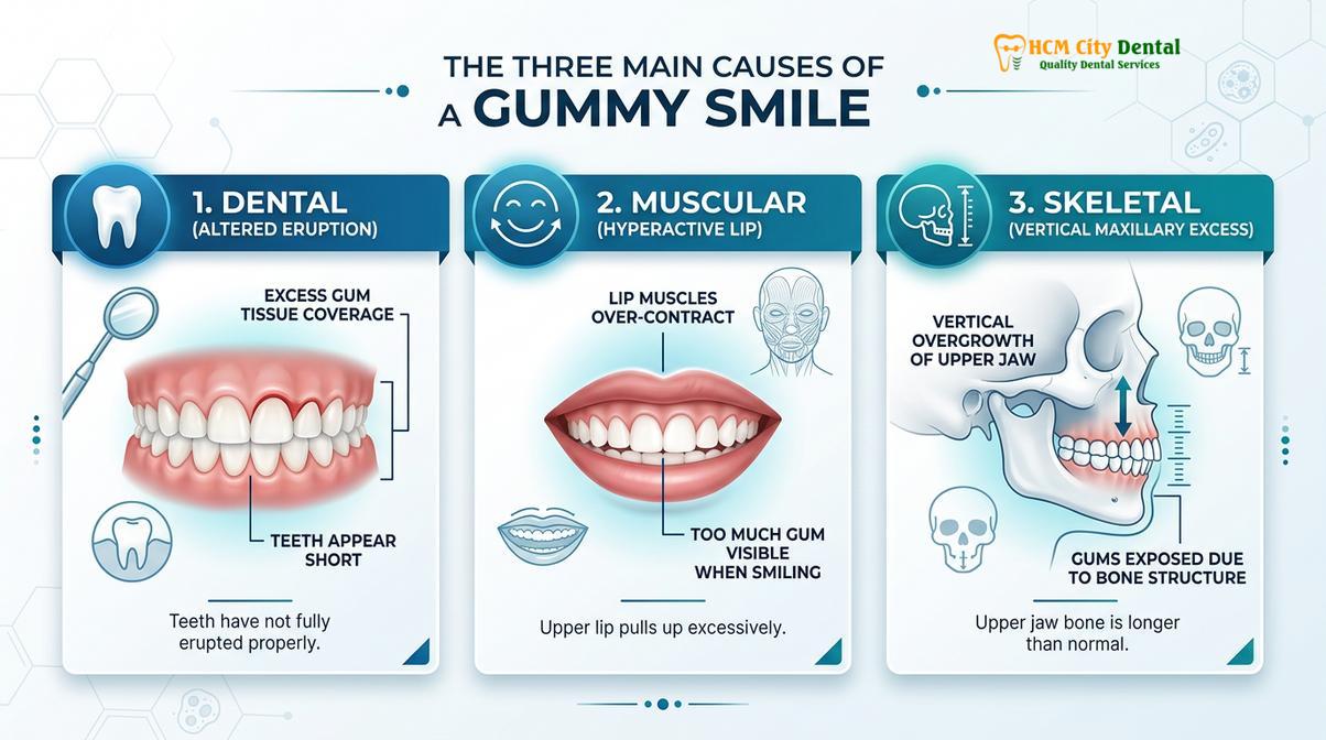
What causes a gummy smile? A gummy smile, clinically known as excessive gingival display, is primarily caused by delayed passive eruption of teeth, hyperactive upper lip muscles, or vertical maxillary excess. Identifying the precise anatomical origin is essential for determining the most effective, minimally invasive corrective treatment.
Clinical Summary:
A gummy smile results from a complex interplay of dental, muscular, and skeletal factors. Accurate diagnosis requires evaluating the biological width, gingival zenith points, and lip mobility. Treatments range from laser gingivectomy using advanced diode lasers to orthognathic surgery for severe skeletal discrepancies, ensuring both aesthetic harmony and long-term periodontal health.
Key Takeaways:
- Excessive gingival display is typically defined as exposing more than 3 to 4 millimeters of gum tissue during a full, dynamic smile.
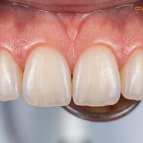
- Altered passive eruption occurs when gum tissue fails to recede naturally, covering the dental crowns and making teeth appear abnormally short.
- Hyperactive lip elevator muscles can pull the upper lip too high, revealing excess gums even if tooth and jaw proportions are completely normal.
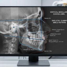
- Vertical maxillary excess (VME) is a skeletal condition where the upper jaw overgrows downward, often requiring collaborative surgical intervention.
- Modern treatments utilize advanced soft-tissue lasers for bloodless contouring, preserving the critical supracrestal tissue attachment for optimal healing.
Defining the Gummy Smile: Aesthetics vs. Clinical Definitions
A gummy smile is clinically diagnosed when more than three to four millimeters of continuous gum tissue is exposed during a dynamic smile, disrupting facial symmetry.
The perception of a beautiful smile is deeply rooted in the harmony and proportion of three primary components: the teeth, the lip framework, and the gingival scaffold. In clinical dentistry, a gummy smile is formally referred to as excess gingival display. While exposing 1 to 2 millimeters of pink gingival tissue is widely considered a sign of youthfulness and vitality, exposure exceeding 3 to 4 millimeters often crosses the threshold into an aesthetic concern for many patients[1]. According to guidelines from the Vietnam Odonto-Stomatology Association (VOSA), evaluating this display requires careful consideration of both resting lip posture and maximum smile dynamics to ensure accurate classification[6].
Understanding the etiology of a gummy smile requires a transition from static to dynamic facial analysis. A patient may exhibit perfect dental proportions when their face is at rest, but the moment they engage in a spontaneous, full smile, the underlying anatomical discrepancies become glaringly apparent. The “pink aesthetic score” is a metric used by cosmetic dentists to evaluate the health, contour, and volume of the gums in relation to the white aesthetics of the teeth. When this score is imbalanced due to an overabundance of visible tissue, a comprehensive diagnostic workup is initiated to pinpoint the exact structural cause.
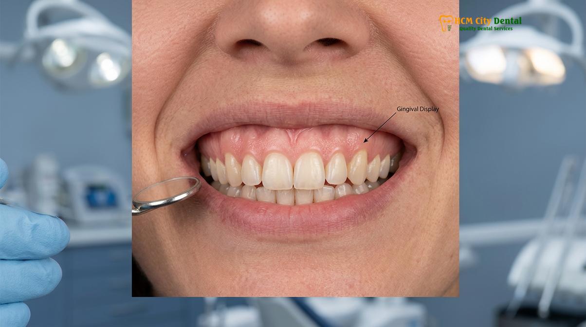
It is crucial to differentiate between a true anatomical gummy smile and transient gingival enlargement caused by acute inflammation. Poor oral hygiene, plaque accumulation, and certain systemic medications can induce gingival hyperplasia—a condition where the gums swell and overgrow the teeth. This inflammatory state mimics a structural gummy smile but is entirely reversible with professional dental scaling, improved home care, and medication management. Therefore, the first step in any aesthetic evaluation is ensuring absolute periodontal health before considering cosmetic interventions.
How Dentists Measure Gum Display During Active Smiling
Dental professionals utilize digital smile design and periodontal probing to measure lip mobility, clinical crown length, and the underlying supracrestal tissue attachment.
Diagnosing the root cause of a gummy smile is a meticulous process that blends artistic vision with precise mathematical measurements. Modern cosmetic dentistry relies heavily on Digital Smile Design (DSD), a protocol that utilizes high-resolution videography and photography to capture the patient’s smile in motion. Because a forced, static smile in a dental chair rarely reflects the patient’s true dynamic expressions, video analysis allows the clinician to freeze frames at the point of maximum lip elevation, providing an accurate assessment of the gingival display.
During the clinical examination, the dentist evaluates several critical parameters. The first is the clinical crown length—the visible portion of the tooth. The ideal width-to-length ratio of a central incisor is approximately 75% to 80%. If the teeth appear square rather than rectangular, it strongly suggests that excess gum tissue is masking the true anatomical crown. To confirm this, the dentist performs a procedure known as “bone sounding” under local anesthesia. A periodontal probe is gently inserted into the gingival sulcus to locate the alveolar bone crest. This measurement helps the dentist map the biological width—the minimum supracrestal tissue attachment required to protect the underlying bone from bacterial invasion and chronic inflammation[2].
| Diagnostic Parameter | Normal Clinical Range | Gummy Smile Indicator | Primary Diagnostic Tool |
|---|---|---|---|
| Gingival Display | 1.0 mm – 2.0 mm | > 3.0 mm to 4.0 mm | Dynamic Videography / DSD |
| Upper Lip Mobility | 6.0 mm – 8.0 mm | > 8.0 mm (Hypermobile) | Digital Calipers |
| Upper Lip Length (Rest) | 20 mm (F) / 22 mm (M) | < 20 mm (Short Lip) | Facial Measurement |
| Incisor Width/Length Ratio | 75% – 80% | > 85% (Square appearance) | Periodontal Probe |
| Supracrestal Attachment | ~ 2.0 mm – 3.0 mm | Altered bone crest position | Bone Sounding / CBCT |
Another vital aspect of the measurement phase is the evaluation of the gingival zenith points. The zenith is the most apical (highest) point of the gingival tissue scallop surrounding the tooth. In a highly aesthetic smile, the zenith of the central incisors and canines should be slightly distal (toward the back) to the long axis of the tooth, while the lateral incisors should have a centered zenith that sits slightly lower. In patients with a gummy smile, these zenith points are often blunted, asymmetrical, or positioned too far incisally (toward the biting edge), disrupting the natural flow of the smile arc.
Delayed Passive Eruption: Gums Failing to Recede Naturally
Delayed passive eruption happens when the gingival margin fails to migrate apically toward the cementoenamel junction during adolescence, leaving teeth looking disproportionately short.
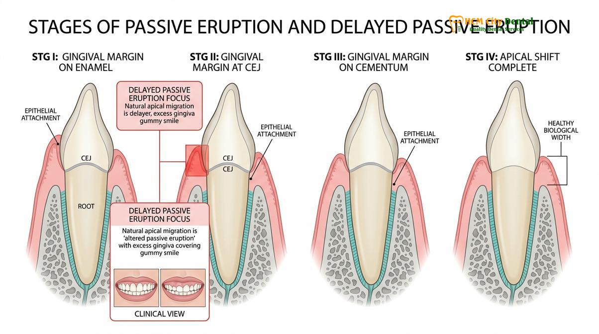
One of the most common dental causes of a gummy smile is a phenomenon known as altered or delayed passive eruption. To understand this condition, one must look at how teeth naturally emerge into the oral cavity. Tooth eruption occurs in two distinct phases: active and passive. Active eruption is the physical movement of the tooth through the jawbone and gum tissue until it meets its opposing tooth in the opposite arch. Once the teeth are in occlusion (biting together), the active phase concludes.
Following this, the passive eruption phase begins. During normal passive eruption, the gum tissue gradually recedes apically (toward the root) over several years, eventually settling at or slightly above the cementoenamel junction (CEJ)—the anatomical line where the enamel of the crown meets the cementum of the root. In cases of delayed passive eruption, this secondary recession simply does not occur. The gingival margin remains stranded high up on the enamel, effectively hiding a significant portion of the beautiful, healthy tooth structure beneath a thick curtain of pink tissue[3].
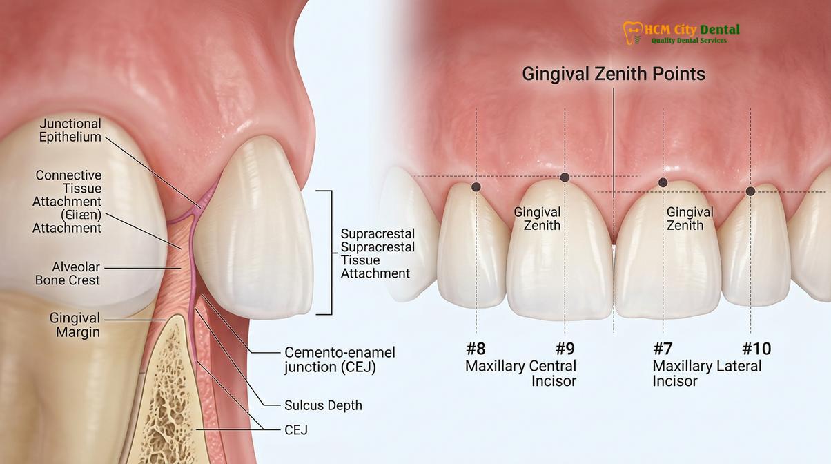
Clinically, delayed passive eruption is categorized using the Coslet classification system, which dictates the surgical approach required for correction. Type 1 indicates an excessively wide band of attached gingiva, while Type 2 indicates a normal or narrow band. Furthermore, Subgroup A means the underlying alveolar bone crest is in a normal position, whereas Subgroup B means the bone crest is abnormally high, sitting directly on the CEJ. Identifying these subgroups is critical because it determines whether the patient needs a simple soft-tissue laser contouring or a more complex functional crown lengthening that involves osteoplasty (bone reshaping).
“When addressing delayed passive eruption, we must meticulously sound the bone to respect the biological width. Removing tissue without understanding the underlying crestal bone position invites chronic inflammation and unpredictable tissue rebound, completely negating the cosmetic results of the procedure.”
If a dentist merely cuts away the excess gum tissue in a Subgroup B case without adjusting the underlying bone, the body will perceive this as a biological violation. The gums will inevitably grow back to re-establish the necessary supracrestal tissue attachment. This is why a thorough periodontal evaluation is the absolute cornerstone of successful gummy smile correction.
Hyperactive Lip Elevators: The Muscular Pull
A hypermobile upper lip contracts excessively during smiling, elevating beyond the standard 6 to 8 millimeters and exposing a significant band of gingival tissue.
Sometimes, the teeth and gums are perfectly proportioned, and the underlying bone is exactly where it should be, yet the patient still presents with a severe gummy smile. In these instances, the culprit is entirely muscular. The elevation of the upper lip during a smile is controlled by a complex network of facial muscles, primarily the levator labii superioris, the levator labii superioris alaeque nasi, and the zygomaticus minor. In a typical patient, these muscles contract just enough to lift the lip 6 to 8 millimeters from its resting position.
However, in patients with a hypermobile upper lip (HUL), these elevator muscles are hyperactive. When the patient smiles, the muscles contract with excessive force, pulling the lip upward by 10 millimeters or more. This dramatic elevation pulls the lip curtain back too far, exposing the gums regardless of how perfectly sized the teeth are. This muscular hyper-contractility is often a genetic trait and can be exacerbated if the patient also possesses a short upper lip anatomy.
Clinical Case Overview: Muscular Gummy Smile Correction
A 28-year-old female patient visited HCMC Dental Clinic in Ho Chi Minh City presenting with a chief complaint of excessive gum display. Clinical examination revealed perfectly proportioned central incisors and normal bone crest levels. However, dynamic videography showed an upper lip mobility of 11mm. The diagnosis was a hyperactive levator muscle. The patient was initially treated with a neuromodulator injection, which temporarily relaxed the muscle and instantly dropped the lip line. Delighted with the preview, she later opted for surgical lip repositioning to achieve a permanent aesthetic result without altering her dental anatomy.
Treating a muscular gummy smile requires a completely different approach than treating dental causes. Soft tissue laser contouring would be ineffective and potentially harmful, as removing gum tissue from already perfectly sized teeth would result in abnormally long, unappealing dental crowns. Instead, therapies must target the muscle itself. Neuromodulators offer a highly effective, albeit temporary, diagnostic and therapeutic solution. For patients seeking a permanent solution, surgical lip repositioning (or lip lowering surgery) involves removing a strip of mucosa from the inside of the upper lip and suturing it lower. Clinical studies have shown that surgical lip repositioning offers a high degree of patient satisfaction and predictable longevity for muscular-induced gingival display[5].
Vertical Maxillary Excess: Skeletal Overgrowth
Vertical maxillary excess is a skeletal developmental anomaly where the upper jaw grows too far downward, resulting in a gummy appearance regardless of lip or tooth size.
The most complex and structurally profound cause of a gummy smile is vertical maxillary excess (VME). Unlike delayed passive eruption (a dental issue) or a hypermobile lip (a muscular issue), VME is a fundamental skeletal discrepancy. During childhood and adolescent facial development, the upper jaw (maxilla) may experience an abnormal downward growth pattern. This hyperdivergent growth results in a condition often referred to as “long face syndrome.”
Patients with vertical maxillary excess typically present with a visibly elongated lower third of the face. Because the entire upper jaw has grown further down than normal, the teeth and gums are positioned lower relative to the upper lip. Consequently, even when the face is completely at rest, these patients may exhibit lip incompetence—an inability to close their lips together without straining the chin muscles. When they smile, the excessive downward projection of the jaw forces a massive amount of gum tissue into view, often exposing the entire alveolar ridge[4].
The etiology of VME is multifactorial, involving both genetic predispositions and environmental factors. Chronic childhood habits, such as prolonged thumb sucking or pacifier use, can alter the resting posture of the tongue and the muscular balance of the face, encouraging downward maxillary growth. Furthermore, chronic mouth breathing forces the child to keep their mouth open constantly. This open-mouth posture removes the natural outward pressure of the tongue against the palate, leading to a narrow, high-vaulted, and vertically elongated upper jaw.
When to Consult a Specialist for a Gummy Smile
Seeking professional evaluation is recommended when excessive gum display affects your self-confidence, causes functional lip incompetence, or is accompanied by chronic gingival inflammation.
While a gummy smile is often viewed purely as a cosmetic concern, it can sometimes indicate underlying functional or periodontal issues that warrant professional attention. If you notice that your gums are frequently red, swollen, or bleed easily when brushing, the excessive display may be exacerbated by gingival hyperplasia or periodontal disease. In such cases, consulting a dental specialist is crucial to address the active infection before considering any aesthetic modifications.
Dr. Nguyen Van Cuong, a leading specialist at HCMC Dental Clinic, emphasizes that a gummy smile is rarely just a surface-level issue. He notes that comprehensive periodontal probing and digital smile design are essential to rule out underlying skeletal or functional problems. Under his guidance, patients receive tailored treatment plans that prioritize long-term oral health alongside beautiful aesthetic improvements.
“Accurate diagnosis is the dividing line between a temporary cosmetic fix and a lifelong, harmonious smile. We must look beyond the gums and evaluate the entire facial framework, including muscle dynamics and skeletal positioning, to deliver truly predictable results.”
Additionally, if you experience difficulty closing your lips at rest (lip incompetence) or suffer from chronic mouth breathing, these functional symptoms strongly suggest a skeletal component like vertical maxillary excess. Early intervention, especially in younger patients whose jaws are still developing, can help guide proper facial growth and potentially mitigate the need for complex orthognathic surgery later in life.

Comprehensive Treatment Protocols and Clinical Technology
Modern corrective procedures range from minimally invasive laser contouring to advanced surgical lip repositioning, tailored to the specific anatomical cause of the gingival display.
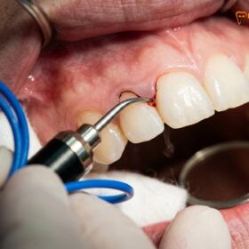
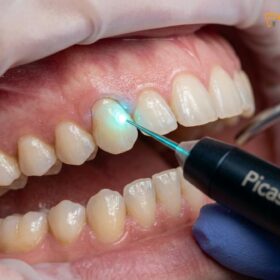
The landscape of cosmetic periodontics has been revolutionized by the advent of advanced clinical technologies. For patients diagnosed with delayed passive eruption, the treatment of choice is often laser gingivectomy. At modern facilities, the integration of diode soft tissue lasers has transformed this procedure into a highly precise and comfortable experience. The specific wavelength of these lasers is highly absorbed by melanin and hemoglobin, making it the perfect tool for soft tissue manipulation.
Unlike traditional scalpels that cause bleeding and require uncomfortable sutures, the laser provides bloodless, sutureless cuts. As the laser energy vaporizes the excess gum tissue, it simultaneously provides instant coagulation and cauterization of the blood vessels. This minimally invasive approach drastically reduces postoperative pain and swelling, allowing for a rapid recovery. Furthermore, the laser beam is inherently sterile, which significantly minimizes the risk of postoperative infection and promotes faster tissue maturation.
If you are ready to uncover the true potential of your smile and address excessive gingival display, expert care is available. Contact HCMC Dental Clinic in Ho Chi Minh City today to schedule a comprehensive digital smile evaluation. Discover how personalized treatments, including advanced Gum Contouring & Gummy Smile tại HCMC Dental Clinic, can restore harmony to your facial aesthetics and boost your confidence.
References
- Journal of Clinical Periodontology. Esthetic crown lengthening outcomes and biological width.
- International Journal of Periodontics & Restorative Dentistry. Diode laser vs scalpel gingivectomy in aesthetic zones.
- Journal of Aesthetic and Restorative Dentistry. Altered passive eruption: diagnosis and treatment planning.
- Journal of Oral and Maxillofacial Surgery. Le Fort I osteotomy stability in vertical maxillary excess.
- Clinical Oral Investigations. Surgical lip repositioning longevity and patient satisfaction.
- Vietnam Odonto-Stomatology Association (VOSA). Guidelines on periodontal aesthetics and gingival display evaluation.