An implant consultation in Saigon provides international patients with a comprehensive clinical evaluation, 3D imaging, and a personalized surgical plan. By leveraging advanced diagnostics, specialists determine bone density and structural viability, ensuring a precise, predictable, and safe foundation for your dental restoration journey in Ho Chi Minh City.
Clinical Summary:
The initial consultation for Dental Implants serves as the critical diagnostic foundation for successful oral rehabilitation. During this session, clinicians utilize high-resolution Cone Beam Computed Tomography (CBCT) and intraoral optical scanners to capture precise anatomical data. This digital information is synthesized to evaluate alveolar bone volume, identify vital neurovascular structures, and formulate a prosthetically driven surgical blueprint. By establishing a clear clinical pathway on day one, patients receive accurate cost estimations, staged treatment timelines, and a comprehensive risk assessment, ensuring that the subsequent surgical phases are executed with maximum safety and predictability.
Key Takeaways:
- Comprehensive diagnostics utilize 3D CBCT imaging to evaluate bone density and anatomical constraints.
- Digital intraoral scanning eliminates the need for traditional, uncomfortable putty impressions.
- Virtual surgical planning software allows for prosthetically driven implant positioning.
- Transparent financial planning and staged scheduling are established during the first visit.
- Systemic health evaluations ensure patient candidacy and mitigate intraoperative risks.
Fast-Tracking Your Dental Treatment in Ho Chi Minh City
Fast-tracking your dental treatment in Ho Chi Minh City involves synchronized clinical workflows that combine initial diagnostics, digital scanning, and surgical planning into a single, highly efficient visit.
For international patients traveling for medical care, time is a critical factor. The concept of fast-track dental consulting has revolutionized how complex oral rehabilitations are managed. In the past, a patient might require multiple separate appointments for initial examinations, radiographic imaging, impression taking, and treatment planning. Today, modern clinical protocols at leading facilities like HCMC Dental Clinic consolidate these vital steps into a streamlined, cohesive day-one experience. This synchronization not only respects the patient’s travel schedule but also minimizes the cognitive and physical fatigue associated with prolonged diagnostic phases.
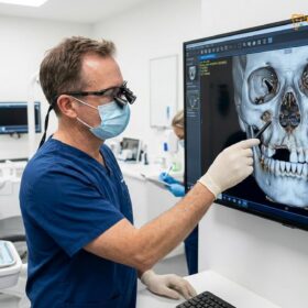
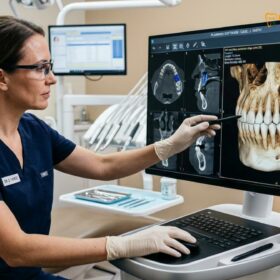
The efficiency of this process relies heavily on the integration of digital dentistry. When a patient arrives in Ho Chi Minh City for their consultation, the clinical team is already prepared to execute a multi-disciplinary assessment. The workflow is designed to transition seamlessly from the administrative intake to the clinical chair, and finally to the digital planning suite. This rapid data acquisition does not compromise clinical rigor; rather, it enhances it by utilizing software that instantly processes anatomical data, allowing the implantologist to visualize the surgical site in real-time[1].
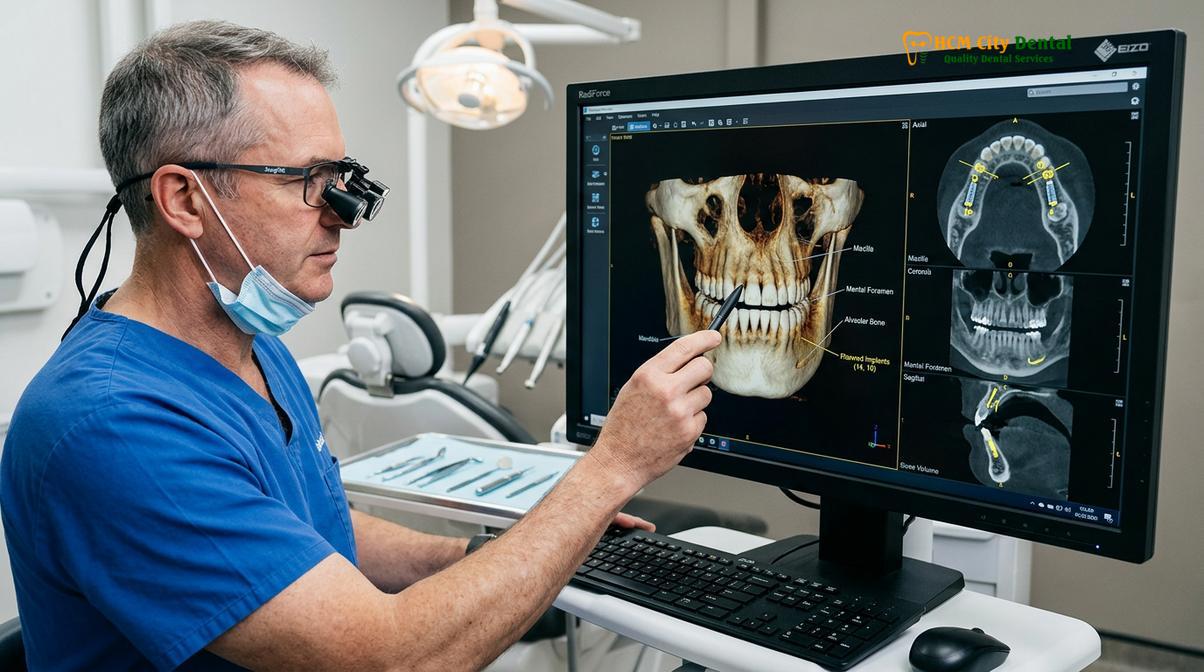
Furthermore, fast-tracking is not merely about speed; it is about optimizing the biological and logistical sequencing of the treatment. By gathering all necessary diagnostic records on the first day, the clinical team can immediately begin fabricating surgical guides and provisional prosthetics. This rapid turnaround is particularly beneficial for patients seeking immediate loading protocols, where temporary teeth are attached to the implants shortly after placement. The ability to compress the diagnostic and planning timeline directly correlates with a faster transition to the active surgical phase, reducing the overall duration of the patient’s stay in Vietnam.
“The essence of a successful international dental consultation lies in our ability to condense weeks of traditional diagnostic work into a single, highly coordinated session. By leveraging digital data acquisition, we provide our patients with immediate, evidence-based clarity regarding their surgical options and timelines.” – Dr. Nguyen Van Cuong
However, it is crucial to understand that fast-tracking the workflow does not mean rushing the biological healing process. The consultation sets the pace for the logistical aspects of the treatment, but the physiological integration of the titanium fixture with the human jawbone—a process known as osseointegration—still requires strict adherence to biological timelines. The consultation serves to map out these timelines accurately, ensuring that the patient has realistic expectations regarding their recovery and the final delivery of their permanent restorations.
Clinical Workflow: Day-1 Consultation, Scanning, and Model Design
The Day-1 clinical workflow seamlessly integrates comprehensive oral examinations, high-resolution radiographic imaging, and digital impression taking to formulate an immediate, evidence-based treatment strategy.
The foundation of any successful implant procedure is laid during the very first clinical encounter. The Day-1 workflow is a meticulously choreographed sequence of diagnostic events designed to leave no anatomical or pathological variable unchecked. The process begins with a comprehensive review of the patient’s medical and dental history. The implantologist must evaluate systemic conditions, pharmacological profiles, and previous dental interventions to establish a baseline of the patient’s overall health. This systemic evaluation is paramount, as conditions such as uncontrolled diabetes or autoimmune disorders can significantly impact the predictability of osseointegration.
Following the medical review, a thorough clinical examination of the oral cavity is conducted. The clinician assesses the health of the remaining dentition, the integrity of the periodontal tissues, and the biomechanics of the patient’s occlusion (bite). The soft tissue architecture—specifically the biotype of the gingiva (thick vs. thin)—is carefully evaluated, as it plays a critical role in the final aesthetic outcome of the implant restoration. The presence of active periodontal disease or endodontic infections in adjacent teeth must be identified and addressed, as a sterile surgical environment is mandatory for implant survival[2].
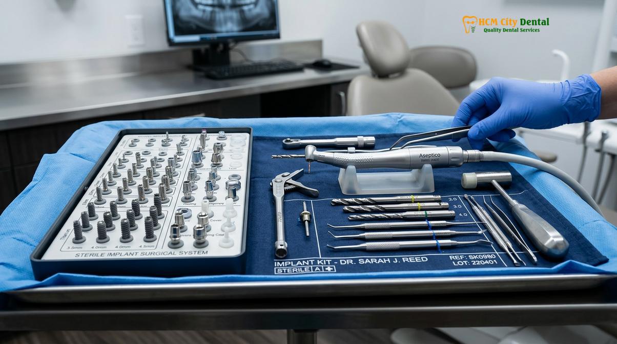
Once the clinical baseline is established, the workflow transitions to digital data acquisition. This is where the true power of modern diagnostics comes into play. The patient undergoes a series of advanced imaging procedures to capture the exact three-dimensional topography of their maxillofacial structures. This data is not merely for visualization; it is the raw material from which the entire surgical and prosthetic plan will be engineered. The integration of radiographic data with surface topography allows the clinical team to create a highly accurate digital twin of the patient’s oral cavity.
With the digital models generated, the clinician and the in-house dental laboratory technicians collaborate to design the preliminary prosthetic outcome. This concept, known as prosthetically driven implantology, dictates that the final position of the tooth crown determines the surgical placement of the titanium root—not the other way around. By designing the ideal tooth position first, the team can reverse-engineer the exact angulation and depth required for the implant fixture. If the digital model reveals that the ideal implant position lacks sufficient bone volume, the clinician can immediately plan for adjunctive procedures, such as structural bone augmentation, ensuring that the patient is fully informed of the required surgical scope on day one.
Sourcing Your Digital Model: CBCT 3D and iTero 5D Scanners
Utilizing advanced CBCT 3D imaging and iTero 5D digital impressions allows clinicians to capture exact anatomical data, assessing bone volume and soft tissue architecture with micrometer precision.
The transition from traditional two-dimensional radiography to three-dimensional volumetric imaging represents one of the most significant advancements in implant dentistry. At the core of this diagnostic revolution is cbct scanning 3d (Cone Beam Computed Tomography). Unlike conventional medical CT scans, which utilize a fan-shaped beam and require high radiation doses, CBCT utilizes a cone-shaped beam that rotates around the patient’s head, capturing hundreds of images in a single sweep. These images are then reconstructed by powerful algorithms into a highly detailed 3D volume.
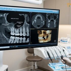
The clinical value of the CBCT scan cannot be overstated. It allows the implantologist to evaluate the alveolar bone in all three planes of space (axial, coronal, and sagittal). The clinician can precisely measure the height, width, and trajectory of the available bone. Furthermore, the software allows for the measurement of bone density using Hounsfield Units (HU), categorizing the bone from D1 (dense cortical bone) to D4 (soft medullary bone). This density assessment is critical for determining the primary stability of the implant and selecting the appropriate surgical drilling protocol. Crucially, the CBCT scan clearly delineates vital anatomical landmarks, such as the inferior alveolar nerve canal in the mandible and the floor of the maxillary sinus in the upper jaw, allowing the surgeon to plan a safe distance from these structures and determine if a maxillary sinus elevation is required[3].
Complementing the radiographic data is the capture of the intraoral surface topography using itero 5d digital impressions. Traditional dental impressions required the patient to bite into trays filled with viscous, often foul-tasting elastomeric materials, which could trigger severe gag reflexes and were prone to dimensional distortion. The iTero 5D scanner eliminates this discomfort by utilizing confocal imaging and Near-Infrared Imaging (NIRI) technology to capture thousands of optical data points per second. The result is a highly accurate, full-color stereolithography (STL) file that perfectly replicates the contours of the teeth, the gingival margins, and the occlusal relationships.
| Diagnostic Modality | Primary Function | Data Output Format | Clinical Application |
|---|---|---|---|
| CBCT 3D Scan | Volumetric analysis of hard tissues | DICOM (Digital Imaging and Communications in Medicine) | Bone density assessment, nerve mapping, sinus evaluation, implant sizing. |
| iTero 5D Scanner | Surface topography of soft tissues and dentition | STL (Stereolithography) / PLY | Prosthetic design, occlusal mapping, surgical guide fabrication, soft tissue profiling. |
| Digital Articulation | Dynamic bite analysis | Virtual Articulator Software | Evaluating excursive movements, preventing occlusal overload on the implant. |
The true magic happens when the DICOM data from the CBCT scan is merged with the STL data from the iTero scanner within specialized implant planning software. This fusion creates a comprehensive digital environment where the clinician can visualize the underlying bone structure simultaneously with the overlying soft tissue and adjacent teeth. This dual-layered digital model is the ultimate diagnostic tool, providing a level of anatomical insight that was previously impossible to achieve without invasive exploratory surgery.
Creating a Virtual Guided Surgery Blueprint
A virtual guided surgery blueprint merges radiographic and intraoral data to simulate the exact implant trajectory, minimizing surgical risks and optimizing the final prosthetic outcome.
Once the digital models are merged, the implantologist begins the process of virtual surgery. This phase is where clinical experience meets digital precision. Using the planning software, the clinician selects a virtual implant from a comprehensive digital library that matches the exact specifications (diameter, length, thread design) of the actual titanium fixture to be used. This virtual implant is then manipulated within the 3D model of the patient’s jawbone to find the optimal position.
The positioning is dictated by several critical factors. First, the implant must be surrounded by a minimum thickness of healthy bone (typically 1.5 to 2.0 mm on the facial aspect) to ensure long-term stability and prevent tissue recession. Second, the trajectory must align perfectly with the planned prosthetic crown to ensure that the chewing forces are directed along the long axis of the implant body. Third, the virtual placement must maintain a strict safety margin (usually 2.0 mm) from vital structures like the mandibular nerve. By simulating the surgery virtually, the clinician can anticipate and resolve potential anatomical conflicts before the patient ever sits in the surgical chair[4].
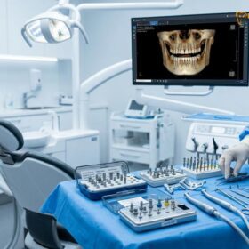
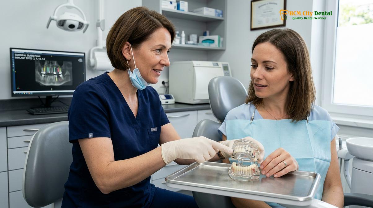
After the ideal virtual position is finalized, the software is used to design a virtual surgical guided template. This template is a custom-fitted appliance that rests on the patient’s remaining teeth or mucosal tissue during the actual surgery. It features precisely angled cylindrical sleeves that correspond exactly to the planned trajectory of the virtual implant. Once the design is complete, the data is sent to a high-resolution 3D printer, which fabricates the physical surgical guide out of a biocompatible, sterilizable resin.
Clinical Case Review: Precision through Guided Surgery
A 55-year-old international patient visited HCMC Dental Clinic in Ho Chi Minh City presenting with a missing mandibular first molar and severe localized bone resorption. Traditional freehand surgery posed a high risk of inferior alveolar nerve injury due to the limited vertical bone height. During the consultation, a virtual guided surgery blueprint was developed. The software allowed the clinical team to select a short, wide-diameter implant and position it with a 2.5mm safety clearance from the nerve canal. The 3D-printed surgical guide ensured that the actual osteotomy (drilling) matched the virtual plan with sub-millimeter accuracy. The use of ultrasonic surgical techniques further preserved the surrounding bone. The procedure was completed flawlessly, demonstrating how digital planning mitigates severe anatomical risks.
The utilization of a surgical guide transforms the implant procedure from a freehand, visually estimated process into a highly controlled, mechanically restricted operation. When the surgeon uses the drills through the guide sleeves, the depth, angulation, and position are physically constrained to match the approved digital blueprint. This level of precision significantly reduces surgical trauma, minimizes the need for extensive soft tissue reflection (flapless surgery), and dramatically decreases postoperative pain and swelling for the patient.
Locking in the Pricing and Staged Scheduling Quickly
Transparent financial planning and staged scheduling ensure that patients understand all associated costs and recovery timelines before any surgical intervention begins.
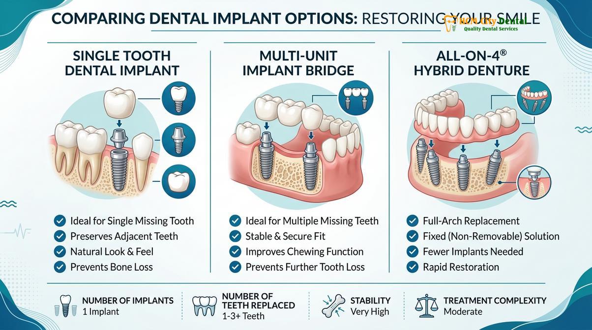
A primary concern for patients seeking dental care abroad is financial transparency and logistical predictability. The comprehensive nature of the day-one consultation allows the clinical team to provide an exact, itemized treatment plan. Because the digital blueprint reveals the precise anatomical conditions, there is no guesswork regarding the necessity of adjunctive procedures. If the CBCT scan indicates that a bone graft or a sinus lift is required to support the implant, this is identified immediately, and the associated costs are included in the initial proposal.
The financial breakdown typically includes the cost of the surgical phase (the titanium fixture and the surgical procedure), the cost of any necessary regenerative materials (bone particulate, collagen membranes), and the cost of the restorative phase (the custom prosthetic abutments and the final ceramic crown). By locking in this pricing during the consultation, patients are protected from unexpected fees or mid-treatment financial surprises. This transparency is a cornerstone of ethical medical tourism and builds essential trust between the patient and the clinical provider.
Important Financial Consideration
Patients should be wary of extremely low-cost implant advertisements that do not require a comprehensive 3D diagnostic consultation first. Often, these “bait-and-switch” quotes only cover the cost of the titanium screw, leaving out the mandatory abutment, the final crown, and any necessary bone grafting. Always demand a fully itemized, all-inclusive treatment plan based on a personalized CBCT analysis before committing to surgery.
Equally important to the financial aspect is the establishment of a staged scheduling timeline. Dental implant therapy is rarely a single-event procedure; it is a biological process that occurs in phases. The consultation provides the patient with a clear roadmap of their treatment journey. The schedule will outline the date of the initial surgery, the required duration of the biological healing phase (typically 3 to 6 months for osseointegration), and the timeline for the final prosthetic delivery. For international patients, this allows for the precise booking of return flights and accommodations, ensuring that their medical travel is as efficient and stress-free as possible.
When to See a Doctor for an Implant Consultation
Determining the right time to seek an implant consultation is crucial for preventing further oral health deterioration. You should schedule a clinical evaluation if you are missing one or more teeth, as the absence of a tooth root leads to progressive atrophy (shrinkage) of the surrounding jawbone. Early intervention is key; the longer a tooth is missing, the more bone is lost, which may eventually necessitate complex and costly bone grafting procedures before an implant can be placed.
Additionally, patients who are currently wearing ill-fitting removable dentures that cause discomfort, speech impediments, or difficulty chewing are prime candidates for an implant consultation. Implants can be used to anchor these prosthetics securely, vastly improving the patient’s quality of life. Furthermore, if you have a failing dental bridge or a tooth that has been deemed unrestorable due to severe decay or advanced periodontal disease, an immediate consultation can help plan for the extraction and simultaneous implant placement, preserving the natural bone architecture.
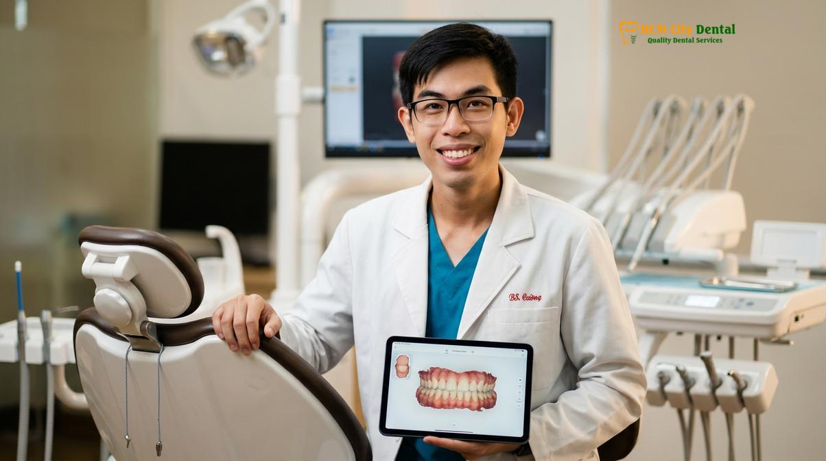
“Patient candidacy is not solely determined by the presence of missing teeth. A successful consultation must evaluate the patient’s systemic ability to heal. We look closely at factors like glycemic control in diabetic patients and bone metabolism, ensuring that when we proceed with surgery, the biological environment is primed for success.” – Dr. Nguyen Van Cuong
However, there are specific clinical contraindications that must be discussed during the consultation. Patients with uncontrolled systemic diseases, those who have undergone recent radiation therapy to the head and neck, or individuals currently taking intravenous bisphosphonates for osteoporosis may be at a higher risk for complications such as implant failure or osteonecrosis of the jaw. A thorough medical evaluation by a qualified implantologist is mandatory to assess these risks and determine if implant therapy is a safe and viable option based on your individualized diagnostic profile[5].
Frequently Asked Questions
How long does the initial consultation and scan take?
The initial implant consultation and comprehensive scanning process typically takes between 60 to 90 minutes. This timeframe allows the clinical team to capture 3D radiographic images, perform intraoral digital impressions, conduct a thorough periodontal examination, and discuss the preliminary surgical blueprint with you in detail. The efficiency of this single-visit diagnostic session is designed to respect your time while ensuring no clinical detail is overlooked.
Can I get my dental scan records emailed to me?
Yes, your digital dental records, including the 3D CBCT imaging files and intraoral scans, can be securely emailed to you. We provide these files in standard medical formats (such as DICOM and STL) so you can retain them for your personal health records or share them with your local dentist. Having access to your own diagnostic data ensures continuity of care, especially for international patients traveling back to their home countries.
Is there a fee for the initial 3D scan and consultation?
Fee structures for the initial 3D scan and consultation vary based on the specific promotional periods and comprehensive packages selected. Currently, many of our international patient pathways include complimentary initial diagnostic imaging when you proceed with the complete surgical treatment plan at our facility. We recommend contacting our patient coordination team directly to receive the most accurate and up-to-date information regarding consultation fees and available diagnostic packages.
What medical conditions might delay my dental implant surgery?
Conditions such as uncontrolled diabetes mellitus, recent cardiovascular events, active periodontal disease, or a history of intravenous bisphosphonate therapy can delay surgery. Your implantologist will require these systemic conditions to be medically managed and stabilized before clearing you for any invasive osseous procedures. A collaborative approach with your primary care physician is often utilized to ensure your body is physiologically prepared for optimal healing and osseointegration.
Do I need to stop taking my medications before the consultation?
No, you should not stop taking any prescribed medications prior to your initial consultation unless explicitly instructed by your primary care physician. During the consultation, the dental specialist will review your complete pharmacological profile to determine if any modifications are necessary before the actual surgical appointment. Certain medications, such as blood thinners or specific immunosuppressants, may require temporary adjustment under medical supervision closer to the date of the surgical intervention.
References
- Journal of Oral Implantology. Diagnostic accuracy of cone-beam computed tomography in implant dentistry. (2021).
- International Journal of Oral and Maxillofacial Implants. Digital workflows in prosthetically driven implant placement. (2020).
- Clinical Implant Dentistry and Related Research. Accuracy of intraoral scanners for full-arch digital impressions. (2019).
- Journal of Prosthetic Dentistry. Biomechanical considerations for immediate loading of dental implants. (2022).
- Periodontology 2000. Systemic risk factors and contraindications in implant therapy. (2018).