A single tooth dental implant is a permanent, surgically placed titanium root that supports a custom prosthetic crown to replace an isolated missing tooth. This advanced clinical solution restores full masticatory function, preserves underlying jawbone density, and seamlessly replicates the natural emergence profile without compromising adjacent healthy teeth.
Clinical Summary:
A single tooth dental implant represents the gold standard in modern restorative dentistry for replacing an isolated missing tooth. The procedure involves surgically embedding a biocompatible titanium fixture into the alveolar bone, which undergoes osseointegration to form a highly stable foundation. Following this critical healing phase, a restorative abutment and a custom-milled porcelain crown are securely attached. This tri-part structure mimics natural dental anatomy, preventing localized bone resorption and maintaining occlusal stability. Clinical success depends on precise 3D radiographic imaging, adequate bone volume, and meticulous surgical execution, offering a highly durable, long-term functional and aesthetic outcome for patients.
Key Takeaways:
- Implants consist of three distinct components: the titanium fixture, the abutment, and the prosthetic crown.
- Osseointegration, the biological fusion between living bone and titanium, is essential for long-term mechanical stability.
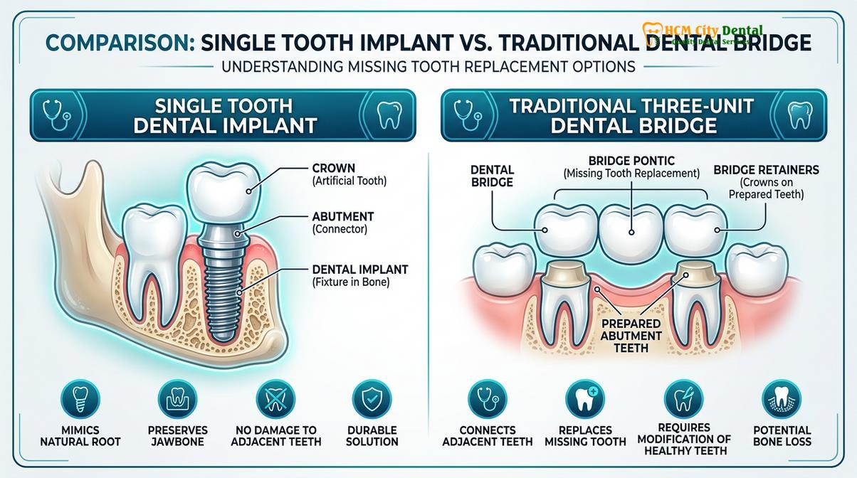
- The procedure preserves adjacent natural teeth, unlike traditional tooth-supported dental bridges that require enamel reduction.
- Treatment timelines typically range from three to six months, depending on individual bone healing capacity and grafting needs.
- Advanced 3D CBCT imaging ensures precise surgical planning, optimizing implant angulation and minimizing intraoperative risks.
Understanding the Single Tooth Dental Implant Structure
A single tooth dental implant is a sophisticated three-part medical device—comprising a titanium fixture, an abutment, and a crown—designed to replicate the exact biomechanical function and aesthetic appearance of a natural human tooth.
When a patient loses a single tooth due to trauma, severe decay, or periodontal disease, the structural integrity of the entire dental arch is compromised. Historically, the primary solution was a traditional three-unit dental bridge, which required the irreversible reduction of healthy adjacent teeth. Today, modern implantology has revolutionized restorative protocols by offering a standalone solution that mimics the anatomy of a natural tooth from the root to the crown. The macro-design of the fixture features a threaded exterior, which provides immediate mechanical stability—known as primary stability—upon insertion into the bone[1].
The Titanium Implant Fixture (The Artificial Root)
The foundation of the entire restoration is the implant fixture, commonly referred to as the titanium implant screw. This component is surgically embedded directly into the alveolar bone of the maxilla (upper jaw) or mandible (lower jaw). Manufactured from highly biocompatible commercially pure titanium or titanium alloys, the fixture is entirely accepted by the human immune system, virtually eliminating the risk of foreign body rejection.
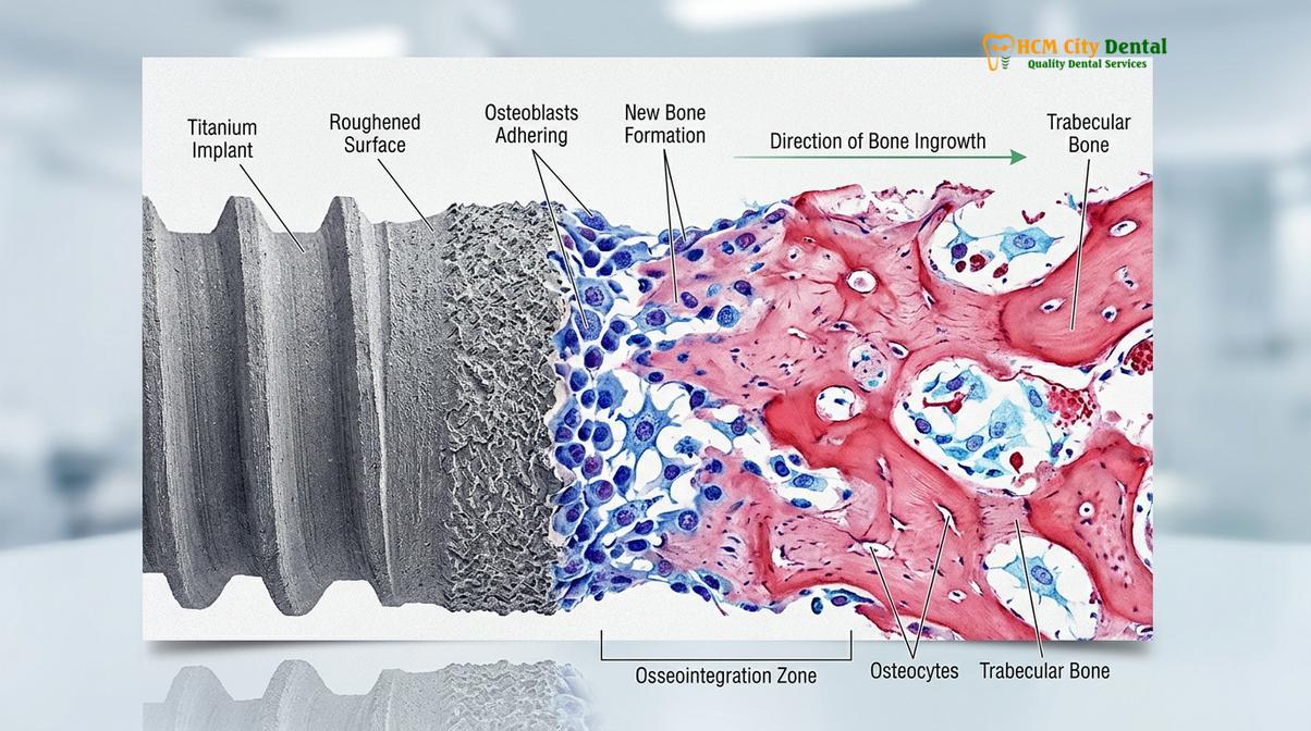
Beyond its macroscopic threads, the micro-surface topography of the titanium implant screw is equally critical. Modern implants undergo specialized surface treatments, such as sandblasting and acid-etching (SLA) or anodic oxidation. These treatments create a microscopically roughened surface that significantly increases the surface area available for bone cells (osteoblasts) to attach. This enhanced surface topography accelerates the biological healing process, allowing for a stronger and faster integration between the metal and the living bone tissue.
The Restorative Abutment (The Connector)
Once the implant fixture has successfully fused with the jawbone, the second component, known as the restorative abutment, is introduced. The abutment serves as the critical transmucosal connector that bridges the gap between the submerged implant fixture and the oral cavity. It is securely screwed into the internal connection of the implant fixture, extending just above the gum line to provide a solid core for the final prosthesis.
Abutments can be fabricated from various materials depending on the clinical requirements. Titanium abutments are renowned for their exceptional fracture resistance and are typically utilized in the posterior regions where masticatory (chewing) forces are highest. Conversely, in the highly visible anterior (front) aesthetic zone, zirconia abutments are frequently preferred. Zirconia, a high-strength white ceramic, prevents the grayish hue of metal from showing through thin gingival tissues, ensuring a highly natural and aesthetically pleasing outcome.
The Prosthetic Crown (The Visible Tooth)
The final component of the implant triad is the prosthetic crown. This is the visible portion of the restoration that interacts with opposing teeth during chewing and speaking. Modern implant crowns are typically milled from advanced ceramics, such as monolithic zirconia or lithium disilicate, utilizing computer-aided design and computer-aided manufacturing (CAD/CAM) technology.
The design of the crown is meticulously customized to match the exact shade, translucency, and anatomical contours of the patient’s adjacent natural teeth. Furthermore, the emergence profile—the way the crown transitions from the circular abutment through the gum tissue to form the shape of a tooth—is carefully sculpted to ensure soft tissue integration and optimal aesthetics[5]. A properly contoured emergence profile not only ensures a seamless aesthetic integration but also facilitates effective oral hygiene, preventing plaque accumulation at the implant-mucosa interface.
The Surgical Phase: Placing the Titanium Post
The surgical placement of the implant fixture is a precision-driven procedure requiring comprehensive 3D radiographic planning to ensure optimal positioning, angulation, and primary stability within the alveolar bone.
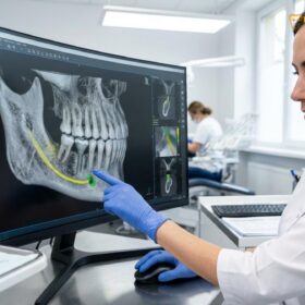
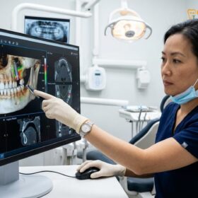
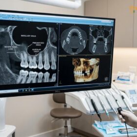
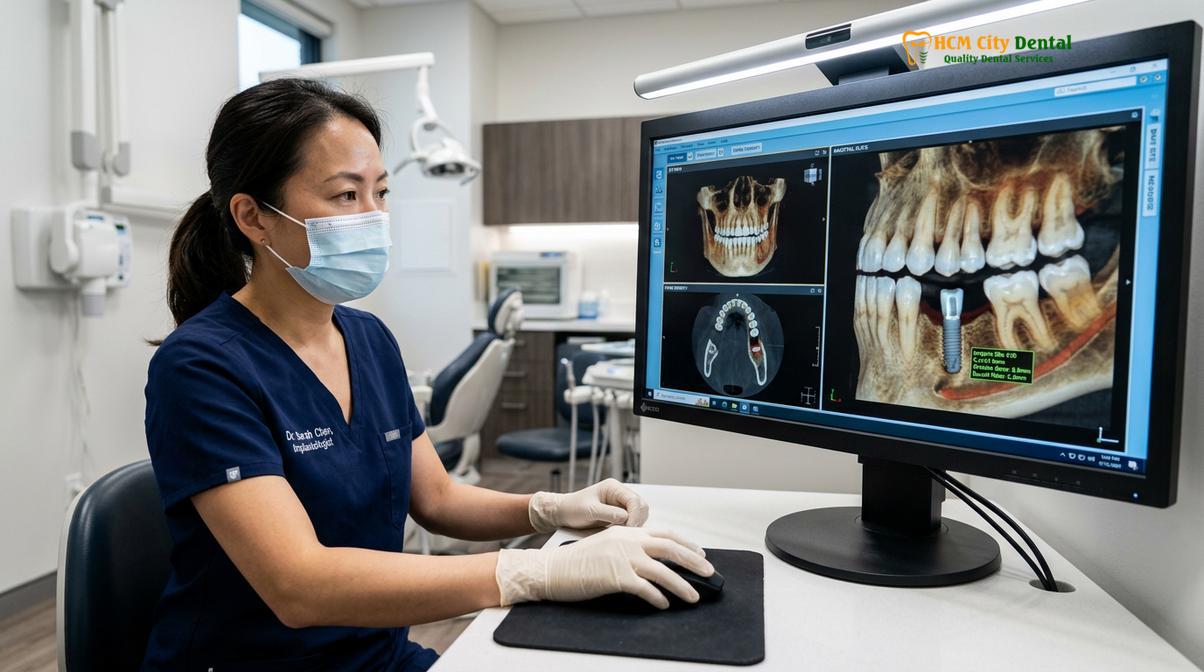
The surgical phase of a single tooth dental implant is a highly orchestrated medical procedure that demands meticulous planning and execution. The success of the entire restoration hinges on the accurate three-dimensional placement of the titanium implant screw within the available bone volume. Prior to any surgical intervention, a comprehensive clinical and radiographic evaluation is mandatory. This involves the acquisition of a Cone Beam Computed Tomography (CBCT) scan, which provides a distortion-free, 3D representation of the patient’s maxillofacial anatomy.
Pre-Surgical 3D Planning and Virtual Placement
Utilizing the CBCT data, the implantologist can precisely measure the height, width, and density of the alveolar bone at the edentulous (missing tooth) site. The scan also highlights critical anatomical landmarks that must be strictly avoided during surgery, such as the inferior alveolar nerve in the lower jaw and the maxillary sinus cavity in the upper jaw. Through specialized implant planning software, the surgeon performs a “virtual surgery,” selecting the exact diameter and length of the implant fixture and determining its ideal trajectory to align with the planned prosthetic crown.
In many advanced clinical settings, this virtual plan is translated into a 3D-printed surgical guide. This custom-made template fits securely over the patient’s remaining teeth and features a precise metallic sleeve that directs the surgical drills. Guided surgery significantly enhances the accuracy of the osteotomy (bone preparation), minimizes surgical trauma, and allows for a minimally invasive flapless approach in suitable candidates. Patients often inquire about the differences between premium implant systems during this planning phase, such as comparing Straumann vs Nobel Biocare Implants, to ensure they receive the most appropriate fixture for their specific bone density.
The Osteotomy and Implant Insertion Protocol
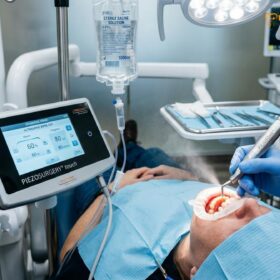
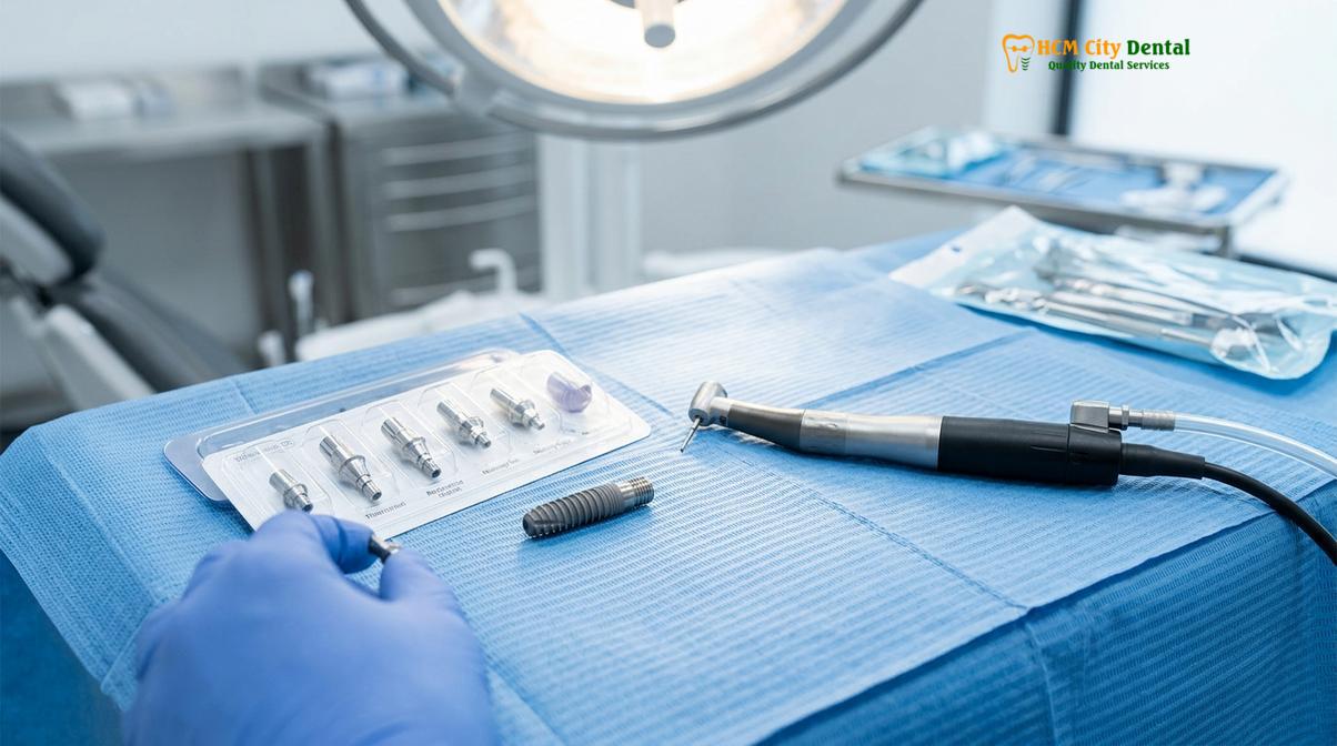
The surgical procedure is typically performed under profound local anesthesia, ensuring the patient remains entirely comfortable and pain-free. If a traditional open-flap approach is utilized, a small incision is made in the gingival tissue to expose the underlying crestal bone. The osteotomy is then initiated using a series of precisely calibrated surgical drills. The drilling sequence begins with a small pilot drill to establish the depth and angulation, followed by sequentially wider drills to gradually expand the site to the exact dimensions of the selected implant.
During the drilling process, copious irrigation with sterile chilled saline is continuously applied. This is a critical step to prevent thermal necrosis (heat-induced death) of the bone cells, as bone tissue is highly sensitive to elevated temperatures. Once the osteotomy is fully prepared, the titanium implant screw is slowly threaded into the bone using a calibrated torque wrench or a surgical motor.
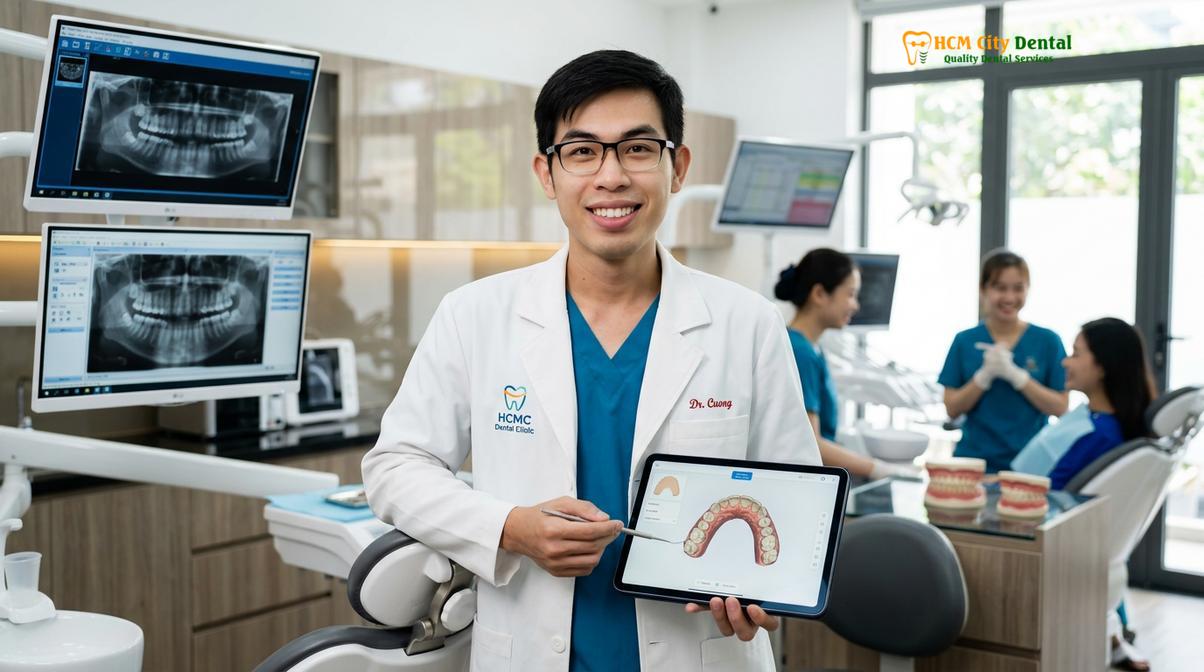
Dr. Nguyen Van Cuong, a leading specialist in restorative dentistry, emphasizes that achieving adequate primary stability during this initial surgical phase is the most critical biomechanical predictor for long-term implant survival. For patients seeking comprehensive insights, Dr. Cuong provides expert guidance on dental implants to ensure optimal clinical outcomes. His approach prioritizes minimally invasive techniques to preserve surrounding bone and soft tissue architecture.
The Healing Interval: Why Osseointegration is Critical
Osseointegration is the vital biological process where living bone cells attach directly to the titanium surface, locking the implant permanently into the jaw architecture and providing secondary stability.
Following the surgical placement of the implant fixture, the treatment enters a critical dormant phase known as the healing interval. While the implant may feel mechanically tight immediately after surgery (primary stability), this initial friction naturally decreases over the first few weeks as the traumatized bone immediately surrounding the implant undergoes resorption. It is during this vulnerable window that the biological phenomenon of osseointegration must successfully occur to establish long-term secondary stability[2].
The Biological Cascade of Bone Healing
Osseointegration is not merely a mechanical locking; it is a dynamic cellular cascade. Immediately after the implant is inserted, a blood clot forms around the titanium threads. Blood platelets release growth factors that stimulate angiogenesis (the formation of new blood vessels) and attract osteoprogenitor cells to the site. Over the ensuing weeks, these cells differentiate into osteoblasts—the specialized cells responsible for synthesizing new bone matrix.
The osteoblasts migrate directly onto the roughened micro-surface of the titanium implant screw, depositing a collagen matrix that eventually mineralizes into woven bone. This immature woven bone is highly cellular but lacks structural density. Over a period of three to six months, the woven bone is gradually remodeled and replaced by highly organized, load-bearing lamellar bone. This mature bone intimately adapts to the implant threads, creating a direct structural and functional connection between living bone and the artificial surface.
“The success of implant dentistry relies entirely on the predictability of osseointegration. Without this direct bone-to-implant contact, the fixture cannot withstand the immense biomechanical forces generated during mastication, leading to inevitable micro-movement and eventual fibrous encapsulation rather than true integration.”
Factors Influencing the Healing Timeline
The duration of the osseointegration phase varies significantly depending on the anatomical location and the patient’s individual physiology. Implants placed in the anterior mandible (lower front jaw), which is composed of dense cortical bone, may integrate in as little as three months. Conversely, implants placed in the posterior maxilla (upper back jaw), where the bone is naturally softer and more trabecular, often require four to six months of undisturbed healing.
Patient compliance during this phase is paramount. Micro-movement of the implant exceeding 150 micrometers during the early healing weeks can disrupt the fragile cellular attachment, leading to soft tissue encapsulation (failure) instead of bone integration. Therefore, patients are strictly advised to adhere to a soft diet and avoid chewing directly over the surgical site. Furthermore, smoking is a major risk factor; the vasoconstrictive effects of nicotine severely impair blood flow to the healing bone, significantly increasing the risk of implant failure.
Expected Timeline and Recovery Milestones
The journey to a fully restored smile via a single tooth dental implant follows a structured timeline, ensuring each biological and mechanical phase is completed safely and effectively.
Understanding the expected timeline helps patients prepare for the restorative journey. While immediate loading (placing a temporary crown on the same day as surgery) is possible in highly specific, ideal cases, the standard protocol involves a phased approach. This phased methodology aligns with the clinical standards outlined by the Vietnam Ministry of Health (MOH)[6], prioritizing patient safety and long-term predictability over rapid aesthetics.
| Treatment Phase | Typical Duration | Clinical Objective |
|---|---|---|
| Initial Consultation & 3D Imaging | Day 1 | Assess bone volume, identify anatomical landmarks, and plan the surgical trajectory. |
| Surgical Placement | 1 to 2 Hours | Insert the titanium fixture into the jawbone to achieve primary mechanical stability. |
| Osseointegration (Healing Phase) | 3 to 6 Months | Allow osteoblasts to fuse with the implant surface, establishing secondary stability. |
| Abutment & Crown Placement | 2 to 3 Weeks | Take digital impressions, fabricate the custom prosthesis, and attach the final crown. |
It is important to note that if a patient requires preliminary procedures, such as a bone graft or a sinus lift to build sufficient bone volume, the overall timeline will be extended by several months to allow the grafted material to mature before the implant can be safely placed.
The Restorative Phase: Custom Abutment and Crown Placement
Following successful bone integration, the restorative phase focuses on designing and attaching the final prosthetic components—the abutment and crown—to restore full occlusal function and natural aesthetics.
Once the clinical and radiographic assessments confirm that osseointegration is complete and the implant has achieved robust secondary stability, the patient transitions to the restorative phase. This phase transforms the integrated titanium root into a fully functional single tooth replacement. If the implant was submerged beneath the gum tissue during the healing phase, a minor second-stage surgery is performed to uncover the implant and attach a temporary healing abutment, which shapes the gingival tissue for two to three weeks.
Digital Impressions and Prosthetic Design
The fabrication of the final prosthesis begins with capturing the precise three-dimensional position of the implant in relation to the surrounding teeth and opposing arch. Modern restorative workflows increasingly utilize digital intraoral scanners rather than traditional gooey impression materials. A specialized scanning post, known as a scan body, is temporarily attached to the implant. The digital scanner captures the exact geometry of the scan body, creating a highly accurate 3D virtual model of the patient’s mouth.
This digital file is transmitted to a dental laboratory, where skilled technicians use CAD software to design the custom abutment and the final crown. The choice between a screw-retained crown (where the crown and abutment are one piece screwed directly into the implant) and a cement-retained crown (where the abutment is screwed in and the crown is cemented over it) depends on the implant’s angulation and aesthetic requirements[3]. Screw-retained restorations are often preferred as they eliminate the risk of excess cement causing peri-implant inflammation.
Final Delivery and Occlusal Adjustment
During the final delivery appointment, the custom abutment and crown are securely attached to the implant fixture. The clinician meticulously checks the interproximal contacts (how the crown touches adjacent teeth) to ensure food does not become impacted. Equally important is the evaluation of the occlusion (the patient’s bite). The implant crown must be precisely adjusted so that it does not bear excessive force during chewing, as implants lack the shock-absorbing periodontal ligament found in natural teeth.
Clinical Case Study: Restoring a Smile in Saigon
Real-world clinical applications demonstrate the transformative impact of a single tooth dental implant on a patient’s oral function and confidence.
Consider the case of a 34-year-old patient who visited HCMC Dental Clinic in Ho Chi Minh City after suffering a traumatic injury that resulted in the loss of a maxillary central incisor. The sudden loss of a highly visible front tooth caused significant aesthetic distress and functional impairment. Upon clinical examination and CBCT analysis, it was determined that the buccal bone plate remained intact, making the patient an excellent candidate for an immediate implant placement protocol.
The surgical team carefully extracted the remaining root fragments and immediately placed a premium titanium implant fixture into the extraction socket. A temporary, non-loading crown was affixed to maintain the gingival architecture during the healing phase. After four months of successful osseointegration, the patient returned to receive a custom-milled zirconia abutment and a highly translucent all-ceramic crown. The final restoration seamlessly blended with the adjacent natural dentition, fully restoring the patient’s smile and confidence.
When to Consult a Specialist and Important Considerations
While dental implants boast a remarkably high success rate, recognizing the signs of potential complications and adhering to strict maintenance protocols are essential for long-term viability.
Patients must understand that while the titanium implant itself cannot decay, the surrounding gingival tissues and alveolar bone remain susceptible to bacterial infection. A condition known as peri-implantitis—an inflammatory process affecting the soft and hard tissues surrounding an implant—can lead to rapid bone loss and eventual implant failure if left untreated[4]. Symptoms such as persistent bleeding when brushing, localized swelling, purulent exudate (pus), or a feeling of looseness in the implant crown warrant immediate clinical evaluation.
“Early intervention is paramount when dealing with peri-implant complications. Recognizing the subtle signs of mucosal inflammation can prevent the progression to irreversible bone loss, safeguarding the structural integrity of the implant restoration.”
To mitigate these risks, patients are strongly advised to maintain impeccable daily oral hygiene, utilizing interdental brushes or water flossers to clean the critical junction where the implant meets the gum line. Routine professional cleanings and radiographic monitoring every six months are non-negotiable components of implant aftercare. For individuals considering this life-changing procedure, exploring comprehensive dental implants at HCMC Dental Clinic provides a clear pathway to restoring oral health. Additionally, understanding the financial investment is crucial; patients can review how much dental implants cost in Vietnam to make informed decisions regarding their restorative care.
If you are missing a single tooth and wish to explore permanent, natural-looking restorative options, contact HCMC Dental Clinic in Ho Chi Minh City today to schedule a comprehensive 3D implant consultation with our specialized clinical team.
References
- Journal of Oral Implantology. Biomechanical principles of single-tooth implant restorations and crestal bone maintenance. (2021).
- International Journal of Oral and Maxillofacial Implants. Osseointegration and primary stability: A clinical review. (2020).
- Clinical Implant Dentistry and Related Research. Screw-retained versus cement-retained implant crowns: A systematic review. (2019).
- Journal of Clinical Periodontology. Diagnosis and management of peri-implant mucositis and peri-implantitis. (2022).
- Journal of Prosthetic Dentistry. Emergence profile design and soft tissue integration in anterior single implants. (2018).
- Vietnam Ministry of Health (MOH). National Guidelines on Oral and Maxillofacial Surgery and Dental Implantology. (2023).