A dark line at the gumline around a porcelain-fused-to-metal (PFM) crown is typically caused by the underlying metal margin becoming exposed due to gum recession. While often a purely cosmetic issue, it can sometimes indicate marginal leakage or decay requiring clinical evaluation and potential replacement with metal-free alternatives.
Clinical Summary:
Porcelain-fused-to-metal (PFM) crowns rely on a durable metal substructure for strength, but this design inherently risks aesthetic compromise over time. As gingival tissues naturally recede due to aging, brushing habits, or periodontal changes, the dark metal collar of the crown becomes visible, creating an unsightly black line at the tooth’s neck. Modern prosthodontics addresses this limitation through the use of advanced all-ceramic materials, such as Zirconia and Lithium Disilicate (E.max). Combined with precise digital intraoral scanning technology (iTero, Medit i700), these metal-free alternatives ensure perfect marginal adaptation, exceptional biocompatibility, and lifelike translucency, completely eliminating the risk of dark gingival shadowing.
Key Takeaways:
- The dark line is primarily the metal base of a PFM crown exposed by receding gums.
- While usually an aesthetic concern, it warrants clinical examination to rule out marginal decay or cement washout.
- Replacing old restorations with Zirconia or E.max completely eliminates the risk of future metal exposure.
- Digital scanning (iTero, Medit i700) ensures highly accurate, comfortable crown fabrication without messy traditional impressions.
- Modern dental tourism allows for complete, high-quality smile makeovers to be completed in just 3 to 5 days.
What Causes the Dark Line Around Dental Crowns?
The dark line is primarily caused by the exposure of a PFM crown’s metal margin due to gum recession, though optical shadowing and marginal staining can also contribute to the discoloration.
When patients look in the mirror and notice a grey or black halo surrounding the base of their dental restoration, it is a classic symptom associated with older prosthodontic techniques. To understand why this occurs, one must examine the anatomical relationship between the dental crown, the underlying tooth structure, and the surrounding gingival (gum) tissue. The junction where the crown meets the prepared tooth is known as the margin. In an ideal clinical scenario, this margin is placed slightly subgingivally (below the gumline) to hide any transition lines and create a seamless emergence profile [1].
However, the oral environment is dynamic. Over time, the position of the gingival crest can migrate apically (towards the root of the tooth). When this happens, the previously hidden margin becomes visible. If the restoration is a traditional metal-based crown, the exposed margin will reveal the dark, opaque collar of the metal substructure. This is one of the most common porcelain fused to metal crown issues reported by patients seeking cosmetic dental revisions.
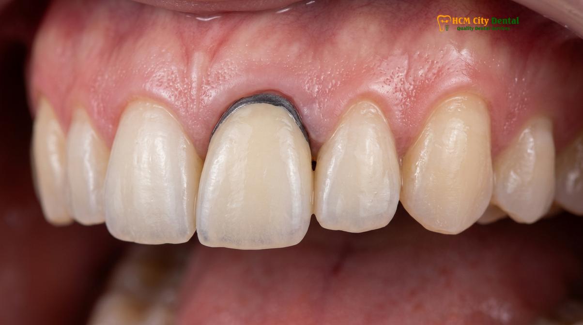
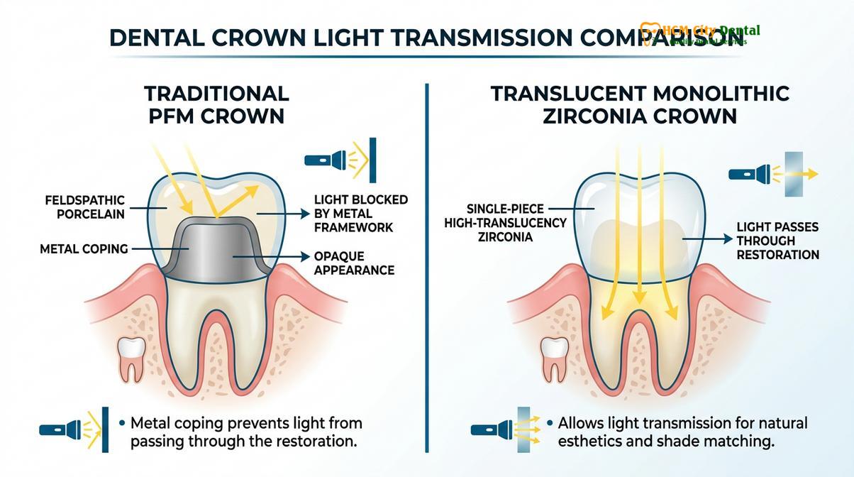
Beyond actual metal exposure, an optical phenomenon known as the “umbrella effect” can also cause a dark line. Because the metal core of a PFM crown completely blocks light transmission, light cannot travel down through the root of the tooth as it does in natural dentition. This lack of light diffusion casts a shadow on the surrounding thin gum tissue, making the gums themselves appear grey or bruised, even if the metal margin has not yet been physically exposed by recession. Furthermore, microscopic gaps at the margin can accumulate dietary stains from coffee, tea, or tobacco, exacerbating the dark appearance at the cervical third of the tooth.
Porcelain-Fused-to-Metal (PFM) Mechanics
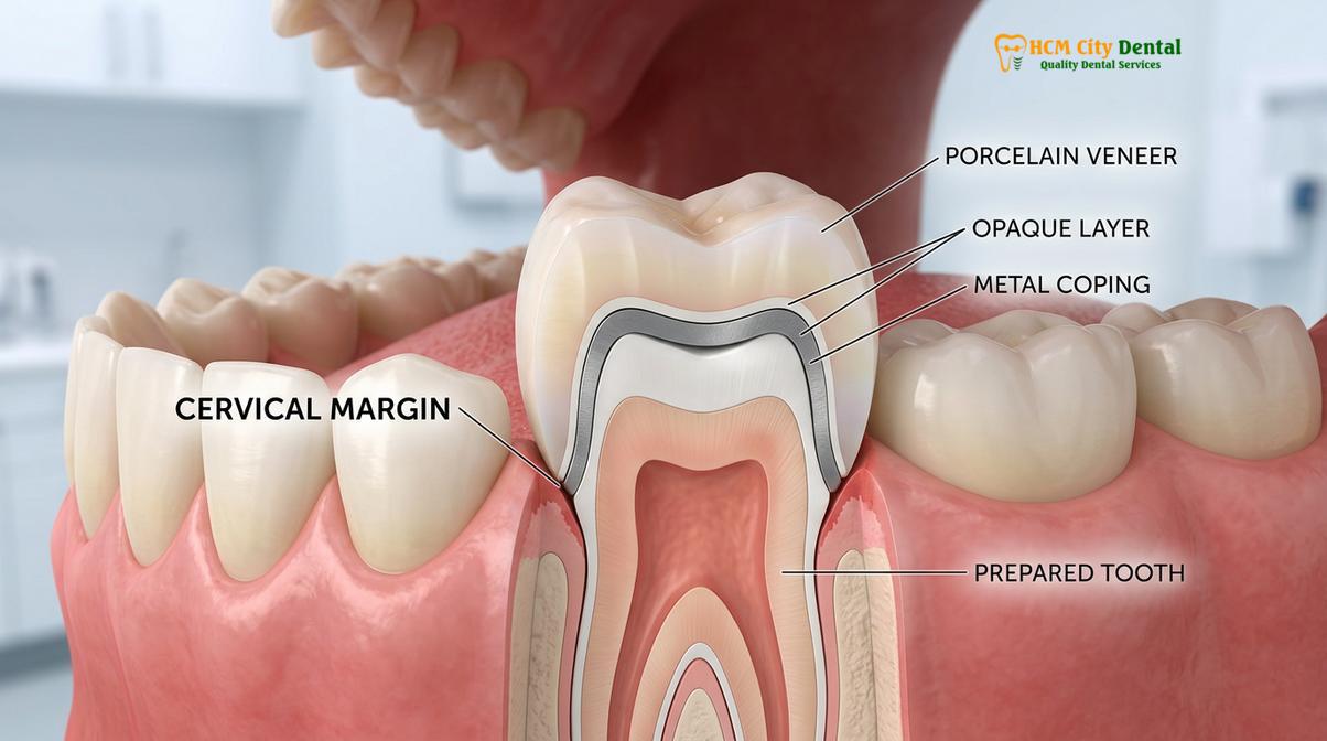
PFM crowns combine a durable cast-metal core with an overlaid porcelain veneer, requiring an opaque masking layer that limits natural light transmission at the cervical margin.
For decades, the porcelain-fused-to-metal crown was the gold standard in restorative dentistry. It offered a reliable compromise: the exceptional fracture resistance of a cast metal alloy combined with the tooth-colored aesthetics of feldspathic porcelain. To fully grasp why these crowns eventually show a dark line, it is necessary to understand their layered fabrication process and the biomechanical requirements of tooth preparation.
The foundation of a PFM crown is the metal coping. This thin thimble of metal (often composed of noble alloys like gold and palladium, or base metals like nickel and chromium) fits directly over the prepared tooth abutment. Because metal is inherently dark and unappealing, the dental ceramist must apply a highly opaque layer of porcelain directly over the metal to mask its color. Without this opaque layer, the grey metal would bleed through the final restoration. Over this opaque layer, translucent dentin and enamel porcelains are stacked and fired in a high-temperature vacuum furnace to mimic the anatomy of a natural tooth [2].
“While PFM restorations have a long history of clinical success and mechanical reliability, their inherent optical limitations at the gingival margin make them less ideal for highly demanding aesthetic zones compared to modern monolithic ceramics.”
The critical challenge lies at the margin. To create a seamless transition without making the tooth look bulky or over-contoured, the dentist must remove a significant amount of tooth structure (typically 1.5 to 2.0 millimeters of reduction) to create a deep shoulder or heavy chamfer margin. Even with adequate reduction, the ceramist often has very little space at the absolute edge of the crown to layer the opaque, dentin, and enamel porcelains. Consequently, the very edge of the crown—the collar—is frequently left as bare metal or covered with only a microscopically thin layer of opaque porcelain. When placed below the gumline, this is invisible. But the moment the tissue shifts, the dark line becomes glaringly obvious.
Gum Recession and Metal Exposure
As periodontal tissues migrate apically over time due to aging, aggressive brushing, or inflammation, the previously hidden metal collar of a PFM crown becomes visibly exposed.
Gingival recession is the primary catalyst that transforms a well-hidden PFM margin into a visible aesthetic defect. The gums are delicate soft tissues that respond to various mechanical, bacterial, and physiological stimuli. Understanding the etiology of gum recession around crown margins is essential for both preventing the issue and planning successful replacement therapies.
One of the most frequent causes of recession is mechanical trauma from aggressive toothbrushing. Using a hard-bristled toothbrush or applying excessive lateral force can physically abrade the delicate gingival margin, causing it to retreat and resulting in metal exposure gumline. Additionally, the patient’s inherent periodontal biotype plays a significant role. Patients with a “thin scalloped” biotype have very delicate, almost translucent gum tissue that is highly susceptible to recession following any minor trauma or restorative intervention. In contrast, a “thick flat” biotype is more robust and resistant to apical migration.
Biological factors also contribute heavily to recession. If a crown margin is placed too deeply below the gumline during the initial tooth preparation, it can violate the “biologic width”—the natural attachment apparatus of the gum to the tooth. When the biologic width is invaded, the body responds with chronic localized inflammation, leading to unpredictable bone loss and subsequent gum recession as the tissue attempts to re-establish a healthy distance from the restorative margin. Furthermore, poor oral hygiene can lead to plaque accumulation at the crown margin, triggering gingivitis and periodontitis, which actively destroy the supporting periodontal ligaments and alveolar bone.
Metal-Free Alternatives for a Natural Smile
Advanced ceramics like Zirconia HT and E.max lithium disilicate provide superior strength and lifelike translucency, completely eliminating the risk of dark gingival lines.
The evolution of dental material science has provided prosthodontists with remarkable alternatives that render the aesthetic compromises of PFM crowns obsolete. Today, patients looking to replace pfm with zirconia or other advanced ceramics can achieve restorations that are virtually indistinguishable from natural teeth, even if future gum recession occurs.
Zirconia HT (High Translucency): Zirconia (Yttria-stabilized tetragonal zirconia polycrystal) is a white, crystalline oxide of zirconium. It is milled from a solid block using advanced CAD/CAM (Computer-Aided Design and Computer-Aided Manufacturing) technology. Historically, early zirconia was extremely strong but highly opaque, making it suitable only for posterior (back) teeth. However, modern Zirconia HT, such as Cercon HT from Germany, has been engineered to offer exceptional flexural strength (often exceeding 1000 MPa) while allowing significant light transmission [3]. Because it is entirely metal-free and tooth-colored throughout its entire thickness, a zirconia crown will never display a dark line, regardless of tissue changes.
Lithium Disilicate (E.max): For the anterior (front) teeth, where aesthetic demands are highest, lithium disilicate glass-ceramic, commonly known by the brand name E.max (Ivoclar, Liechtenstein), is the material of choice. E.max offers unparalleled lifelike translucency, opalescence, and fluorescence that perfectly mimics natural enamel [4]. While its flexural strength (around 400-500 MPa) is lower than zirconia, it is more than sufficient for anterior bite forces when properly bonded to the underlying tooth structure.
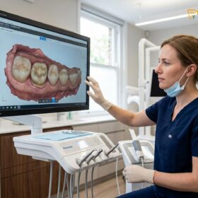
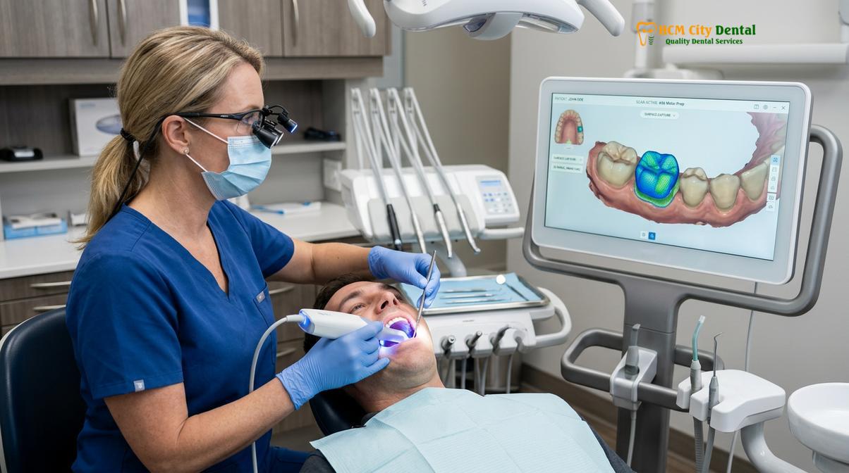
The fabrication of these modern restorations relies heavily on digital dentistry. At advanced facilities, messy traditional silicone impressions have been entirely replaced by state-of-the-art digital scanning technology. Using the iTero or Medit i700 Intraoral Scanners, the dentist captures a highly precise 3D topographical map of the prepared tooth in seconds. This digital file is transmitted instantly to the laboratory, ensuring maximum patient comfort and microscopic precision for the final crown design.
| Material Feature | PFM (Porcelain-Fused-to-Metal) | Zirconia HT (Cercon HT) | Lithium Disilicate (E.max) |
|---|---|---|---|
| Aesthetics & Translucency | Moderate (Opaque core blocks light) | High (Excellent light transmission) | Exceptional (Mimics natural enamel) |
| Risk of Dark Gumline | High (Metal margin exposure) | None (100% metal-free) | None (100% metal-free) |
| Flexural Strength | High (Metal core) | Very High (>1000 MPa) | Moderate (400-500 MPa) |
| Primary Clinical Indication | Older restorations, heavy bruxism | Posterior crowns, dental bridge structures | Anterior cosmetic smile makeovers |
How to Replace Old PFM Crowns
The replacement workflow involves careful removal of the old restoration, refinement of the tooth preparation, digital impression scanning, and precise cementation of a new all-ceramic crown.
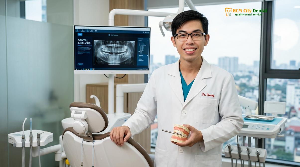
Replacing an aging PFM crown that exhibits a dark line is a highly predictable and routine procedure in modern prosthodontics. The clinical workflow is designed to maximize tissue health, ensure structural integrity of the underlying tooth, and deliver a flawless aesthetic result. Dr. Nguyen Van Cuong emphasizes that the success of a replacement crown relies heavily on meticulous soft tissue management and precise digital protocols.
The procedure begins with the administration of profound local anesthesia to ensure complete patient comfort. The old PFM crown is then carefully sectioned using specialized diamond and carbide burs and gently removed. Once the underlying tooth structure is exposed, the dentist conducts a thorough clinical evaluation. Any old, degraded cement is cleaned away, and the tooth is inspected for secondary decay or structural cracks. If the tooth has previously undergone a root canal and lacks sufficient coronal structure, a new composite core buildup or a fiber post may be placed to provide a solid foundation for the new restoration.
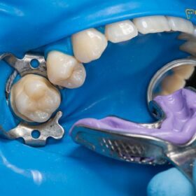
Next, the tooth preparation is refined. Because all-ceramic materials like Zirconia and E.max require different margin designs (typically a smooth, rounded shoulder or heavy chamfer) compared to PFM crowns, the dentist will carefully reshape the margin. To ensure the digital scanner captures the exact finishing line, a gentle retraction cord or paste is temporarily placed into the gingival sulcus to push the gum tissue slightly away from the tooth. The Medit i700 or iTero scanner is then used to capture the 3D digital impression. A temporary crown, custom-milled or fabricated from bis-acrylic resin, is cemented with temporary cement to protect the tooth and maintain gingival contours while the final restoration is being made.
For international patients and expats, dental tourism has revolutionized access to premium restorative care. Through a Direct Lab Partnership with a leading laboratory, clinics can offer an Express 3-5 Day Turnaround. This allows patients to undergo complete smile makeovers or receive multiple crowns and bridges within a single week, without compromising on quality or material certification.
A 45-year-old patient presented at HCMC Dental Clinic in Ho Chi Minh City complaining of a prominent dark line above her upper right central incisor (tooth #8), which had been restored with a PFM crown 12 years prior. Clinical examination revealed 2mm of gingival recession and a healthy, intact underlying tooth structure. Dr. Nguyen Van Cuong carefully removed the old restoration, refined the margin, and utilized the Medit i700 for a flawless digital impression. An E.max (Ivoclar) lithium disilicate crown was fabricated and bonded in just 4 days. The final result demonstrated seamless integration with the natural gingival contours, restoring the patient’s confidence with a highly translucent, metal-free smile.
Understanding the financial investment is crucial for patients considering restorative upgrades. By utilizing authentic, certified materials from Germany and Liechtenstein, patients receive world-class quality at highly accessible price points. According to the latest clinic fee schedule, the pricing structure (incorporating a 40% pre-arrival discount for WhatsApp bookings) is highly competitive:
- PFM Crown: Walk-in: ~$140 (3.5M VND) | WhatsApp pre-booking discount: From $84 (2.1M VND).
- Zirconia Crown (Cercon): Walk-in: ~$200 (5.0M VND) | WhatsApp pre-booking discount: From $120 (3.0M VND).
- Zirconia HT Crown (Cercon HT): Walk-in: ~$260 (6.5M VND) | WhatsApp pre-booking discount: From $156 (3.9M VND).
- E.max Crown (Ivoclar): Walk-in: ~$280 (7.0M VND) | WhatsApp pre-booking discount: From $168 (4.2M VND).
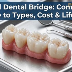
- 3-Unit Bridge (Zirconia HT): Walk-in: ~$480 (12.0M VND) | WhatsApp pre-booking discount: From $288 (7.2M VND).
- 4-Unit Bridge (Zirconia HT): Walk-in: ~$640 (16.0M VND) | WhatsApp pre-booking discount: From $384 (9.6M VND).
- Inlay / Onlay (Sứ Lab): Walk-in: ~$180 (4.5M VND) | WhatsApp pre-booking discount: From $108 (2.7M VND).
To further support international and local patients, comprehensive treatments are backed by a global warranty policy with remote follow-up support via WhatsApp, utilizing strict photo and video assessment protocols. Additionally, 0% interest monthly credit card installment plans are available to make premium dental care financially stress-free.
When to See a Doctor
While a dark line at the gumline is frequently a benign aesthetic annoyance, it is imperative to differentiate between simple metal exposure and active pathological processes. Patients should not assume that a dark margin is harmless without a professional clinical assessment. The oral cavity is a harsh environment, and even the most meticulously crafted dental crowns are subject to wear, cement degradation, and bacterial infiltration over time.
You should schedule an immediate consultation if you experience any of the following symptoms associated with your crowned tooth: localized pain when chewing, sensitivity to hot or cold temperatures that lingers, a foul taste or odor emanating from the specific tooth, or visible swelling and redness of the adjacent gum tissue. These are classic signs of marginal microleakage, where the cement seal between the crown and the tooth has washed out, allowing bacteria to penetrate and cause secondary decay beneath the restoration [5]. Because the crown covers the tooth, this decay is often hidden from the naked eye and can progress rapidly toward the dental pulp, potentially necessitating a root canal or even extraction if left untreated.
“A thorough clinical and radiographic examination is essential to differentiate between benign gingival recession and active marginal microleakage that threatens the underlying tooth structure. Early intervention can save the abutment tooth and allow for a predictable restorative replacement.”
Furthermore, if the dark line appears around a dental implant crown, it requires specific evaluation. Implant restorations can be either screw-retained or cement-retained. A dark shadow around an implant could indicate peri-implantitis (bone loss around the implant fixture) or exposure of the titanium implant abutment. A prosthodontist will utilize tactile examination with an explorer, vitality testing, and high-resolution digital radiographs to accurately diagnose the underlying cause of the discoloration and recommend the appropriate clinical intervention.
Frequently Asked Questions
Why is there a black line at my gumline?
The black line is typically the exposed metal base of a porcelain-fused-to-metal (PFM) crown. As your gums naturally recede over time due to aging, brushing habits, or periodontal changes, the hidden metal margin becomes visible, creating a dark, unnatural shadow along the neck of the tooth.
Can a dark line under a crown be fixed?
Yes, the most effective way to fix a dark line is to replace the old PFM restoration with a modern, metal-free all-ceramic crown. Materials like Zirconia or E.max completely eliminate the metal substructure, ensuring a seamless, natural-looking gumline that remains highly aesthetic even if minor tissue recession occurs in the future.
Are metal-free crowns better than PFM?
Metal-free crowns are generally superior in terms of aesthetics and biocompatibility. They provide lifelike translucency, eliminate the risk of dark gingival lines, and modern materials like Zirconia offer flexural strength that rivals or exceeds traditional metal-based restorations, making them suitable for both anterior and posterior teeth.
Does a dark line mean my crown is failing?
Not necessarily. A dark line is often a purely cosmetic issue caused by gum recession exposing the metal margin. However, it can sometimes indicate marginal leakage, cement washout, or underlying tooth decay, which requires a professional clinical evaluation with radiographs to rule out structural failure.
How long do PFM crowns last before showing a dark line?
PFM crowns can last 10 to 15 years or more, but a dark line may appear much sooner—often within 3 to 5 years—if you have thin gum tissue, practice aggressive brushing, or develop periodontal disease that accelerates gingival recession and exposes the restorative margin.
If you are experiencing aesthetic concerns with old dental work or wish to explore modern, metal-free restorative options, we encourage you to schedule a comprehensive evaluation at HCMC Dental Clinic. Our team is dedicated to utilizing advanced digital workflows to restore the natural beauty and optimal function of your smile.
References
- Journal of Prosthetic Dentistry. Aesthetic evaluation of the peri-implant soft tissue and crown margins. (2021).
- International Journal of Prosthodontics. Long-term survival of porcelain-fused-to-metal crowns. (2019).
- Journal of Prosthodontic Research. Flexural strength and optical properties of monolithic zirconia. (2022).
- Journal of Esthetic and Restorative Dentistry. Translucency and marginal adaptation of lithium disilicate. (2020).
- American Dental Association. Clinical guidelines for the diagnosis and management of dental caries at restoration margins. (2018).