The Complete Gum Health Guide: Bleeding Gums & Gingivitis

Comprehensive gum health guide at HCMC Dental Clinic. Dr. Cuong explains bleeding gums, gingivitis treatments, and prevention tips. Free consultation.
Healthy gums are the foundation of a healthy smile. Without strong, disease-free gingival tissue and supporting alveolar bone, even the healthiest teeth are at risk of mobility and loss. Periodontal (gum) disease is a silent, progressive condition that affects over 70% of adults globally. Because early stages are often painless, many patients are unaware of the bacterial destruction occurring beneath the gumline until irreversible bone loss has occurred.
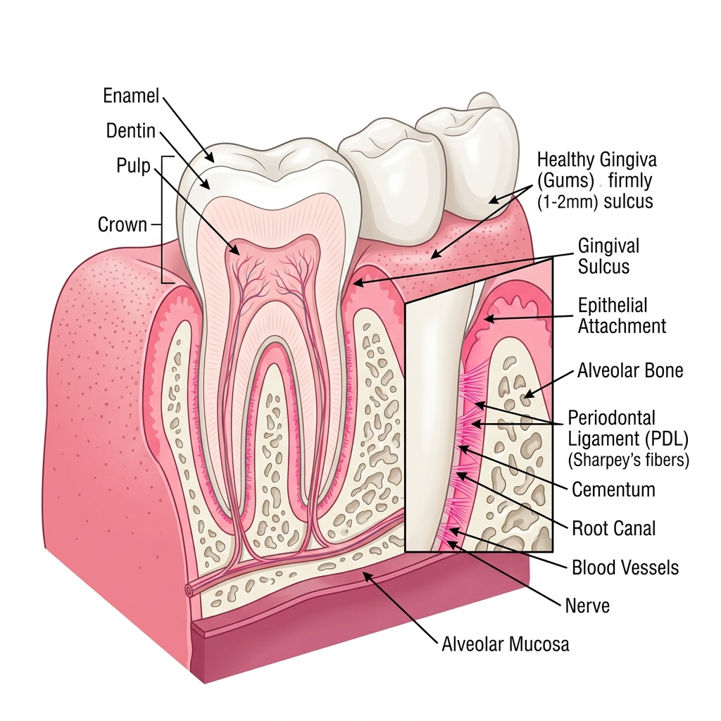
1. Introduction: The Periodontium & Gum Health
The periodontium refers to the specialized tissues that both surround and support the teeth, maintaining them in the maxillary and mandibular bones. It consists of four principal components: the gingiva (gums), the alveolar bone (the jawbone surrounding the roots), the cementum (the calcified substance covering the tooth root), and the periodontal ligament (the connective tissue fibers that attach the cementum to the alveolar bone).
A healthy periodontium is characterized by firm, coral-pink gums that do not bleed upon probing or brushing. The gingival sulcus—the natural crevice between the tooth and the gum margin—typically measures between 1 to 3 millimeters in depth. When oral bacteria accumulate along the gumline, they initiate an inflammatory response that can compromise this delicate biological seal. If the attachment is breached, bacteria migrate deeper, leading to periodontal pockets and the gradual destruction of the supporting bone structure.
2. The Stages of Gum Disease
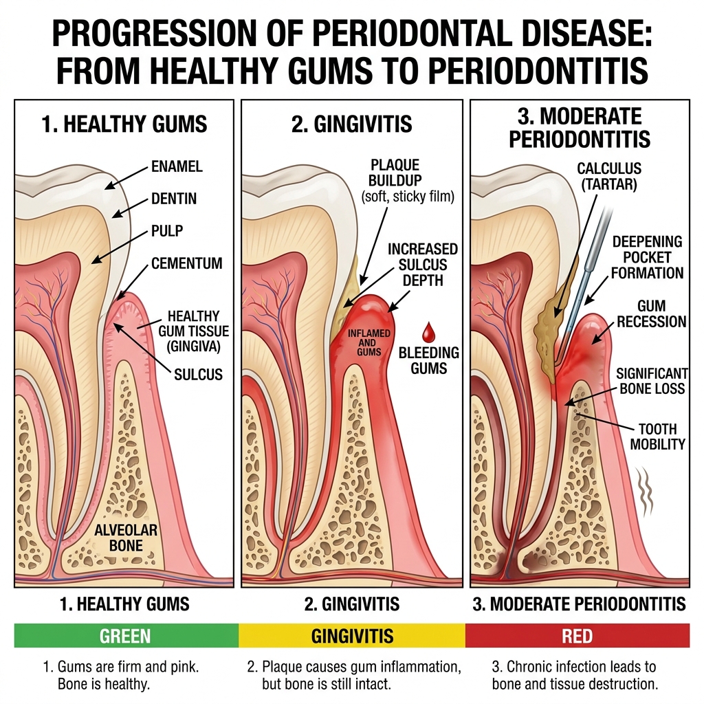
Periodontal disease is not a single event but a continuum of inflammatory stages. Understanding these stages is critical because early intervention can completely reverse the condition, whereas late-stage treatment focuses on managing irreversible damage.
2.1. Gingivitis (Reversible Plaque-Induced Inflammation)
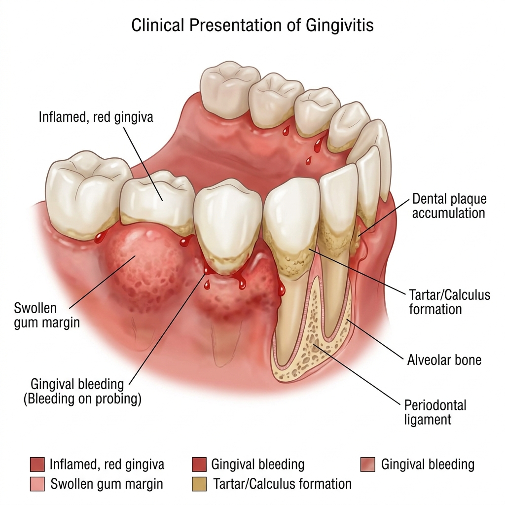
Gingivitis is the earliest and most common stage of gum disease. It is triggered by the accumulation of dental plaque—a sticky biofilm composed of millions of bacteria. When plaque is not adequately removed through daily brushing and flossing, the bacteria release toxins that irritate the gingival tissue. The body responds by increasing blood flow to the area, delivering immune cells to fight the infection. This inflammatory response causes the gums to become red, swollen, and prone to bleeding during routine hygiene.
Critically, gingivitis does not involve the loss of alveolar bone or periodontal ligament attachment. Because the foundational structures are intact, gingivitis is entirely reversible with professional scaling and strict adherence to a home care protocol.
2.2. Mild to Moderate Periodontitis
If gingivitis is left untreated, the bacterial biofilm matures and calcifies into dental calculus (tartar). The chronic presence of these calcified deposits drives the bacteria deeper into the gingival sulcus. The persistent immune response, combined with bacterial collagenases (enzymes that break down tissue), begins to destroy the periodontal ligament and the crestal alveolar bone.
The gingival sulcus deepens into a “periodontal pocket,” typically measuring 4 to 6 millimeters. As the pockets deepen, they create an anaerobic (oxygen-free) environment that favors highly virulent, tissue-destroying bacteria. Symptoms at this stage include persistent bad breath (halitosis), gum recession, and occasional dull aching. Treatment requires specialized deep cleaning (Scaling and Root Planing) to debride the roots and halt disease progression.
2.3. Advanced Periodontitis
In advanced periodontitis, the destruction of the supporting bone is severe. Pocket depths often exceed 7 millimeters, making it impossible for patients to clean them at home. The extensive loss of alveolar bone means that teeth lose their structural foundation. They may become highly mobile, drift out of alignment, or become sensitive as root surfaces are heavily exposed due to severe gum recession.
Patients with advanced periodontitis may experience recurrent acute periodontal abscesses, characterized by severe pain and pus exudation from the gums. At this terminal stage, aggressive surgical intervention—such as flap surgery, bone grafting, or extractions followed by dental implants—is often necessary to eliminate the infection and restore oral function.
3. Why Gums Bleed: Etiology & Systemic Links
Bleeding gums are an alarm bell indicating active inflammation and ulceration of the pocket lining. Healthy gums should never bleed during normal brushing or flossing.
3.1. Plaque & Biofilm Calcification
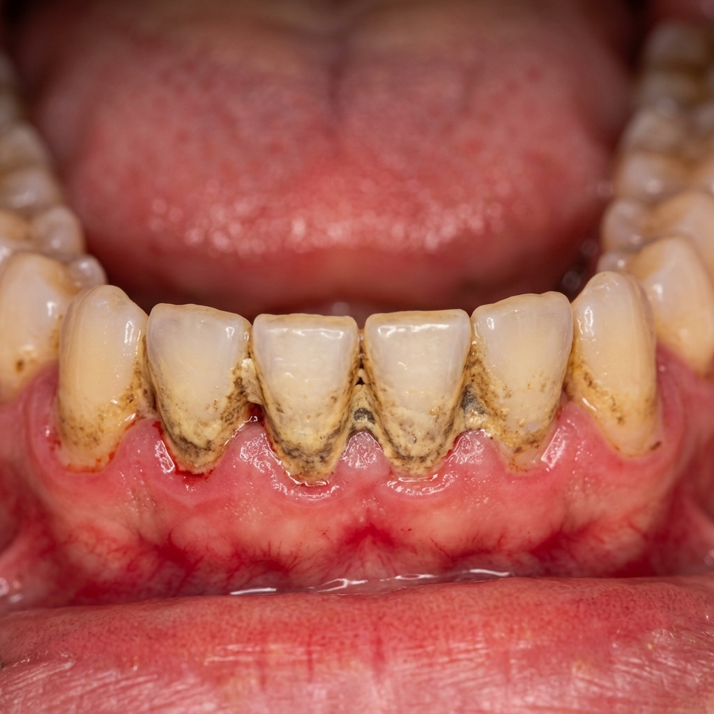
The primary etiologic agent of gum bleeding is the bacterial biofilm. Within 48 hours of forming, soft plaque begins to absorb calcium and phosphate minerals from saliva, hardening into calculus. Calculus has a rough, porous surface that acts as a scaffold for even more plaque accumulation. This calcified matrix cannot be removed with a toothbrush; it requires professional ultrasonic scaling. The constant mechanical irritation and bacterial toxin release from subgingival calculus cause the pocket epithelium to become ulcerated, leading to bleeding on probing.
3.2. Pregnancy Gingivitis
During pregnancy, significant fluctuations in estrogen and progesterone levels alter the body’s response to dental plaque. The elevated hormones increase vascular permeability in the gum tissues, making them highly reactive to even minuscule amounts of bacteria. This condition, known as pregnancy gingivitis, typically peaks in the second trimester and presents with extremely red, swollen, and easily bleeding gums. Maintaining rigorous oral hygiene and attending professional cleanings are essential to prevent this transient condition from advancing to true periodontitis.
3.3. Systemic Connections
Periodontal disease is profoundly interconnected with systemic health. The highly vascularized, ulcerated pockets in periodontitis provide a direct portal of entry for oral bacteria into the bloodstream (bacteremia). Below is a detailed clinical analysis of how periodontal disease affects cardiovascular, endocrine, and gestational health.
3.3.1. Cardiovascular Disease (Atherosclerosis & Stroke)
Chronic periodontitis creates a continuous reservoir of highly virulent Gram-negative anaerobic bacteria, specifically the “Red Complex” pathogens: Porphyromonas gingivalis, Treponema denticola, and Tannerella forsythia. These bacteria easily pass through the ulcerated lining of deep periodontal pockets during chewing, brushing, or scaling, causing transient bacteremia.
Research has demonstrated that P. gingivalis possesses specialized fimbriae (surface proteins) that allow it to actively invade endothelial cells lining the coronary arteries. Once inside the arterial wall, the bacteria trigger a local inflammatory response, prompting the expression of adhesion molecules that recruit macrophages. These macrophages ingest LDL cholesterol and transform into foam cells—the fundamental building blocks of fatty atherosclerotic plaques. Over time, this accelerates plaque buildup, narrowing the arteries. Furthermore, chronic periodontal inflammation stimulates the liver to release high levels of C-Reactive Protein (CRP), fibrinogen, and Interleukin-6 (IL-6) into the bloodstream. These markers increase blood viscosity and promote platelet aggregation, directly elevating the risk of acute myocardial infarction (heart attack) and cerebrovascular stroke.
3.3.2. Diabetes Mellitus (The Bidirectional Link)
The relationship between diabetes and periodontitis is one of the most thoroughly documented bidirectional links in medicine. Uncontrolled diabetes acts as a major accelerant of periodontal destruction. Chronic hyperglycemia (high blood sugar) leads to the non-enzymatic glycation of proteins, producing Advanced Glycation End-products (AGEs) that accumulate in the periodontal tissues. When AGEs bind to their receptors (RAGE) on inflammatory cells, it triggers a hyper-inflammatory state. Macrophages release excessive amounts of destructive cytokines, such as Tumor Necrosis Factor-Alpha (TNF-α), Interleukin-1 Beta (IL-1β), and collagenases, which aggressively destroy the periodontal ligament and alveolar bone. Additionally, high blood sugar impairs polymorphonuclear leukocyte (PMN) chemotaxis, compromising the first line of defense against subgingival plaque.
Conversely, active periodontitis severely impairs blood glucose control. The constant entry of periodontal bacteria and inflammatory cytokines (TNF-α, IL-6) into the bloodstream creates state-wide, low-grade systemic inflammation. Systemic TNF-α directly interferes with insulin signaling by promoting the serine phosphorylation of Insulin Receptor Substrate-1 (IRS-1), which blocks the normal transport of glucose into muscle and fat cells. This exacerbates insulin resistance. Clinical trials show that treating periodontitis with thorough Scaling and Root Planing (SRP) can reduce systemic HbA1c levels in diabetic patients by up to 0.4% to 0.6%—an effect comparable to adding a secondary class of oral anti-diabetic medication.
3.3.3. Adverse Pregnancy Outcomes (Gestational Diabetes & Preterm Birth)
The microvascular changes of pregnancy gingivitis, if neglected, can transition into active periodontitis, which poses a serious threat to the developing fetus. Deep periodontal pockets contain high concentrations of lipopolysaccharides (LPS) and inflammatory cytokines. If these endotoxins enter maternal circulation, they can cross the placental barrier and reach the amniotic fluid.
The presence of lipopolysaccharides and cytokines within the placental-fetal unit triggers a localized immune response. Placental macrophages secrete elevated levels of Prostaglandin E2 (PGE2) and TNF-α. PGE2 is a powerful physiological trigger for uterine contractions, while TNF-α and other matrix metalloproteinases can cause premature rupture of the amniotic membranes. This premature spike in inflammatory signaling can initiate early labor and lead to preterm births and low birth weight. Furthermore, the systemic inflammatory response from periodontitis can exacerbate gestational insulin resistance, increasing the mother’s risk of developing gestational diabetes.
3.3.4. Rheumatoid Arthritis & Respiratory Pathologies
Recent rheumatological research has highlighted a direct biochemical link between periodontal pathogens and Rheumatoid Arthritis (RA). Porphyromonas gingivalis is the only known prokaryotic organism that produces the enzyme peptidylarginine deiminase (PPAD). This enzyme converts arginine residues in proteins into citrulline (citrullination). When these citrullinated proteins enter the systemic circulation, the immune system fails to recognize them, triggering the production of Anti-Citrullinated Protein Antibodies (ACPAs). ACPAs are the primary biomarkers and drivers of joint destruction in rheumatoid arthritis. Treating periodontitis directly decreases systemic citrullination and ACPA titers, significantly reducing joint stiffness and inflammation in RA patients.
Furthermore, the chronic aspiration of subgingival plaque bacteria is a documented cause of respiratory infections. In patients with compromised lung clearance (such as the elderly or patients with COPD), aspirating periodontal pathogens like Fusobacterium nucleatum can lead to severe aspiration pneumonia and chronic exacerbation of obstructive pulmonary diseases, highlighting that poor gum health directly impacts airway safety. Clinical studies have shown that regular periodontal scaling and plaque control can reduce the risk of respiratory flare-ups and hospitalizations by up to 30% in these vulnerable populations.
4. Clinical Treatments for Periodontal Disease
Treating periodontal disease aims to arrest the inflammatory process, reduce pocket depths to cleanable levels, and regenerate lost tissues where possible. The approach is dictated by the severity of the clinical attachment loss.
4.1. Scaling and Root Planing (SRP)
Often referred to as a “deep cleaning,” SRP is the gold standard, non-surgical therapy for active periodontitis.
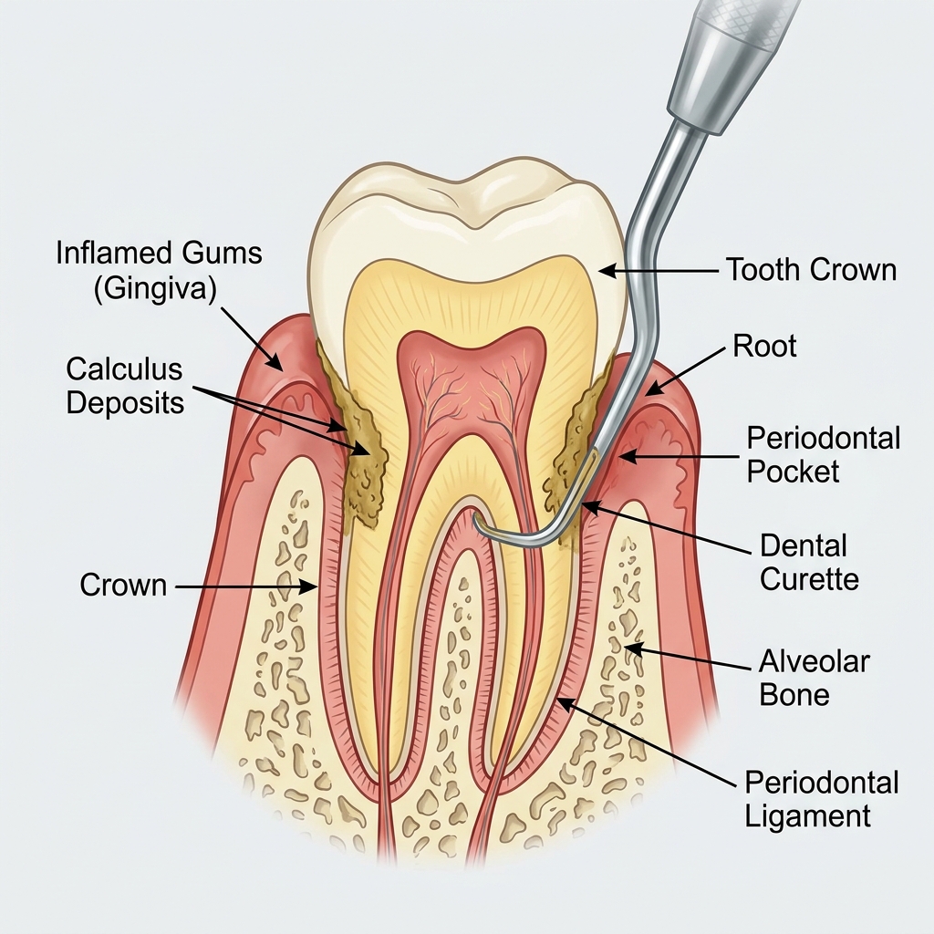
The procedure begins with the administration of local anesthesia to ensure absolute patient comfort. Using precision ultrasonic scalers, the clinician vibrates and flushes away the heavy calcified calculus deposits from above and below the gumline (Scaling). Hand instruments (curettes) are then meticulously used to smooth the root surfaces (Root Planing). Smoothing the roots serves a dual purpose: it removes cementum that has been impregnated with bacterial endotoxins, and it provides a glass-like surface that discourages new bacterial adherence, allowing the gingival tissues to heal and reattach.
4.2. Laser-Assisted Periodontal Therapy
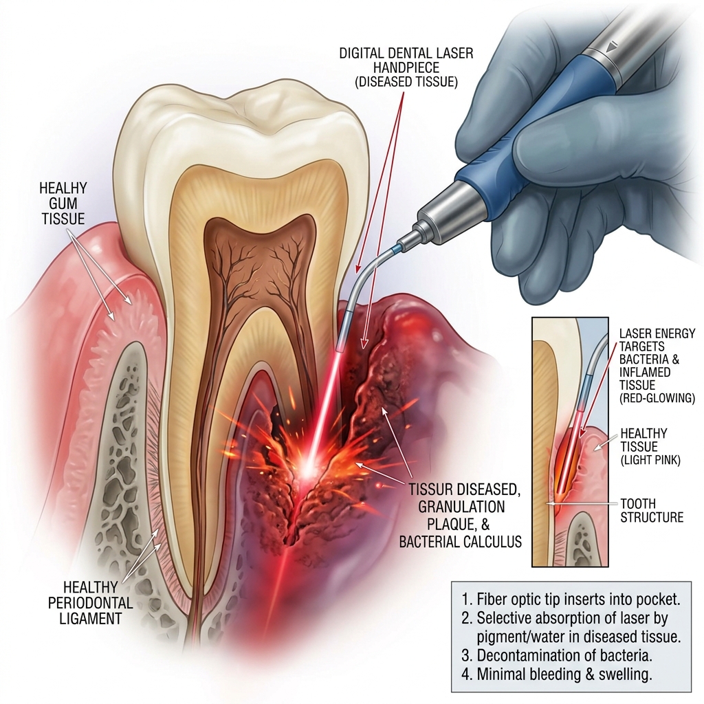
At HCMC Dental, we frequently adjunct SRP with diode laser therapy. The precise wavelength of the dental laser is highly absorbed by pigmented bacteria and inflamed, diseased tissue. By passing the fine laser tip into the periodontal pocket, the clinician can achieve high-level bacterial decontamination (bactericidal effect) and selective vaporization of the diseased epithelial lining, leaving the healthy connective tissue intact. This modern protocol promotes faster healing, minimizes postoperative discomfort, and stimulates favorable tissue response.
4.3. Regenerative Gum Surgery
When severe bone loss has resulted in deep vertical defects, non-surgical therapy is insufficient. In these scenarios, a periodontal flap surgery is performed to fold back the gums, providing direct visual access to thoroughly clean the root surfaces and reshape the jagged bone.
To repair the damage, regenerative techniques are employed. This involves placing bone graft material (often allograft or xenograft) into the bony defect and covering it with a Guided Tissue Regeneration (GTR) membrane. The membrane acts as a physical barrier, preventing fast-growing gum tissue from filling the void, thus allowing the slower-growing bone cells to populating and regenerate the lost alveolar support.
4.4. Supportive Periodontal Therapy (SPT) & Maintenance
Supportive Periodontal Therapy (SPT) is a structured, customized recall program designed to monitor and stabilize the periodontal status of patients who have completed active non-surgical or surgical therapy. Unlike routine prophylactic cleaning, SPT is a therapeutic intervention aimed at preventing disease recurrence.
SPT visits are scheduled every 3 to 4 months instead of the standard 6 months. During an SPT appointment, our clinicians perform:
- Periodontal Probing & Charting: Measuring pocket depths at six points around every tooth to detect any disease recurrence early.
- Site-Specific Micro-Debridement: Using fine ultrasonic inserts to clean active pocket areas where plaque and calculus have begun forming.
- Plaque and Bleeding Indexing: Quantifying the percentage of sites showing plaque or bleeding to assess oral hygiene efficacy.
Studies show that patients who skip their SPT schedule experience a 500% increase in bone resorption and attachment loss over 5 years compared to those who strictly attend maintenance appointments.
Recolonization Kinetics of Periodontal Pathogens: The clinical necessity of the 3-to-4-month SPT interval is rooted in subgingival microbiotic dynamics. Following a thorough scaling and root planing procedure, the subgingival pocket environment is debrided, and the microflora shifts from a pathogenic Gram-negative anaerobic profile to a healthy Gram-positive facultative profile. However, microscopic remnants of pathogenic bacteria inevitably remain within the microscopic irregularities of the root surfaces and pocket soft tissues. These remaining pathogens begin to replicate. It takes approximately 9 to 12 weeks (roughly 90 days) for virulent species such as Porphyromonas gingivalis and Treponema denticola to complete their recolonization cycle and re-establish a mature, structured subgingival biofilm capable of triggering host tissue destruction. By scheduling maintenance every 12 weeks, the clinician mechanically disrupts these pathogenic colonies before they can reach the critical threshold required to reactivate bone resorption.
5. Daily Home Care Protocol
Periodontal therapy is a partnership between the clinician and the patient. Without exceptional daily home care, periodontal disease has a 100% relapse rate. The goal of oral hygiene is to disrupt the bacterial biofilm every 12 to 24 hours, preventing it from maturing into pathogenic colonies or calcifying into calculus.
- The Modified Bass Brushing Technique: This is the most critical brushing method for gum health. Angle the bristles of a soft-bristled toothbrush at 45 degrees towards the gumline. Apply gentle pressure so the bristles slightly enter the gingival sulcus, and use small vibratory/circular motions. Sweep the brush away from the gums. Do not use a hard scrubbing motion, as this causes traumatic gum recession.
- Electric Toothbrushes: High-quality sonic or oscillating-rotating electric toothbrushes are highly recommended. They are clinically proven to remove up to 300% more plaque along the gumline than manual brushing.
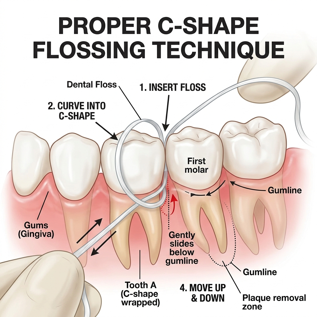
- Interdental Cleaning: A toothbrush cannot reach the spaces between teeth, which is exactly where periodontitis most frequently originates. Daily use of string floss or interdental brushes is mandatory. The floss must be curved in a ‘C’ shape around the side of each tooth and gently slid beneath the gumline.
- Water Flossers (Oral Irrigators): Devices like a Waterpik are excellent adjuncts, especially for patients with deep pockets, dental bridges, or orthodontic appliances. The pulsating water stream flushes out unattached bacteria and debris from deep within the periodontal pockets, disrupting the anaerobic environment.
6. Cost Index & Treatment Packages in Vietnam
International patients often seek periodontal care at HCMC Dental due to the significant cost savings without compromising on sterile standards or advanced laser technologies. The tables below reflect standard estimates.
| Periodontal Treatment | Estimated Cost (Vietnam) | Avg. Cost (USA/Australia) |
|---|---|---|
| Routine Dental Cleaning & Polish | $20 – $30 USD | $100 – $150 USD |
| Scaling & Root Planing (per quadrant) | $40 – $60 USD | $250 – $400 USD |
| Laser Pocket Disinfection (add-on) | $20 – $50 USD | $150 – $300 USD |
| Periodontal Flap Surgery (per quadrant) | $150 – $300 USD | $800 – $1,500 USD |
| Soft Tissue / Gum Grafting (per tooth) | $200 – $400 USD | $900 – $1,800 USD |
*Note: Costs are approximate estimates for international comparison. Complex regenerative procedures involving bone grafts and membranes will incur additional biomaterial costs. Please contact us for a specific, customized treatment plan.
7. Frequently Asked Questions (FAQ)
Why do my gums bleed only when I floss?
Bleeding only during flossing indicates highly localized interproximal gingivitis. Plaque is accumulating between your teeth where your brush cannot reach. Consistent, daily flossing will usually resolve the inflammation and stop the bleeding within 7 to 10 days.
Can receded gums grow back?
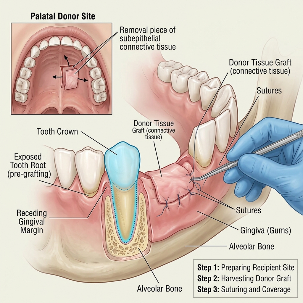
No, once the gingival tissue has receded and the underlying bone is lost, it will not naturally regenerate. However, recession can be surgically repaired using soft tissue grafting techniques to cover the exposed root and protect the tooth.
Is scaling and root planing painful?
At HCMC Dental, we ensure you are completely comfortable. Local anesthesia is administered prior to the procedure so you will not feel any sharp pain, only vibration and mild pressure. Post-operative sensitivity is managed with specialized toothpaste and mild analgesics.
How often should I get a dental cleaning if I have periodontitis?
Patients diagnosed with periodontitis are placed on a “periodontal maintenance” schedule, which requires professional cleanings every 3 to 4 months. The standard 6-month interval is insufficient because virulent periodontal pathogens can repopulate a pocket to destructive levels in just 9 to 12 weeks.
Does mouthwash cure gum disease?
No. Over-the-counter mouthwashes cannot penetrate the biofilm matrix or remove calcified tartar. While therapeutic mouthwashes (like Chlorhexidine) are excellent chemical adjuncts to reduce bacterial load, they do not replace the mechanical removal of plaque and calculus through brushing, flossing, and professional scaling.
Can I lose my teeth from gum disease?
Yes. In fact, advanced periodontitis is the leading cause of adult tooth loss worldwide. As the alveolar bone resorbs, the teeth lose their anchor and become mobile. Early diagnosis and intervention are critical to preserving your natural dentition.
Is gum disease contagious?
The specific bacteria that cause periodontitis can be transmitted through saliva (e.g., sharing utensils or kissing). While exposure to the bacteria does not guarantee you will develop the disease, it can alter your oral microbiome. Maintaining excellent personal oral hygiene is your best defense.
What is the link between smoking and periodontitis?
Smoking is one of the most significant risk factors for severe periodontal disease. Nicotine acts as a vasoconstrictor, reducing blood flow to the gums, which masks the early warning sign of bleeding and severely impairs the body’s immune response and healing capacity. Smokers are far more likely to experience bone loss and implant failure.