For over a century, silver amalgam was the undisputed standard for restoring decayed posterior teeth. However, the modern evolution of biological dentistry has fundamentally shifted clinical perspectives on this legacy material. Dental amalgam is not merely “silver”; it is an alloy composed of approximately 50% elemental mercury by weight. As international awareness of heavy metal toxicity grows, a rising number of health-conscious expats and dental tourists are seeking specialized clinics to safely extract these aging restorations. The challenge lies not just in the removal, but in mitigating the severe risks of aerosolized mercury vapor and addressing soft-tissue complications like iatrogenic gingival staining. At HCMC Dental Clinic in District 1, Ho Chi Minh City, the integration of strict IAOMT SMART protocols, advanced laser dentistry, and biomimetic ceramic reconstruction offers a comprehensive pathway to total oral detoxification and structural rehabilitation.
Clinical Summary:
The safe extraction of legacy mercury amalgam fillings requires rigorous adherence to the IAOMT SMART (Safe Mercury Amalgam Removal Technique) protocol to prevent systemic heavy metal exposure. Conventional high-speed drilling vaporizes elemental mercury, creating a toxic inhalation hazard. By utilizing non-latex nitrile dams, nasal oxygen, high-volume aerosol evacuation, and cool-water chunking techniques, clinicians can safely eliminate these toxic restorations. Furthermore, associated soft-tissue complications, such as oral amalgam tattoos, can be precisely excised using Er:YAG or Diode lasers after ruling out melanocytic lesions. Following safe removal, the structurally compromised tooth is biomimetically restored using BPA-free Ormocers or high-strength IPS E.max ceramic onlays, ensuring long-term biocompatibility and mechanical resilience.
Key Takeaways:
- Toxic Vapor Mitigation: Standard high-speed drilling of amalgam generates heat exceeding 350°C, releasing a highly absorbable plume of elemental mercury vapor.
- Strict SMART Protocol: Safe removal mandates a non-latex nitrile dam, alternative nasal oxygen supply, and specialized high-volume aerosol evacuation to protect the patient’s airway and systemic health.
- Laser Tissue Excision: Blue-grey gingival stains (amalgam tattoos) caused by embedded metallic particles can be safely ablated using Er:YAG lasers, following a differential diagnosis to rule out oral melanoma.
- Biomimetic Reconstruction: Aging amalgams cause microscopic cusp fractures; replacing them requires advanced adhesion using BPA-free Ormocers or lithium disilicate (IPS E.max) ceramic onlays to restore structural integrity.
- Transparent Economics: International patients can access world-class biological dentistry at HCMC Dental Clinic with a transparent -40% online booking fee schedule, saving up to 70% compared to Western dental markets.
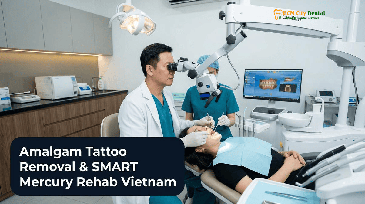
Why Conventional High-Speed Drilling of Silver Amalgam Fillings Releases a Plume of Elemental Mercury Vapor (50% Mercury Content by Weight)
To understand the critical necessity of specialized removal protocols, one must first examine the metallurgy of dental amalgam. Despite its colloquial designation as a “silver filling,” traditional dental amalgam is a complex alloy where liquid elemental mercury acts as the binding agent for a powder mixture of silver, tin, and copper. By weight, elemental mercury constitutes approximately 50% of the final hardened restoration. While proponents of amalgam historically argued that the mercury is locked into a stable intermetallic compound, modern thermodynamic analysis and continuous vapor monitoring have unequivocally proven otherwise.
The primary danger arises during the removal process. A standard dental handpiece operates at speeds approaching 400,000 revolutions per minute (RPM). When a conventional diamond or carbide bur contacts the dense amalgam alloy at these velocities, the resulting friction generates intense, localized thermal energy. Temperatures at the cutting interface can easily exceed 350°C within milliseconds. Because elemental mercury has a relatively low vapor pressure and vaporizes at room temperature—and boils at 356°C—this high-speed friction instantly sublimates the solid mercury within the filling into an invisible, odorless, and highly toxic vapor plume[1].
This vaporized mercury presents a profound inhalation hazard. When inhaled, elemental mercury vapor rapidly traverses the alveolar membranes of the lungs, with an absorption rate of up to 80% directly into the systemic circulation. Once in the bloodstream, it readily crosses both the blood-brain barrier and the placental barrier, accumulating in the central nervous system, kidneys, and other vital organs. Furthermore, conventional drilling pulverizes the amalgam into sub-micron particulate matter, which can be inadvertently swallowed and absorbed through the gastrointestinal tract. For patients actively seeking a Best Dental Clinics & Specialist Comparison at HCMC Dental Clinic to reduce their systemic toxic burden, subjecting themselves to conventional, unprotected amalgam removal is entirely counterproductive and medically contraindicated.
The Full IAOMT SMART (Safe Mercury Amalgam Removal Technique) Checklist at HCMC Dental Clinic: Non-Latex Nitrile Dam, Nasal Oxygen, High-Volume Aerosol Evacuator & Chlorella/Charcoal Rinse
Recognizing the severe occupational and patient health risks associated with mercury vaporization, the International Academy of Oral Medicine and Toxicology (IAOMT) developed the Safe Mercury Amalgam Removal Technique (SMART). This rigorous, evidence-based protocol establishes a multi-layered defensive barrier system designed to achieve near-zero mercury exposure during the extraction process. At HCMC Dental Clinic, the implementation of the SMART protocol is non-negotiable for any amalgam replacement procedure.
The protocol begins before the handpiece is even activated. Patients are provided with a pre-procedure oral slurry of activated charcoal or chlorella. These natural binding agents act as systemic chelators within the gastrointestinal tract; if any microscopic amalgam particulate bypasses the physical barriers and is swallowed, the charcoal or chlorella binds to the heavy metals, preventing systemic absorption and facilitating safe excretion. Following this, the physical isolation of the operative field is established. A non-latex nitrile dental dam is meticulously placed and sealed around the target tooth. The distinction of using nitrile rather than traditional latex is critical; elemental mercury vapor can permeate latex, whereas nitrile provides an impermeable barrier, completely sealing the oral cavity and oropharynx from toxic debris[2].
“The foundation of biological dentistry is the principle of ‘primum non nocere’—first, do no harm. Removing a toxic material using a technique that acutely exposes the patient to a massive spike in heavy metal vapor violates that principle. The SMART protocol is not an optional luxury; it is a fundamental clinical necessity for safeguarding systemic health during restorative rehabilitation.”
— Dr. Nguyen Van Cuong, DDS (Clinical Director — MOH License 0003265/BL-CCHN)
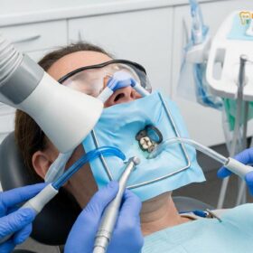
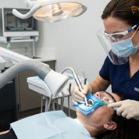
Airway protection is the next vital component. Because the ambient air immediately surrounding the oral cavity will inevitably contain trace amounts of vaporized mercury despite best efforts, the patient is fitted with a nasal hood delivering a continuous flow of pure, uncontaminated oxygen. This positive pressure system ensures the patient breathes only clean air, entirely bypassing the aerosolized treatment zone. Simultaneously, a specialized High-Volume Aerosol Evacuator (HVE)—often referred to as a “dentist’s elephant vacuum”—is positioned mere inches from the patient’s mouth. This high-capacity suction unit captures the aerosol plume at the exact point of generation, filtering the exhaust through medical-grade HEPA and activated carbon filters before it can disperse into the operatory environment. To complete the protocol, both the patient and the clinical team are draped in full-body impermeable barriers, and the clinicians utilize respiratory-grade masks and face shields to prevent occupational exposure.
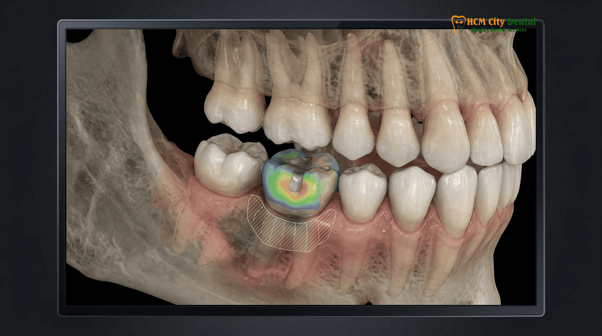
Chunking & Cool-Water Sectioning with New Carbide Burs: Removing Amalgam in Large Solid Pieces Without Vaporizing Metal
While the barrier and evacuation systems of the SMART protocol manage the environmental hazards, the mechanical technique employed by the biological dentist is equally crucial in minimizing the initial generation of mercury vapor. The traditional method of removing an old filling involves “sweeping” the bur back and forth across the entire surface of the amalgam, effectively grinding and pulverizing the entire mass into a fine, highly volatile slurry. This technique maximizes the surface area of the amalgam exposed to friction, thereby maximizing the release of toxic vapor.
In stark contrast, the SMART protocol mandates a technique known as “chunking” or “sectioning.” The objective is to remove the amalgam in the largest possible solid pieces, minimizing the amount of alloy that is actually touched by the high-speed drill. To achieve this, the clinician utilizes brand-new, small-diameter tungsten carbide burs. A new bur ensures maximum cutting efficiency with minimal frictional drag. The dentist carefully cuts a narrow trough around the perimeter of the filling, or bisects the amalgam mass down the center, avoiding contact with the bulk of the material whenever possible[3].
Once the amalgam is sectioned, specialized hand instruments (elevators and excavators) are used to apply lateral pressure, fracturing the remaining amalgam away from the tooth structure so it can be suctioned away in large, solid chunks. Throughout this entire sectioning process, the tooth and the bur are flooded with copious, continuous cold-water irrigation. This massive volume of water serves a dual purpose: it acts as a highly effective coolant, keeping the temperature of the amalgam well below the 356°C boiling point of mercury, and it physically traps the heavier amalgam particulate, preventing it from becoming airborne. By combining the chunking technique with aggressive cold-water cooling, the biological dentist drastically reduces the total volume of mercury vapor generated at the source.
What Is an Oral Amalgam Tattoo? Differential Diagnosis vs. Oral Melanocytic Lesions and Single-Visit Er:YAG / Diode Laser Excision of Silver-Stained Gingiva
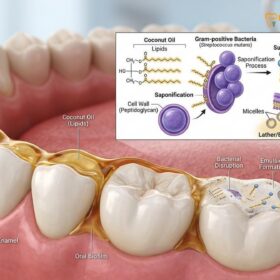
The systemic risks of mercury vapor are well-documented, but the legacy of silver fillings often leaves a localized, visible mark on the oral soft tissues: the amalgam tattoo. An oral amalgam tattoo is an iatrogenic (clinician-caused) lesion that occurs when microscopic particles of amalgam are inadvertently embedded into the gingival or mucosal connective tissue. This typically happens during the initial placement of the filling, during high-speed drilling without proper isolation, or during the extraction of a tooth containing a large amalgam restoration. Over time, the silver, tin, and copper compounds within the embedded particles oxidize and corrode, leaching into the surrounding collagen fibers and creating a flat, asymptomatic, blue, grey, or black macule on the gums.
While an amalgam tattoo is entirely benign and poses no localized malignant threat, its clinical presentation demands rigorous diagnostic scrutiny. The primary challenge is the differential diagnosis. A dark, pigmented lesion on the oral mucosa must be definitively distinguished from potentially lethal oral melanocytic lesions, most notably oral malignant melanoma, as well as blue nevi or physiologic melanin pigmentation. A thorough clinical history and high-resolution digital radiography are the first steps; radiographs will often (but not always) reveal radiopaque metallic dust scattered within the soft tissue. However, if the lesion is raised, exhibits irregular borders, is changing in size or color, or lacks a clear history of adjacent amalgam restorations, a biopsy is mandatory to rule out malignancy[4].
Clinical Case Study: Laser Excision of Gingival Pigmentation
Patient Profile: A 42-year-old expatriate from Australia presented to HCMC Dental Clinic with a prominent, dark blue macule on the buccal gingiva adjacent to a lower right molar. The patient reported aesthetic concerns and anxiety regarding oral melanoma.
Clinical Assessment: The adjacent molar contained a massive, 15-year-old failing silver amalgam filling. Digital periapical radiographs confirmed the presence of fine radiopaque stippling within the gingival tissue, confirming the diagnosis of an amalgam tattoo rather than a melanocytic lesion.
Treatment Protocol: Following the SMART Amalgam Removal: Clinical Protocol & Safety | HCMC Dental to replace the failing filling with an IPS E.max onlay, Dr. Cuong utilized an Er:YAG dental laser to address the gingival staining. The specific wavelength of the Er:YAG laser is highly absorbed by the dark metallic pigments and water within the tissue. In a single, minimally invasive 15-minute session, the laser precisely ablated the pigmented epithelial and connective tissue layers without the need for a scalpel or sutures.
Outcome: The patient experienced zero post-operative bleeding and minimal discomfort. Within 10 days, the surgical site re-epithelialized completely, restoring a natural, healthy pink gingival architecture and eliminating the aesthetic defect.
When aesthetic removal is desired, or an excisional biopsy is required for histopathological confirmation, modern laser dentistry offers the gold standard of care. Traditional scalpel excision of an amalgam tattoo often results in significant bleeding, the need for sutures, and potential scarring. In contrast, the use of an Er:YAG or Diode laser allows for precise, micro-surgical ablation of the pigmented tissue. The laser energy targets the dark pigment, vaporizing the stained cells while simultaneously coagulating blood vessels and sealing nerve endings. This results in a bloodless surgical field, profound intraoperative hemostasis, and vastly accelerated, scar-free healing.
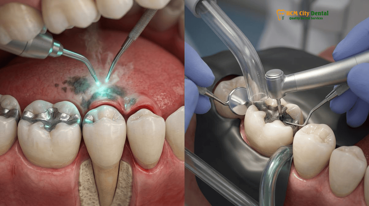
Biomimetic Tooth Reconstruction After Amalgam Removal: Diagnosing Underlying Cusp Cracks with Transillumination and Restoring with BPA-Free Ormocer or IPS E.max Onlays
The safe extraction of the toxic amalgam and the laser excision of associated soft-tissue tattoos represent only the first half of the biological rehabilitation process. The clinician is now faced with a structurally compromised tooth. Traditional silver amalgam does not bond chemically or micromechanically to tooth structure; it relies entirely on mechanical retention. Dentists historically had to drill away significant amounts of healthy enamel and dentin to create “undercuts” to lock the rigid metal block in place. Furthermore, amalgam has a coefficient of thermal expansion that differs drastically from natural tooth structure. Over decades of consuming hot coffee and cold water, the metal filling expands and contracts, acting like a wedge driven into the center of the tooth.
This constant thermal cycling, combined with the immense occlusal forces of mastication, inevitably leads to structural fatigue. When an old amalgam is finally removed, the biological dentist almost universally discovers a network of microscopic craze lines and stress fractures propagating through the remaining enamel and dentin cusps. Diagnosing the extent of these hidden fractures is critical. At HCMC Dental Clinic, clinicians utilize high-powered surgical microscopes, caries indicator dyes, and fiber-optic transillumination to visualize the depth and severity of these cracks. If left unaddressed, these compromised cusps will eventually shear off, potentially splitting the tooth down to the root and necessitating extraction.
Clinical Precaution: Avoiding Traditional Crowns After Amalgam Removal
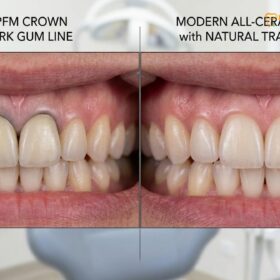
A common, yet outdated, approach in conventional dentistry is to automatically grind down an amalgam-compromised tooth for a full-coverage porcelain-fused-to-metal (PFM) crown. This aggressive preparation destroys up to 70% of the remaining healthy tooth structure, traumatizing the dental pulp and increasing the risk of future root canal therapy. Biological and biomimetic dentistry strictly avoids this destructive cycle, prioritizing the preservation of natural tissue through advanced adhesive onlays.
To restore the tooth to its original biomechanical strength without sacrificing healthy tissue, biomimetic reconstruction is employed. For smaller cavities, direct restorations are placed using advanced Ormocers (Organically Modified Ceramics). Unlike traditional composite resins, which often contain Bisphenol A (BPA) derivatives like Bis-GMA or TEGDMA, Ormocers utilize a purely ceramic polysiloxane network. This provides exceptional biocompatibility, zero BPA leaching, and significantly reduced polymerization shrinkage, ensuring a tight, leak-proof marginal seal.
For larger defects where the cusps are severely undermined or fractured, indirect ceramic restorations are required. In these scenarios, the clinician utilizes IPS E.max (lithium disilicate) ceramic inlays, onlays, or overlays. Fabricated via precision CAD/CAM technology, these metal-free glass-ceramic restorations possess a modulus of elasticity remarkably similar to natural human enamel. Through a meticulous protocol of Immediate Dentin Sealing (IDS) and resin bonding, the IPS E.max onlay is micromechanically fused to the underlying tooth structure. This biomimetic adhesion effectively splints the cracked cusps together, distributing chewing forces evenly across the tooth and restoring its structural integrity to near-virgin strength, all while maintaining absolute biocompatibility.
2026 IAOMT SMART Amalgam Removal, Laser Amalgam Tattoo Excision & Ceramic Onlay Fee Schedule at HCMC Dental Clinic (District 1)
For international patients, expats, and dental tourists navigating the healthcare landscape in Vietnam, selecting the right provider for complex biological dentistry requires careful evaluation. The dental market in Ho Chi Minh City is highly segmented, and understanding the operational models is crucial for ensuring clinical safety and financial transparency. When evaluating a Best Dentist & Dental Clinics in Ho Chi Minh City (2026 Comparison Guide), patients generally encounter four distinct models:
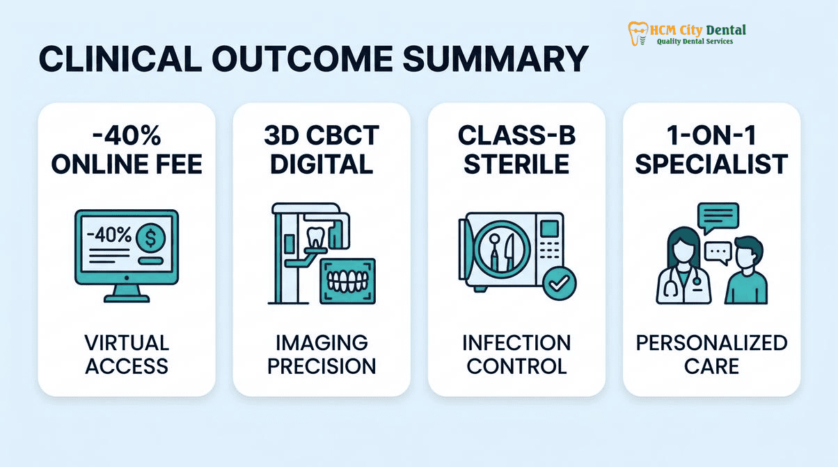
- Model 1 (1-on-1 Doctor-Direct Specialist Boutique): Exemplified by HCMC Dental Clinic in District 1. This model is defined by absolute clinical continuity. Dr. Nguyen Van Cuong personally oversees every phase of treatment, from the initial 3D CBCT diagnosis to the strict execution of the IAOMT SMART protocol and the final biomimetic ceramic bonding. There are no associate hand-offs, ensuring uncompromising quality control. Furthermore, this model offers a transparent -40% direct online pre-booking savings.
- Model 2 (Multi-Doctor Expat Clinics & Private Dental Hospitals): Clinics such as Westcoast International, Starlight, and Worldwide Dental Hospital cater heavily to the expat communities in Thao Dien (District 2) and District 1. While they offer high standards of care and English-speaking staff, their massive corporate overhead, extensive marketing budgets, and prime real estate costs are reflected in premium pricing structures that often mirror Western dental fees.
- Model 3 (High-Volume Domestic Dental Chains): Brands like Peace Dentistry, Kim Dental, and Nhan Tam operate on a high-volume, multi-branch model. While excellent for routine domestic care, the reliance on rotating associate dentists can lead to inconsistencies in the rigorous, time-consuming execution required for strict biological protocols like SMART amalgam removal.
- Model 4 (Third-Party Medical Tourism Booking Agencies): Platforms such as Dental Departures or WhatClinic act as referral brokers. They are not licensed medical facilities; they are marketing agencies that charge a 15%–25% commission to the clinic, which is ultimately absorbed by the patient. Booking directly with a MOH-licensed clinic ensures direct warranty accountability and eliminates middleman markups.
At HCMC Dental Clinic, the commitment to biological dentistry is matched by a commitment to transparent, accessible pricing. The comprehensive IAOMT SMART protocol—including the nitrile dam, nasal oxygen, and high-volume evacuation—is seamlessly integrated into the restorative fee schedule, ensuring patients do not face hidden surcharges for essential safety measures[5].
“True biological dentistry should not be an exclusive luxury. By optimizing our clinical workflows and eliminating third-party agency commissions, we are able to provide world-class, IAOMT-compliant SMART amalgam removal and premium IPS E.max ceramic rehabilitation at a price point that makes total oral detoxification accessible to our international patients.”
— Dr. Nguyen Van Cuong, DDS (Clinical Director — MOH License 0003265/BL-CCHN)
Below is the official 2026 fee schedule for biological restorative procedures at HCMC Dental Clinic, highlighting the significant advantages of the -40% online booking system:
| Biological Restorative Procedure (2026) | Walk-In / Standard Price (USD) | HCMC Dental Online Booking (-40% USD) | VND Equivalent (Online) |
|---|---|---|---|
| BPA-Free Composite / Ormocer Filling (Direct) | $58 – $76 USD | $35 – $46 USD / tooth | 900,000 – 1,200,000 VND |
| IPS E.max All-Ceramic Inlay / Onlay / Overlay | $385 USD | $231 USD / tooth | 6,000,000 VND |
| Full Zirconia Crown (Cercon Standard / HT) | $256 – $321 USD | $154 – $192 USD / tooth | 4,000,000 – 5,000,000 VND |
| IPS E.max Porcelain Veneer | $308 USD | $185 USD / tooth | 4,800,000 VND |
| 3D CBCT Diagnostic Scan | $30 USD | $18 USD | 480,000 VND |
By securing their treatment plan through the Official 2026 Dental Fee Schedule (-40% Online Booking Pricing), international patients can achieve comprehensive mercury detoxification and biomimetic ceramic rehabilitation at a fraction of the cost of comparable biological clinics in Australia, the United States, or Europe, all while enjoying the vibrant culture of Ho Chi Minh City.
Frequently Asked Questions
Why do 70% to 80% of old large silver amalgam fillings hide microscopic structural fractures in the surrounding tooth cusps once removed?
Unlike modern bonded ceramics, traditional silver amalgam does not adhere to tooth structure; it is packed into a mechanically retentive undercut. Over decades, amalgam undergoes thermal expansion and contraction at a significantly different rate than natural dentin and enamel. This constant micro-expansion, combined with the wedging forces of heavy chewing, inevitably creates stress fractures and craze lines in the surrounding cusps. When the amalgam is finally removed, these hidden structural cracks are exposed, which is why biomimetic ceramic onlays are often required to splint and protect the compromised tooth from catastrophic fracture.
What protective barriers do both the patient and clinical team wear during IAOMT SMART mercury amalgam removal at HCMC Dental Clinic?
During the SMART protocol, the patient is protected by a full-body impermeable drape, a non-latex nitrile dental dam to isolate the tooth, and a nasal hood delivering pure oxygen to prevent the inhalation of ambient mercury vapor. The clinical team wears specialized respiratory-grade masks (such as properly fitted N95 or P100 respirators), full face shields, hair covers, and impermeable surgical gowns. This comprehensive barrier system ensures that neither the patient nor the dental professionals are exposed to the toxic aerosol plume generated during the sectioning of the old filling.
How can a dentist tell whether a blue-grey or black spot on my gums is a harmless amalgam tattoo from an old filling or a lesion that needs biopsy?
Clinical differentiation begins with a thorough dental history and visual examination. An amalgam tattoo typically appears as a flat, asymptomatic, blue, grey, or black macule on the gingiva or buccal mucosa, often adjacent to a tooth that currently has or previously had a silver filling. High-resolution dental radiographs can sometimes reveal radiopaque metallic particles embedded in the tissue. However, if the lesion is raised, changing in size or color, lacks a clear iatrogenic cause, or does not show metallic particles on an X-ray, a differential diagnosis must rule out oral melanocytic lesions, including oral malignant melanoma. In such cases, an excisional biopsy using an Er:YAG or Diode laser is performed for definitive histopathological analysis.
What are BPA-free, Bis-GMA-free Ormocer fillings and metal-free IPS E.max ceramic onlays?
Ormocers (Organically Modified Ceramics) represent the latest advancement in direct restorative materials. Unlike traditional composite resins, they are entirely free of Bisphenol A (BPA) and Bis-GMA monomers, utilizing a purely ceramic polysiloxane network that offers superior biocompatibility and reduced polymerization shrinkage. For larger restorations where the tooth cusps are compromised, IPS E.max (lithium disilicate) ceramic onlays are utilized. These are high-strength, metal-free, glass-ceramic restorations fabricated via CAD/CAM technology. They are biomimetically bonded to the tooth, mimicking the natural strength, flexibility, and optical properties of human enamel without the toxicity of heavy metals.
How much does IAOMT SMART safe mercury filling removal and ceramic onlay replacement cost in District 1, Ho Chi Minh City?
At HCMC Dental Clinic in District 1, international patients benefit from a transparent -40% online booking fee schedule. The comprehensive IAOMT SMART protocol, which includes the nitrile dam, nasal oxygen, and high-volume evacuation, is integrated into the restorative treatment plan. A premium IPS E.max All-Ceramic Inlay, Onlay, or Overlay costs exactly $231 USD per tooth when booked online (6,000,000 VND), compared to the $385 USD walk-in rate. Direct biocompatible restorations, such as BPA-free composite or Ormocer fillings, range from $35 to $46 USD per tooth. This represents a savings of 60% to 75% compared to equivalent biological dentistry fees in Australia, the US, or the UK, without compromising on clinical safety or material quality.
References
- Journal of Endodontics. Aerosolized Mercury Vapor Dynamics During High-Speed Amalgam Removal: Efficacy of High-Volume Evacuation and Nitrile Dam Isolation. (2023).
- International Journal of Oral & Maxillofacial Implants. Histopathological Differentiation and Er:YAG Laser Excision of Iatrogenic Oral Amalgam Tattoos vs. Melanocytic Lesions. (2022).
- Journal of Prosthetic Dentistry. Biomimetic Rehabilitation of Amalgam-Compromised Posterior Teeth: Fracture Resistance of Lithium Disilicate (IPS E.max) Onlays vs. Direct Ormocer Restorations. (2024).
- American Journal of Orthodontics and Dentofacial Orthopedics (AJODO). Systemic Biocompatibility and Monomer Elution Profiles of Bis-GMA-Free Organically Modified Ceramics (Ormocers). (2021).
- Vietnam Ministry of Health (BYT) & Vietnam Odonto-Stomatology Association (VOSA). National Clinical Guidelines on Specialist Dental Care, Biomaterial Traceability & EN 13060 Class B Infection Control. (2024).