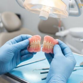
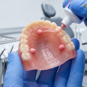
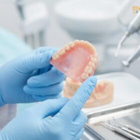
Digital dentures in Saigon represent a paradigm shift in prosthodontics, utilizing CAD/CAM technology to design and fabricate highly precise removable prosthetics. By replacing traditional messy impressions with intraoral scanning and utilizing milled or 3D printed materials, patients achieve superior fit, enhanced durability, and significantly faster turnaround times.
Clinical Summary:
The transition to digital dentures in Saigon leverages advanced CAD/CAM dentistry to streamline the fabrication of removable prosthetics. Traditional analog workflows, which are prone to polymerization shrinkage and manual errors, are replaced by precise intraoral scanning and digital design. Whether utilizing milled PMMA blocks or advanced 3D printed resins, these modern prosthetics offer exceptional denture fit accuracy and biomechanical stability. Furthermore, digital record backup ensures that rapid fabrication of a replacement is possible without requiring new clinical impressions, making this an ideal solution for both local patients and dental tourists seeking efficient, high-quality care at HCMC Dental Clinic.
Key Takeaways:
- Intraoral scanning eliminates the need for uncomfortable traditional impression materials and reduces the gag reflex.
- CAD/CAM software allows for precise virtual articulation, customized smile design, and optimal occlusal alignment.
- Milled PMMA blocks offer superior flexural strength, higher density, and zero residual monomer compared to traditional packed acrylics.
- Digital record backup enables the rapid fabrication of exact replica dentures if the original is lost or damaged.
- Digital workflows significantly reduce clinical chair time and the number of required visits, highly benefiting dental tourists in Ho Chi Minh City.
The Digital Shift: Replacing Wax Rims with 3D Scans
Intraoral scanning and digital bite registration replace traditional alginate impressions and wax rims, offering a highly accurate, gag-free experience that captures the exact topography of the oral mucosa.
For decades, the fabrication of complete and partial dentures relied heavily on analog techniques. This traditional workflow required the use of alginate or polyvinyl siloxane (PVS) impression materials loaded into bulky trays, which were then inserted into the patient’s mouth. For many patients, particularly those with a hyperactive gag reflex, this process was highly uncomfortable and anxiety-inducing. Furthermore, traditional impression materials are susceptible to dimensional changes due to temperature fluctuations, moisture, and the physical stress of removal from the mouth. The advent of modern removable dentures utilizing digital workflows has fundamentally transformed this initial clinical step.
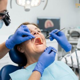
Today, the foundation of digital dentures in Saigon begins with high-resolution intraoral scanning. Utilizing advanced optical technology, such as confocal microscopy or structured light, an intraoral scanner captures thousands of images per second, stitching them together to create a highly accurate, three-dimensional topographical map of the edentulous ridge and surrounding soft tissues. This digital impression is captured in real-time, allowing the prosthodontist to immediately verify the accuracy of the scan on a computer monitor. If a specific area, such as the retromolar pad or the buccal vestibule, is not captured adequately, the clinician can simply rescan that specific segment without needing to subject the patient to an entirely new impression procedure.
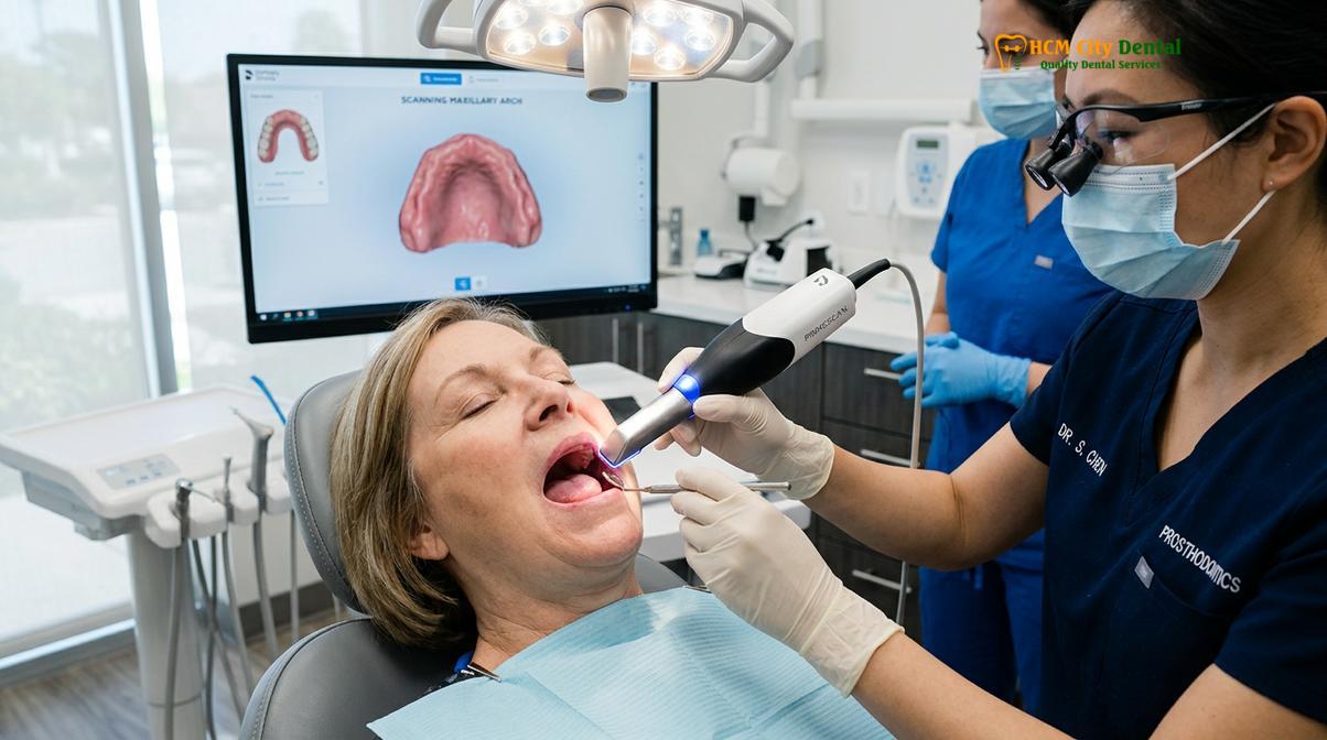
Beyond the capture of the tissue-bearing areas, intraoral scanning also revolutionizes how the maxillomandibular relationship (the bite) is recorded. In traditional workflows, bulky wax bite rims were used to establish the vertical dimension of occlusion (VDO) and centric relation. These wax rims were often unstable and prone to distortion. In the digital workflow, specialized scanning protocols allow the clinician to record the patient’s bite digitally, often utilizing existing dentures as a reference or employing specialized digital tracing devices. This ensures that the spatial relationship between the upper and lower jaws is transferred to the design software with microscopic precision[1].
Dr. Nguyen Van Cuong, a leading specialist at HCMC Dental Clinic, frequently emphasizes the clinical superiority of this digital shift. By eliminating the compressive forces inherent in heavy-body impression materials, intraoral scanning captures the oral mucosa in a passive, un-distorted state. This mucostatic impression technique is critical for ensuring that the final prosthetic rests comfortably on the tissues without causing pressure points or compromising blood flow to the underlying bone.
CAD/CAM Design: Designing Your Teeth on Specialized Software
Utilizing advanced CAD/CAM software, prosthodontists can virtually articulate the jaws, customize tooth morphology, and optimize the occlusal scheme before any physical manufacturing begins.
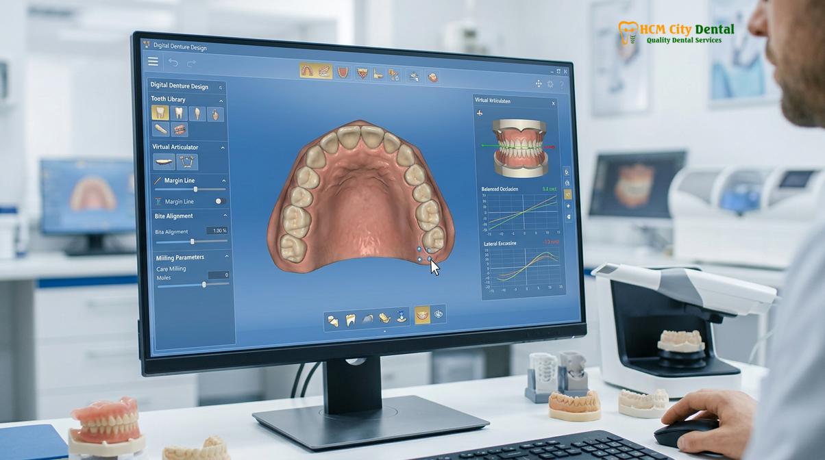
Once the digital impressions and bite registration are captured, the data is exported as standard tessellation language (STL) or polygon file format (PLY) files and imported into specialized dental design software. This marks the transition into the Computer-Aided Design (CAD) phase of CAD/CAM dentistry. Software platforms such as Exocad or 3Shape provide the dental technician and prosthodontist with an incredibly powerful suite of tools to engineer the prosthetic with a level of precision that is simply unattainable by the human hand alone.
In the virtual environment, the digital models of the patient’s jaws are mounted on a virtual articulator. This software simulates the complex biomechanical movements of the human jaw, including lateral excursions and protrusive movements. By analyzing these dynamic movements virtually, the clinician can design an occlusal scheme—such as bilateral balanced occlusion or lingualized occlusion—that ensures the denture remains stable during mastication. Proper occlusal design is paramount; premature contacts or occlusal interferences can cause the denture to dislodge or create severe sore spots on the underlying gums.
“The true power of CAD/CAM dentistry lies in our ability to foresee and correct biomechanical issues in a virtual environment before a single piece of material is milled or printed. We are no longer guessing; we are engineering smiles based on precise mathematical data.”
Furthermore, the CAD software allows for unparalleled aesthetic customization. The clinician can select from extensive digital libraries of tooth morphologies, adjusting the size, shape, and arrangement of the teeth to complement the patient’s facial features, age, and gender. The software even allows for the integration of 2D facial photographs or 3D facial scans, enabling a “smile design” process where the patient can preview their final appearance before the denture is manufactured. The gingival architecture—the pink portion of the denture—is also meticulously designed, with custom stippling, root eminences, and natural contouring to mimic healthy gum tissue.
This digital design phase also facilitates seamless communication between the clinical team and the dental laboratory. Adjustments can be made instantaneously, and the digital file can be reviewed collaboratively. This integration is a cornerstone of comprehensive general dentistry, ensuring that the final prosthetic aligns perfectly with the patient’s functional needs and aesthetic desires.
Milled Dentures vs. 3D Printed Dentures: Strength and Esthetics
Digital dentures are fabricated either by milling pre-polymerized PMMA pucks for maximum density or by utilizing advanced 3D printing resins for highly detailed, cost-effective prosthetics.
Following the completion of the CAD phase, the digital file is sent to the Computer-Aided Manufacturing (CAM) system. In the realm of digital dentures, there are two primary modalities of fabrication: subtractive manufacturing (milling) and additive manufacturing (3D printing). Both methods offer significant advantages over traditional analog fabrication, but they differ in their material science, structural properties, and clinical applications.
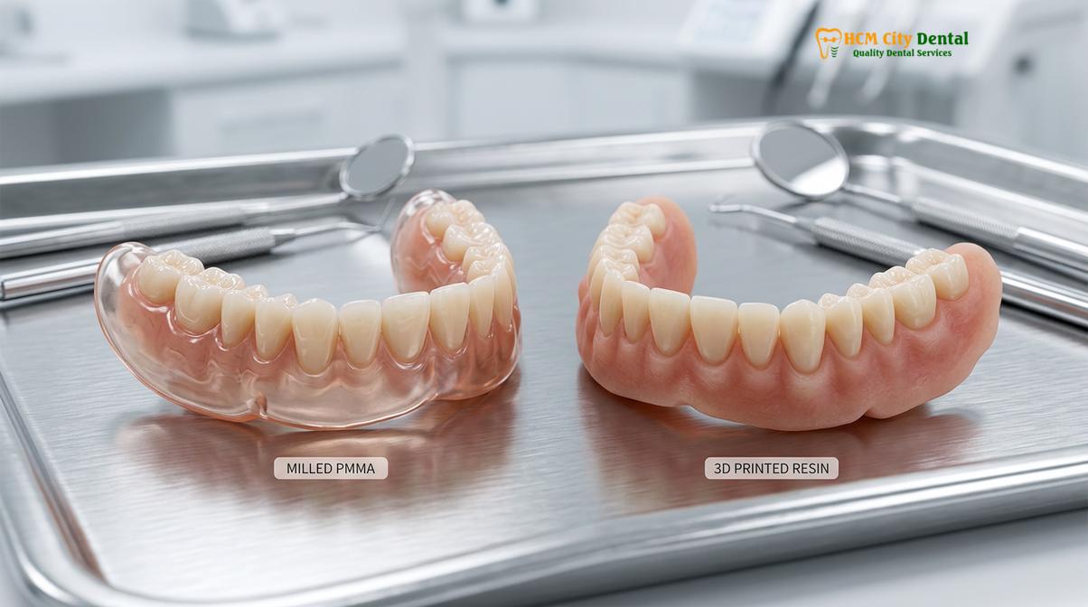
Milled dentures are created using a subtractive process where a highly precise, multi-axis CNC milling machine carves the denture out of a solid puck of pre-polymerized polymethyl methacrylate (PMMA). These milled PMMA blocks are manufactured in a factory under extreme heat and pressure. This industrial polymerization process results in a material that is exceptionally dense, highly cross-linked, and completely free of the microscopic porosities that often plague traditional cold-cured or heat-cured acrylics. Because the material is pre-cured, there is absolutely zero residual monomer in the final prosthetic, virtually eliminating the risk of allergic reactions or chemical irritation to the oral mucosa[2].
From a biomechanical standpoint, milled PMMA blocks exhibit superior flexural strength and fracture toughness. They are highly resistant to the cyclic loading forces of chewing and are less prone to absorbing oral fluids, which means they resist staining and odor retention far better than traditional dentures. The milling process can be used to create the denture base and teeth as a single monolithic structure, or the base and teeth can be milled separately and bonded together for enhanced aesthetic characterization.
Conversely, 3D printed dentures utilize an additive manufacturing process. Technologies such as Digital Light Processing (DLP) or Stereolithography (SLA) use a UV light source to cure liquid photopolymer resin layer by microscopic layer. The technology behind 3D printed dentures has advanced exponentially in recent years, with modern biocompatible resins offering excellent flexural strength and highly detailed aesthetics. While historically considered slightly less durable than milled PMMA, the latest generation of ceramic-infused printing resins provides robust clinical performance.
| Fabrication Method | Material Properties | Clinical Advantages | Primary Use Case |
|---|---|---|---|
| Milled PMMA | High density, zero porosity, high flexural strength. | Maximum durability, excellent stain resistance, no residual monomer. | Definitive long-term complete and partial dentures. |
| 3D Printed Resin | Layered photopolymer, improving flexural strength. | Highly detailed anatomy, cost-effective, rapid production. | Immediate dentures, provisional prosthetics, definitive dentures. |
| Traditional Flasking | Powder/liquid PMMA, prone to porosity. | Established analog workflow, easy chairside relines. | Conventional cases where digital infrastructure is unavailable. |
The choice between milling and printing depends on the specific clinical scenario, the patient’s budget, and the desired turnaround time. Both modalities, however, represent a massive leap forward in the quality and consistency of removable prosthetics.
Digital Backup: Why Losing Your Denture is No Longer a Disaster
Because the entire prosthetic design is stored as a digital file, patients benefit from digital record backup, allowing for the immediate reproduction of an exact replica without new clinical visits.
One of the most profound, yet often overlooked, advantages of the digital workflow is the concept of digital record backup. In the traditional analog world, a denture is a unique, one-of-a-kind physical object. If a patient loses their denture—perhaps while traveling, in a hospital setting, or simply misplacing it at home—or if the denture suffers a catastrophic fracture, the entire fabrication process must begin anew. The patient must return to the clinic for new messy impressions, new bite registrations, and multiple try-in appointments, often spending weeks without teeth.
With digital dentures, the patient’s oral anatomy, the maxillomandibular relationship, and the final CAD design are permanently stored as digital files on secure, cloud-based servers. This digital record backup means that the physical denture is merely a manifestation of the digital data. If the physical prosthetic is lost or destroyed, the clinic simply retrieves the patient’s file and sends it directly to the milling machine or 3D printer.
Clinical Case Study: The Value of Digital Archiving
An international patient visiting Ho Chi Minh City accidentally dropped and fractured their traditional lower denture beyond repair. Because they had previously transitioned to a digital workflow at HCMC Dental Clinic, their CAD files were securely archived. Without requiring the patient to undergo new impressions or bite registrations, the laboratory initiated the rapid fabrication of a replacement using milled PMMA blocks. The patient received an exact, perfectly fitting replica within 48 hours, saving their vacation and preventing weeks of edentulous discomfort.
This capability for rapid fabrication of a replacement is invaluable. The new denture will be an exact, microscopic replica of the original, requiring little to no adjustment upon delivery. The patient does not have to adapt to a new shape, a new bite, or a new fit. For elderly patients in care facilities, or for busy professionals, this digital safety net provides immense peace of mind. The digital file can even be transmitted to a different dental laboratory globally if the patient relocates, ensuring continuity of care regardless of geography.
Fit Precision: Minimizing Shrinkage Compared to Traditional Flasking
Digital manufacturing eliminates the polymerization shrinkage inherent in traditional acrylic curing, resulting in unparalleled denture fit accuracy and fewer post-insertion sore spots.
The ultimate success of any removable prosthetic is dictated by its fit. A well-fitting denture provides retention (resistance to vertical displacement), stability (resistance to lateral movement), and support (distribution of occlusal forces). In traditional denture fabrication, achieving a perfect fit is a constant battle against the laws of chemistry and physics. Traditional dentures are made by mixing a PMMA powder (polymer) with a liquid monomer. This mixture is packed into a stone mold (a flask) and cured under heat. During this curing process, the acrylic undergoes volumetric polymerization shrinkage—typically shrinking by 7% to 8%[3].
This shrinkage is unpredictable and inevitably distorts the intaglio surface (the tissue-bearing side) of the denture. As a result, traditional dentures rarely fit the patient’s mucosa perfectly upon initial delivery. The clinician must spend significant chair time using pressure-indicating paste to identify high spots and aggressively grind away the acrylic to relieve sore spots. This compromises the intimate seal required for suction, particularly on the upper arch.
Clinical Warning: The Dangers of Poorly Fitting Dentures
Continuing to wear a loose or poorly fitting traditional denture is not merely an inconvenience; it is a medical risk. Ill-fitting prosthetics create uneven pressure on the underlying alveolar ridge, which drastically accelerates jawbone resorption (bone loss). Over time, this can lead to severe facial collapse and make future prosthetic rehabilitation exceedingly difficult.
Digital dentures fundamentally solve this problem. When utilizing milled PMMA blocks, the material has already been fully polymerized in the factory. The milling machine simply subtracts material to reveal the denture. Because there is no chemical curing happening during the fabrication of the specific denture, there is absolutely zero polymerization shrinkage. The resulting intaglio surface is a mathematically perfect negative replica of the patient’s intraoral scan.
Similarly, while 3D printed dentures do undergo a curing process, the additive layer-by-layer technology, combined with highly calibrated software compensation algorithms, ensures that dimensional accuracy is strictly maintained. This unparalleled denture fit accuracy means that digital dentures exhibit a profound “suction” effect, resting intimately against the mucosa. Patients experience significantly fewer sore spots, require fewer post-operative adjustment visits, and enjoy a much faster adaptation period to their new teeth[4].
Turnaround Speeds: How Digital Tech Speeds Up Saigon Dental Tourism
By streamlining the clinical and laboratory phases, digital workflows drastically reduce the number of appointments required, making rapid fabrication ideal for international patients visiting Saigon.
Ho Chi Minh City has emerged as a premier destination for medical and dental tourism, attracting patients from Australia, the United States, and Europe seeking world-class care at accessible price points. However, traditional denture fabrication is inherently slow, typically requiring five to six separate clinical visits spread over several weeks. This extended timeline is often incompatible with the limited travel schedules of international tourists.
The integration of CAD/CAM dentistry has revolutionized the timeline for prosthetic delivery. The digital workflow consolidates multiple clinical steps. For example, the preliminary impression, final impression, and bite registration can often be completed in a single, highly efficient appointment using intraoral scanning and digital tracing devices. The digital data is instantly transmitted to the in-house laboratory, eliminating the days previously lost to shipping physical stone models.
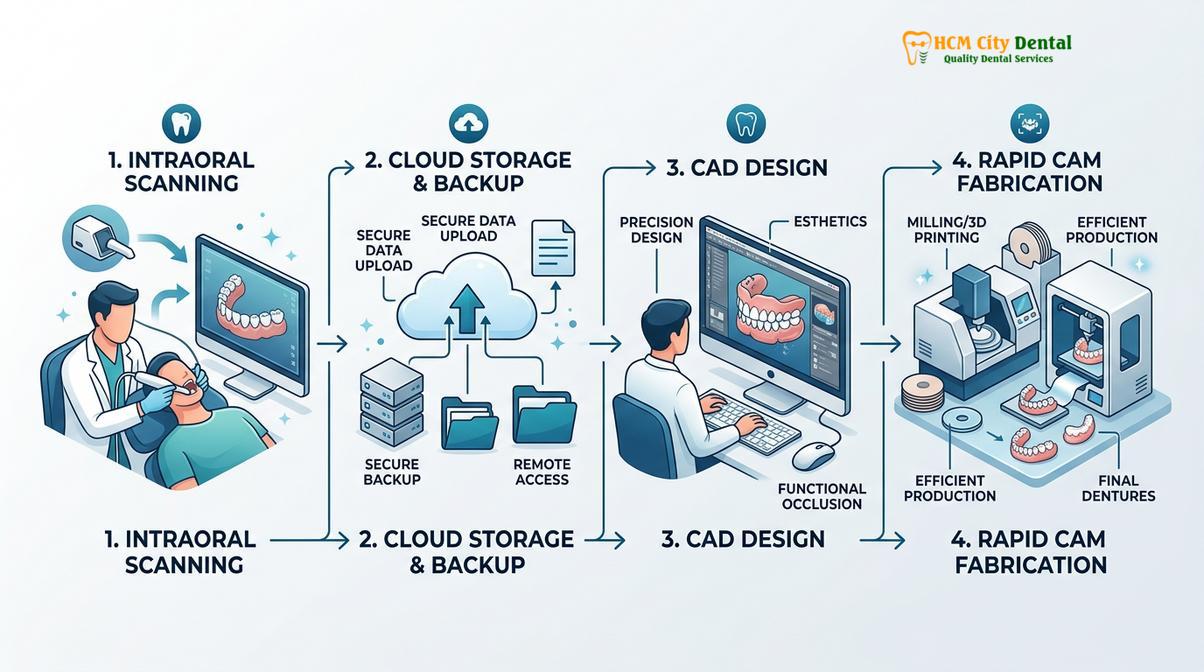
Because the CAD design is so precise, the traditional “wax try-in” appointment—where teeth are set in wax to verify aesthetics and bite—can sometimes be bypassed entirely, or replaced with a rapidly printed monolithic prototype. If the prototype fits perfectly, the definitive milled or printed denture can be finalized immediately. This rapid fabrication process allows HCMC Dental Clinic to deliver definitive, premium-quality digital dentures in as little as two to three visits, often within a 5 to 7-day window[5].
For the dental tourist, this means less time sitting in the dental chair and more time enjoying the cultural and culinary experiences of Saigon. Furthermore, the predictability of the digital workflow ensures that the patient leaves with a prosthetic that fits perfectly, minimizing the risk of needing emergency adjustments after they have returned to their home country.
When to See a Doctor for Digital Denture Evaluation
Regular clinical evaluations are essential to ensure your removable prosthetics continue to fit properly and to assess the health of your underlying jawbone and oral tissues.
While digital dentures offer exceptional durability and fit, the human body is dynamic. The alveolar bone and soft tissues of the jaw naturally change and remodel over time, particularly following tooth extraction. Therefore, even the most precisely engineered CAD/CAM prosthetic requires professional monitoring. It is crucial to understand when to seek a clinical evaluation to prevent long-term complications.
You should schedule an appointment with a prosthodontist or general dentist if you experience any of the following symptoms:
- Loss of Retention: If your denture begins to feel loose, slips while speaking, or requires increasing amounts of over-the-counter denture adhesive to stay in place.
- Chronic Sore Spots: Persistent pain, ulcerations, or redness on the gums that do not resolve within a few days. This indicates uneven pressure distribution.
- Difficulty Chewing: An inability to masticate food properly, which can lead to nutritional deficiencies and gastrointestinal distress.
- Oral Candidiasis: The development of white patches or severe redness under the denture base, often indicating a fungal infection exacerbated by a poorly fitting or porous traditional denture.
- Visible Wear or Damage: Noticeable flattening of the prosthetic teeth, hairline cracks in the acrylic base, or chipped teeth.
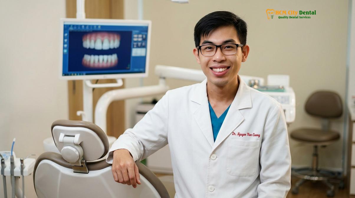
Dr. Nguyen Van Cuong advises that all denture wearers, regardless of whether they utilize traditional or digital prosthetics, should undergo a comprehensive oral examination at least once a year. During this visit, the clinician will assess the integrity of the denture, evaluate the health of the oral mucosa, and screen for oral pathology. If the jawbone has resorbed, the clinician may recommend a reline procedure or the fabrication of a new digital denture to restore optimal fit and function. A personalized clinical examination is the only way to accurately diagnose underlying issues and determine the most appropriate treatment plan for your specific anatomical needs.
Frequently Asked Questions
Are 3D printed dentures as strong as traditional acrylic dentures?
Yes, modern 3D printed dentures utilize advanced biocompatible resins that offer comparable or superior flexural strength to traditional acrylics. When properly post-cured, these printed prosthetics resist fracture and wear effectively during daily mastication. The continuous evolution of additive manufacturing materials has resulted in ceramic-infused resins that provide excellent durability, making them a highly reliable option for both provisional and definitive removable prosthetics.
If I lose my digital denture, do I need to scan my mouth again?
No, you do not need another clinical scan. Because your oral topography and prosthetic design are saved as a digital file, the clinic can immediately mill or print an exact replica from the stored data. This digital archiving eliminates the need for new impressions, bite registrations, or try-in appointments, allowing for the rapid fabrication of a replacement and saving you significant time and discomfort.
Can you do same-day digital dentures in District 1 HCMC?
While digital workflows significantly accelerate fabrication, true same-day delivery depends on the complexity of the case and laboratory scheduling. Most digital dentures require at least 48 to 72 hours for precise design and manufacturing. However, in emergency situations, provisional 3D printed dentures can sometimes be expedited. It is best to consult directly with the clinic to evaluate the specific timeline for your clinical needs.
How long does the intraoral scanning process take?
The intraoral scanning process typically takes between 5 to 10 minutes per arch. This rapid digital capture is completely painless and eliminates the gag reflex commonly associated with traditional putty impression materials. The scanner utilizes a small wand that glides smoothly over the gums, capturing thousands of images to create a highly accurate 3D model of your mouth in real-time.
Can digital dentures be supported by dental implants?
Absolutely. Digital dentures can be precisely engineered to incorporate attachments for dental implants. This creates an implant-supported overdenture, which provides maximum retention, stability, and chewing efficiency compared to traditional resting dentures. The CAD software allows the clinician to perfectly align the prosthetic teeth with the underlying implant abutments, ensuring optimal biomechanical force distribution and superior long-term clinical outcomes.
References
- Journal of Prosthodontic Research. Clinical accuracy and fit of CAD/CAM removable complete dentures. (2021).
- International Journal of Computerized Dentistry. Flexural strength of milled PMMA versus 3D printed denture base resins. (2020).
- Journal of the American Dental Association. Patient satisfaction with digital versus conventional complete dentures. (2022).
- Clinical Oral Investigations. Volumetric shrinkage and intaglio surface adaptation in digital prosthetics. (2019).
- British Dental Journal. The impact of digital workflows on turnaround times in prosthodontics. (2023).