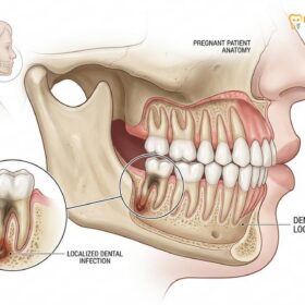
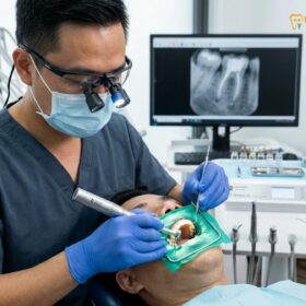
A root canal CBCT scan is an advanced three-dimensional imaging technique that captures highly detailed, cross-sectional views of a tooth’s internal anatomy and surrounding alveolar bone. Unlike traditional two-dimensional X-rays, this cutting-edge technology allows endodontists to accurately diagnose hidden infections, locate complex nerve canals, and identify microscopic root fractures with unprecedented precision.
By utilizing a targeted, small field-of-view, a 3D dental scan provides a distortion-free map of the pulpal system. This level of diagnostic clarity is essential for planning complex root canal procedures, minimizing procedural risks, and ensuring the highest possible success rate for saving your natural tooth.
Clinical Summary:
Cone beam computed tomography (CBCT) has fundamentally revolutionized endodontic diagnostics by eliminating the anatomical superimposition and geometric distortion inherent in conventional 2D radiography. By utilizing a small field of view (FOV), endodontic CBCT provides high-resolution, three-dimensional mapping of the pulpal floor, accessory canals, and periapical tissues. This precision imaging is critical for diagnosing persistent apical periodontitis, identifying elusive MB2 canals in maxillary molars, and detecting vertical root fractures before irreversible structural damage occurs. For complex cases, a 3D dental scan ensures predictable treatment planning, minimizes procedural complications, and significantly improves the long-term prognosis of endodontic therapy.
Key Takeaways:
- CBCT imaging provides a 3D, distortion-free view of the tooth, eliminating the overlapping anatomical structures seen in standard 2D X-rays.
- Small field-of-view (FOV) scans minimize radiation exposure while delivering the ultra-high resolution required to visualize microscopic endodontic anatomy.
- 3D scans are clinically essential for locating hidden MB2 canals, complex C-shaped root systems, and unpredictable apical bifurcations.
- Early detection of silent apical periodontitis and subtle vertical root fractures is significantly higher with CBCT compared to traditional radiography.
- The technology strictly adheres to ALARA (As Low As Reasonably Achievable) principles, ensuring maximum patient safety during diagnostic procedures.
Diagnostic Limitations of Traditional 2D Panoramic X-Rays
Traditional 2D X-rays compress complex three-dimensional dental structures into a flat image, often obscuring critical anatomical details and early signs of infection. This superimposition limits the diagnostic accuracy required for complex endodontic treatments.
For decades, conventional two-dimensional radiography—including periapical and panoramic X-rays—has been the cornerstone of dental diagnostics. While these imaging modalities remain highly valuable for routine dental check-ups, detecting interproximal caries, and assessing general periodontal health, they possess inherent physical limitations when it comes to the intricate micro-anatomy of the root canal system. The primary limitation of 2D imaging is the phenomenon of anatomical superimposition. When a three-dimensional object, such as a multi-rooted molar and its surrounding dense cortical bone, is compressed into a single two-dimensional plane, the resulting image is a composite shadow. Structures that lie in the buccolingual plane (front-to-back) overlap one another, creating visual noise that can easily mask critical endodontic pathology.
In the context of endodontics, this flattening effect means that a dentist can only accurately assess the tooth in the mesiodistal (side-to-side) and apicocoronal (top-to-bottom) dimensions. The buccolingual dimension is entirely lost. Consequently, if a tooth has two canals superimposed directly over one another in the path of the X-ray beam, they will appear as a single canal on the final radiograph. This is a frequent cause of missed anatomy, particularly in premolars and molars, leading to incomplete disinfection and subsequent treatment failure. Furthermore, traditional 2D X-rays are subject to geometric distortion and magnification errors. Depending on the angle of the X-ray tube head and the placement of the digital sensor, the roots may appear artificially elongated or foreshortened, complicating the accurate measurement of working lengths required for precise endodontic therapy.
Another significant limitation of 2D radiography is its inability to detect early-stage periapical bone lesions. The human jawbone consists of a dense outer layer called cortical bone and a spongy, porous inner layer called cancellous bone. When a dental infection spreads from the root tip into the surrounding bone, it typically begins destroying the cancellous bone first. Because the dense cortical plates remain intact during the early stages of infection, they block the X-ray beam, effectively hiding the bone loss. Studies have shown that a periapical lesion must erode a significant portion of the cortical bone before it becomes visible as a dark spot (radiolucency) on a standard 2D X-ray[1]. This means that a patient could be suffering from an active, painful infection that appears completely “normal” on a conventional radiograph.
“The transition from two-dimensional radiography to three-dimensional cone beam computed tomography represents the most significant diagnostic paradigm shift in the history of endodontics, allowing clinicians to finally see the true anatomical reality of the tooth rather than a distorted shadow.”
Because of these inherent physical limitations, relying solely on 2D imaging for complex endodontic cases or retreatments is often insufficient. When a patient presents with persistent pain, swelling, or a history of failed root canal treatments, the diagnostic clarity provided by a 3D scan becomes not just beneficial, but clinically essential for determining the correct course of action and preventing unnecessary surgical interventions.
Cone Beam Computed Tomography (CBCT): The 3D Endodontic Standard
CBCT technology utilizes a rotating X-ray source to capture hundreds of distinct images, reconstructing them into a highly accurate 3D model of the tooth. This standard of care allows for precise spatial measurement and comprehensive anatomical mapping.
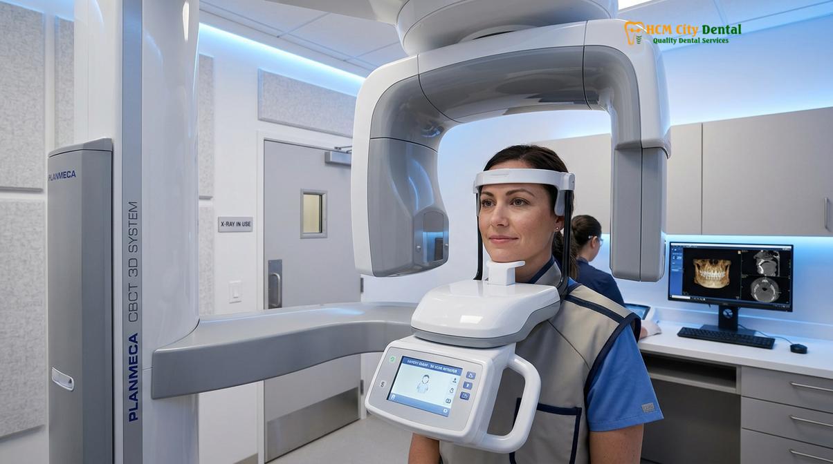
Cone beam computed tomography (CBCT) is a highly advanced medical imaging technique specifically adapted for the maxillofacial region. Unlike traditional medical CT scans that use a fan-shaped X-ray beam and capture images in multiple slices, a dental CBCT utilizes a cone-shaped X-ray beam. During the scanning process, the patient remains stationary while the X-ray source and a flat-panel solid-state detector rotate 360 degrees around the patient’s head. In a matter of seconds, the machine captures hundreds of individual projection images from various angles. Powerful computer algorithms, utilizing filtered back-projection techniques, then reconstruct these raw images into a highly accurate, high-resolution three-dimensional volumetric dataset.
The fundamental building block of a CBCT image is the voxel, or volumetric pixel. Unlike the flat, 2D pixels on a computer screen, voxels are three-dimensional cubes of data. In endodontic CBCT imaging, the voxels are isotropic, meaning they are perfectly equal in all three dimensions (length, width, and depth). This isotropic nature ensures that the 3D reconstruction is geometrically accurate and free from the magnification or distortion errors that plague 2D X-rays. Endodontic scans typically utilize extremely small voxel sizes—often ranging from 75 to 90 micrometers. This ultra-high spatial resolution allows the endodontist to visualize microscopic details, such as the delicate curvature of a root canal, the presence of calcified pulp stones, and the exact location of the apical foramen (the opening at the tip of the root).
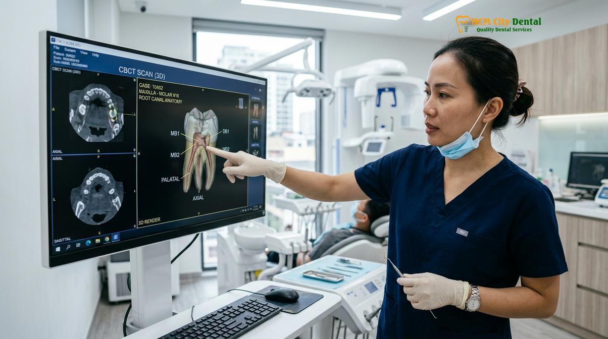
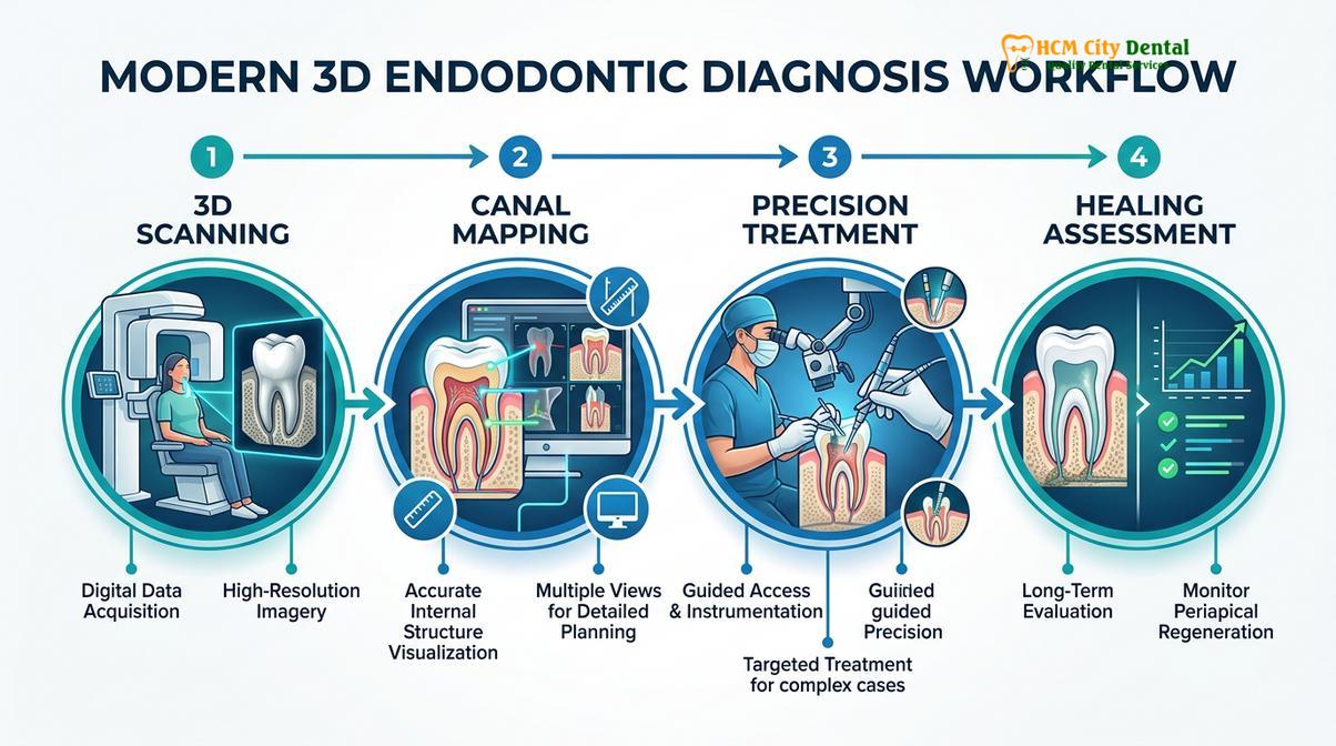
Once the 3D volume is generated, the endodontist can use specialized software to navigate through the tooth in three distinct anatomical planes: the axial plane (viewing the tooth from top to bottom), the coronal plane (viewing from front to back), and the sagittal plane (viewing from side to side). This multi-planar reconstruction allows the clinician to virtually “slice” through the tooth at any angle, examining the internal anatomy millimeter by millimeter. This capability is invaluable for mapping the pulpal floor, determining the exact number and morphology of the root canals, and planning the safest, most conservative access cavity preparation.
In modern endodontics, the standard of care dictates the use of a small field-of-view (FOV) for root canal diagnostics. The field of view refers to the anatomical area captured by the scan. While a large FOV might capture the entire skull, a small FOV (typically 4×4 cm or 5×5 cm) focuses exclusively on the specific tooth in question and its immediate surrounding bone. Utilizing a small FOV is critical for two main reasons. First, it significantly increases the spatial resolution of the image, providing the crisp, detailed views necessary for micro-endodontics. Second, it drastically reduces the amount of scattered radiation, resulting in a clearer image while simultaneously minimizing the patient’s radiation exposure.
| Diagnostic Parameter | Traditional 2D Periapical X-ray | 3D Endodontic CBCT Scan |
|---|---|---|
| Dimensionality | 2D (Mesiodistal and Apicocoronal only) | 3D (Axial, Coronal, and Sagittal planes) |
| Anatomical Superimposition | High (Structures overlap and obscure details) | None (Distortion-free cross-sectional views) |
| Spatial Resolution | Moderate (Limited by 2D compression) | Ultra-High (Isotropic voxels, 75-90 µm) |
| Detection of Early Bone Loss | Poor (Requires significant cortical bone destruction) | Excellent (Detects early cancellous bone changes) |
| Primary Clinical Use Case | Routine screening, working length estimation | Complex anatomy, retreatments, surgical planning |
The integration of cone beam computed tomography into clinical practice has elevated the predictability and success rates of advanced endodontic procedures. By providing a clear, comprehensive roadmap of the internal tooth structure before the procedure even begins, CBCT allows the clinician to navigate complex anatomy with confidence, ensuring that no canal is left untreated and no infection is left behind.
Identifying Complex Root Canal Anatomy: C-Shaped Canals and MB2
A 3D scan is crucial for mapping unpredictable internal tooth structures, such as the elusive MB2 canal in upper molars or C-shaped canal systems in lower molars. Identifying these hidden pathways prevents treatment failure and persistent infection.
The human root canal system is rarely a simple, straight tube. It is a highly complex, branching network of main canals, lateral canals, accessory canals, and apical deltas that more closely resembles the root system of a tree. Successfully treating an infected tooth requires the complete debridement and disinfection of this entire network. If a single canal is missed during the procedure, the bacteria residing within it will continue to multiply, leading to persistent inflammation, pain, and eventual failure of the treatment. This is where the diagnostic power of a 3D dental scan HCMC becomes indispensable, particularly when dealing with notoriously complex anatomical variations.
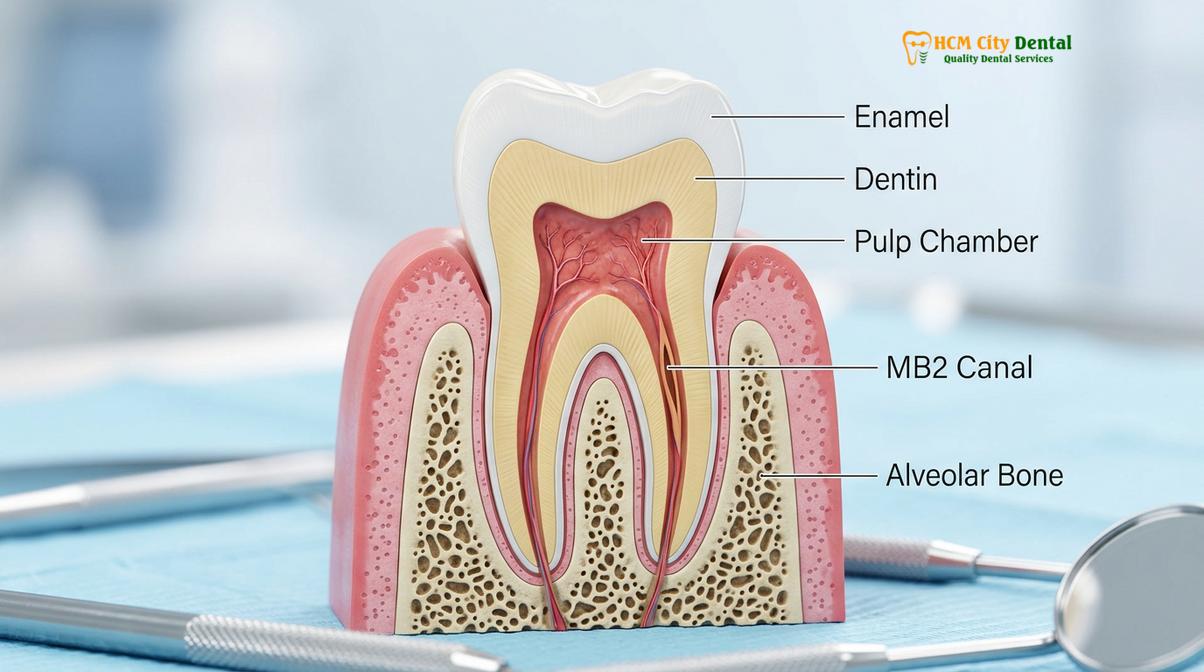
One of the most common and clinically challenging anatomical variations is the presence of a second mesiobuccal canal, commonly referred to as the MB2 canal, in maxillary (upper) first and second molars. Clinical studies utilizing CBCT imaging have demonstrated that the MB2 canal is present in over 70% to 90% of maxillary first molars[3]. Despite its high prevalence, the MB2 canal is frequently missed during conventional root canal therapy. This is primarily because the MB2 canal is often very narrow, heavily calcified, and located in a difficult-to-see position on the pulpal floor, slightly palatal to the main mesiobuccal canal. Furthermore, on a traditional 2D X-ray, the MB2 canal is completely superimposed behind the larger, denser palatal root, rendering it virtually invisible. A high-resolution CBCT scan allows the endodontist to view the tooth in the axial plane, clearly revealing the exact location, trajectory, and degree of curvature of the MB2 canal, ensuring it is located and thoroughly treated.
Clinical Case Study: Resolving Persistent Pain
A patient presented to HCMC Dental Clinic in Ho Chi Minh City experiencing persistent, throbbing pain in an upper right molar that had undergone root canal therapy at a different facility two years prior. Standard 2D X-rays showed what appeared to be a perfectly executed treatment with no obvious signs of failure. However, Dr. Nguyen Van Cuong recommended a targeted 3D dental scan HCMC to investigate further. The axial slices of the CBCT volume clearly revealed an untreated, highly infected MB2 canal that was completely hidden behind the palatal root on the 2D film. Dr. Cuong performed a precise endodontic retreatment, navigating the newly discovered canal under a dental microscope. Following the disinfection and sealing of the MB2 canal, the patient’s symptoms resolved completely within days, and subsequent follow-ups showed excellent bone healing.
Another highly complex anatomical variation that necessitates 3D imaging is the C-shaped canal system, most frequently found in mandibular (lower) second molars. Instead of having distinct, separate root canals, teeth with this morphology feature a continuous, ribbon-like canal system that sweeps in a “C” shape from the mesial to the distal aspect of the root. C-shaped canals are notoriously difficult to clean and shape because they contain numerous fins, webs, and irregular anastomoses (connections) that harbor bacterial biofilms. Melton’s classification system is often used to categorize the various configurations of C-shaped canals, which can change dramatically from the pulp chamber down to the root apex. Attempting to treat a C-shaped canal system based solely on 2D imaging is highly unpredictable. A CBCT scan provides a detailed, cross-sectional map of the entire C-shaped morphology, allowing the clinician to design a customized disinfection protocol, often involving advanced ultrasonic activation and specialized bioceramic sealers, to ensure the entire irregular space is thoroughly cleaned and obturated.
Beyond the MB2 and C-shaped canals, CBCT is invaluable for identifying other anatomical anomalies, such as dens invaginatus (a tooth within a tooth), taurodontism (enlarged pulp chambers), and unpredictable root bifurcations. By providing a clear, three-dimensional blueprint of the tooth’s internal architecture, CBCT empowers the endodontist to perform highly targeted, conservative treatments. This precision minimizes the removal of healthy pericervical dentin—the crucial band of tooth structure near the gumline that provides the tooth with its structural strength—thereby enhancing the long-term survivability of the restored tooth.
Detecting Silent Bone Loss: Apical Periodontitis Under the Radar
CBCT imaging can detect early-stage bone destruction at the root apex long before it becomes visible on conventional X-rays. This early detection of apical periodontitis is vital for preventing systemic infection and tooth loss.
Apical periodontitis is an inflammatory disease of the tissues surrounding the root apex, caused primarily by a bacterial infection within the root canal system. When bacteria and their toxic byproducts exit the tooth through the apical foramen, they trigger a robust immune response in the surrounding alveolar bone. This immune response, while attempting to wall off the infection, inadvertently leads to the localized destruction of bone tissue, creating a periapical lesion (often referred to as a dental granuloma or cyst). Detecting and accurately assessing the size and extent of these lesions is a fundamental aspect of endodontic diagnosis and treatment planning.
As previously discussed, traditional 2D radiography is notoriously insensitive to early periapical bone changes. Because the dense cortical bone plates mask the less dense cancellous bone, a significant amount of bone destruction must occur before a radiolucency becomes apparent on a standard X-ray. This diagnostic delay can allow the infection to progress silently, potentially leading to acute flare-ups, severe pain, facial swelling, or the formation of a sinus tract (a drainage pathway through the gums). In contrast, apical periodontitis imaging utilizing CBCT is highly sensitive to subtle changes in bone density. The 3D volume allows the clinician to visualize the exact size, shape, and spatial location of the periapical lesion in all three dimensions, regardless of the thickness of the overlying cortical bone[2].
The ability to accurately visualize periapical lesions in 3D is particularly critical when the infection is located in close proximity to vital anatomical structures. For example, the roots of maxillary molars and premolars are often situated very close to, or even protruding into, the maxillary sinus cavity. An untreated endodontic infection in these teeth can easily spread into the sinus, causing a condition known as odontogenic sinusitis. On a 2D X-ray, the superimposition of the sinus floor often obscures the root apices, making it difficult to determine if the infection has breached the sinus lining. A CBCT scan clearly delineates the boundary between the root apex, the periapical lesion, and the Schneiderian membrane (the lining of the sinus), allowing the clinician to accurately diagnose the source of the sinus inflammation and plan the appropriate endodontic intervention.
Similarly, in the lower jaw, the roots of mandibular molars can be in close proximity to the inferior alveolar nerve canal, a major nerve pathway that provides sensation to the lower lip and chin. Accurately mapping the spatial relationship between a periapical lesion and the nerve canal is essential for preventing accidental nerve injury during complex endodontic procedures or surgical apicoectomies. CBCT provides the precise, millimeter-accurate measurements required to navigate these high-risk anatomical zones safely.
Furthermore, CBCT is the gold standard for evaluating the healing progress of periapical lesions following root canal therapy. While 2D X-rays may show ambiguous or delayed signs of bone regeneration, a follow-up 3D scan can definitively confirm the reduction in lesion volume and the re-establishment of the periodontal ligament space, providing both the clinician and the patient with concrete evidence of a successful treatment outcome.
Root Fracture Detection: Spotting Micro-Cracks in 3D
Vertical root fractures are notoriously difficult to diagnose clinically, but high-resolution 3D imaging can reveal subtle fracture lines and characteristic patterns of localized bone loss. Accurate diagnosis prevents futile endodontic interventions on unsalvageable teeth.
A vertical root fracture (VRF) is one of the most devastating complications in endodontics. These fractures typically initiate at the root apex and propagate coronally (upward) toward the crown of the tooth. They are most commonly seen in teeth that have previously undergone root canal treatment, particularly those that have been heavily restored with large metal posts or subjected to excessive condensation forces during the obturation (filling) phase. Unlike a cracked crown, which can often be repaired, a true vertical root fracture compromises the structural integrity of the root and creates a direct pathway for bacteria to invade the surrounding periodontal tissues. In the vast majority of cases, a tooth with a confirmed vertical root fracture has a hopeless prognosis and must be extracted to prevent severe, localized bone destruction.
Diagnosing a vertical root fracture is notoriously challenging. The clinical signs and symptoms are often vague and can mimic other endodontic or periodontal conditions. A patient may experience mild pain upon biting, a localized dull ache, or the presence of a deep, narrow periodontal pocket isolated to one specific aspect of the root. Traditional 2D radiography is highly unreliable for detecting VRFs. For a fracture line to be visible on a 2D X-ray, the X-ray beam must pass perfectly parallel to the plane of the fracture. Because most vertical fractures occur in the buccolingual plane, they are almost never visible on standard periapical films. The clinician is often left looking for indirect signs, such as a “J-shaped” or “halo” radiolucency wrapping around the root apex, which is suggestive but not definitive proof of a fracture.
High-resolution CBCT imaging has significantly improved the ability to detect and confirm vertical root fractures. By allowing the clinician to scroll through the tooth in thin, axial slices, a 3D scan can often reveal the direct visualization of the fracture line separating the root segments. Even when the fracture line itself is microscopic and falls below the spatial resolution of the scanner, CBCT excels at identifying the highly specific patterns of bone loss associated with VRFs. The 3D volume will clearly show the localized destruction of the buccal or lingual cortical plate adjacent to the fracture site, providing strong indirect evidence of the crack[4].
Clinical Diagnostic Warning: Beam Hardening Artifacts
While CBCT is the most advanced tool for detecting root fractures, its accuracy can be compromised by the presence of dense radiopaque materials, such as metal posts, amalgam fillings, or dense gutta-percha root fillings. These high-density materials absorb a significant portion of the X-ray beam, creating dark streaks and bright bands known as “beam hardening artifacts” or “scatter.” These artifacts can mimic the appearance of a fracture line or obscure a true fracture entirely. Therefore, interpreting a CBCT scan for a suspected root fracture requires specialized training and clinical expertise to differentiate between true anatomical defects and software-generated artifacts.
Dr. Nguyen Van Cuong emphasizes that the early and accurate diagnosis of a vertical root fracture is crucial for patient care. By utilizing advanced 3D imaging to definitively confirm a fracture, the clinician can avoid performing futile, expensive, and painful endodontic retreatments or exploratory surgeries on a tooth that is ultimately unsalvageable. Instead, the focus can immediately shift to extracting the compromised tooth, preserving the remaining alveolar bone, and planning for a predictable replacement, such as a dental implant.
How We Minimize Radiation Exposure: High-Speed Digital Diagnostics
Modern dental CBCT units employ targeted, small field-of-view scanning and pulsed X-ray technology to drastically reduce radiation exposure. This ensures that the diagnostic benefits far outweigh any minimal risks, adhering strictly to global safety protocols.
A common and entirely understandable concern among patients when discussing 3D imaging is the issue of radiation exposure. It is important to distinguish between the high-dose medical CT scans used in hospitals to image large portions of the body and the highly targeted, low-dose cone beam computed tomography used in modern dental clinics. Dental CBCT technology has been specifically engineered to provide maximum diagnostic information while minimizing radiation dose, strictly adhering to the fundamental principles of radiation safety: ALARA (As Low As Reasonably Achievable) and ALADA (As Low As Diagnostically Acceptable).
The primary method by which endodontic CBCT minimizes radiation exposure is through the use of a small field of view (FOV). As discussed earlier, endodontic diagnosis does not require imaging the entire skull or jaw. By collimating (narrowing) the X-ray beam to a small 4×4 cm or 5×5 cm cylinder, the scanner focuses exclusively on the specific tooth of interest and its immediate surrounding bone. This targeted approach drastically reduces the volume of tissue irradiated, resulting in a significantly lower effective dose compared to large FOV scans or traditional medical CTs. Furthermore, modern CBCT machines utilize pulsed X-ray technology. Rather than emitting a continuous stream of radiation during the 360-degree rotation, the X-ray source pulses on and off rapidly, capturing images in fractions of a second. This means that the actual time the patient is exposed to radiation is only a small percentage of the total scan time.
“In modern endodontics, the judicious use of small field-of-view CBCT imaging provides an unparalleled diagnostic yield. When clinically justified, the minimal radiation exposure is vastly outweighed by the prevention of misdiagnosis, the avoidance of surgical complications, and the successful preservation of the natural dentition.”
To put the radiation dose into perspective, radiation is measured in microsieverts (µSv). We are all exposed to background environmental radiation every day from the sun, the earth, and even the food we eat. The effective dose of a small FOV endodontic CBCT scan is typically in the range of 10 to 50 µSv, depending on the specific machine settings and the patient’s anatomy[5]. This is roughly equivalent to the amount of background radiation an average person receives in just a few days of normal life, or the radiation exposure experienced during a short commercial airline flight. It is also significantly less radiation than a traditional full-mouth series of 2D dental X-rays.
At HCMC Dental Clinic, patient safety is the highest priority. CBCT scans are never used as a routine screening tool. They are prescribed only after a thorough clinical examination and only when the diagnostic information required to safely and effectively treat the patient cannot be obtained through standard 2D radiography. This strict adherence to clinical justification ensures that every patient receives the precise diagnostic imaging they need with the lowest possible risk.
When to See a Doctor for a 3D Endodontic Evaluation
While standard 2D X-rays are sufficient for diagnosing straightforward dental issues, certain clinical scenarios necessitate the advanced diagnostic capabilities of a 3D CBCT scan. You should seek a comprehensive evaluation with an endodontic specialist if you experience any of the following symptoms or situations:
- Persistent Pain After Treatment: If you continue to experience throbbing pain, sensitivity to biting, or discomfort weeks or months after a root canal procedure, it may indicate a missed canal or persistent infection that requires 3D imaging to locate.
- Unexplained Swelling or Sinus Tracts: The presence of localized swelling in the gums, or a small pimple-like bump (sinus tract) that drains pus, is a clear sign of an active periapical infection that needs detailed anatomical mapping.
- History of Dental Trauma: Teeth that have suffered physical trauma (such as a sports injury or an accident) are at a higher risk for complex root fractures or internal resorption, which are best evaluated using high-resolution CBCT.
- Complex Anatomy Diagnosed by a General Dentist: If your general dentist informs you that your tooth has severely curved roots, calcified canals, or unusual anatomy, a referral for a 3D scan is highly recommended before initiating treatment.
- Pre-Surgical Planning: If an apicoectomy (root-end surgery) is being considered, a CBCT scan is mandatory to assess the proximity of the infection to vital structures like the maxillary sinus or major nerve pathways.
If you are experiencing complex dental symptoms, a personalized clinical examination is essential. The expert team at HCMC Dental Clinic in Ho Chi Minh City utilizes state-of-the-art 3D imaging to provide accurate diagnoses and predictable treatment outcomes. Contact the clinic to schedule a comprehensive evaluation and determine if a 3D endodontic scan is the right diagnostic step for preserving your natural smile.
Frequently Asked Questions
Why does my dentist need a 3D CBCT scan for a root canal?
A 3D CBCT scan is needed to visualize complex root anatomy, hidden canals, and early bone infections that traditional 2D X-rays cannot show. This advanced imaging allows your endodontist to map the exact internal structure of your tooth, significantly reducing the risk of missed canals and ensuring a more predictable, successful treatment outcome. By seeing the tooth in three dimensions, the clinician can plan a conservative approach that preserves healthy tooth structure while thoroughly eradicating the infection.
Does a dental CBCT scan expose me to safe radiation levels?
Yes, modern dental CBCT scans use highly targeted, small field-of-view technology that emits very low, safe levels of radiation. The exposure is strictly regulated and adheres to the ALARA (As Low As Reasonably Achievable) principle, meaning the diagnostic benefits of accurately treating a severe dental infection far outweigh the minimal radiation risk involved. The dose from a targeted endodontic scan is comparable to just a few days of natural background radiation.
What is the cost of a CBCT scan at a dental clinic in Saigon?
The cost of a CBCT scan in Saigon varies depending on the specific clinic, the technology used, and the required field of view, but it remains highly affordable compared to international standards. During your initial consultation, the clinical team will provide a transparent fee schedule based on your individualized diagnostic requirements. Investing in a precise 3D diagnosis often saves money in the long run by preventing treatment failures and the need for more expensive retreatment or implant procedures.
Can a CBCT scan detect a failed root canal?
Yes, a CBCT scan is the most effective diagnostic imaging tool for identifying the underlying causes of a failed root canal. It provides a three-dimensional view that can reveal untreated accessory canals, microscopic root fractures, or persistent periapical infections that are completely invisible on standard two-dimensional dental radiographs. This clarity allows the specialist to determine whether the tooth can be saved through retreatment or if extraction is the only viable option.
How long does a dental CBCT scan take?
The actual scanning process for a dental CBCT takes only 10 to 20 seconds, making it a remarkably fast and comfortable experience. You simply remain still while the scanner rotates around your head; the software then takes a few minutes to reconstruct the data into a detailed 3D model for immediate clinical review. The entire diagnostic appointment is efficient, non-invasive, and completely painless.
References
- Journal of Endodontics. The impact of cone beam computed tomography on endodontic diagnosis and treatment planning. (2021).
- International Endodontic Journal. Detection of apical periodontitis using 2D radiography versus 3D CBCT imaging. (2020).
- Journal of the American Dental Association. Prevalence of the MB2 canal in maxillary molars evaluated via CBCT. (2019).
- Clinical Oral Investigations. Diagnostic accuracy of CBCT in detecting vertical root fractures. (2022).
- British Dental Journal. Radiation dose and risk assessment in small field-of-view endodontic CBCT. (2018).