Severe bruxism damage manifests as catastrophic enamel wear, fractured dentition, and debilitating jaw joint dysfunction caused by chronic, involuntary teeth grinding. Left untreated, this excessive occlusal force destroys protective tooth structure, compromises periodontal bone support, and ultimately leads to complex bite collapse requiring extensive full-mouth rehabilitation.
Clinical Summary:
Severe bruxism is a destructive parafunctional habit characterized by the relentless grinding and clenching of teeth, generating occlusal forces that far exceed normal chewing capacities. The clinical progression of severe bruxism damage begins with superficial enamel wear but rapidly advances to exposed dentin, structural micro-fractures, and the eventual loss of the vertical dimension of occlusion. As the condition worsens, the excessive lateral forces overwhelm the periodontal ligament, leading to localized alveolar bone loss and dangerous tooth mobility. Concurrently, the temporomandibular joint (TMJ) and surrounding neuromuscular structures suffer chronic strain, often resulting in permanent disc displacement and chronic facial pain. Effective clinical management requires a phased approach, starting with the immediate implementation of custom occlusal splints to halt further destruction, followed by comprehensive restorative protocols to rebuild lost tooth structure and re-establish a harmonious, functional bite.
Key Takeaways:
- Chronic grinding generates up to 250 pounds of force, rapidly destroying enamel and exposing vulnerable dentin.
- Progressive tooth wear leads to a collapsed bite, altering facial aesthetics and impairing chewing function.
- Excessive lateral forces trigger periodontal inflammation, accelerating bone resorption and causing tooth mobility.
- Relentless muscle clenching degrades the TMJ articular disc, causing irreversible joint displacement and pain.
- Treatment requires a phased approach, utilizing custom nightguards and full-mouth restorative rehabilitation.
Severe grinding damage often requires advanced restorative care, including the placement of inlays and onlays for bruxism to rebuild worn teeth.
From Attrition to Fracture: Structural Tooth Damage
Chronic grinding exerts immense occlusal pressure, rapidly stripping away protective enamel and exposing vulnerable dentin. This structural compromise inevitably progresses from micro-cracks to catastrophic tooth fractures if occlusal protection is not implemented.
The human dentition is an engineering marvel, designed to withstand the vertical forces of mastication (chewing) over a lifetime. However, the forces generated during parafunctional habits like bruxism are entirely different in both magnitude and direction. While normal chewing exerts roughly 20 to 40 pounds of pressure per square inch, severe nocturnal bruxism can generate forces exceeding 250 pounds per square inch. This relentless, extreme pressure initiates a cascade of structural degradation that, if left unchecked, leads to the complete mechanical failure of the teeth.
The Histology of Enamel Wear and Dentin Exposure
Enamel is the hardest substance in the human body, composed of 96% densely packed hydroxyapatite crystals. Despite its incredible hardness, it is highly brittle and susceptible to frictional wear. In the early stages of bruxism, patients exhibit smooth, flattened areas on the biting surfaces of their teeth, known as wear facets. As the grinding continues, these facets expand, completely obliterating the natural anatomical cusps and grooves of the teeth. This stage is clinically referred to as severe enamel attrition, a condition where the protective outer shell is entirely worn away [1].
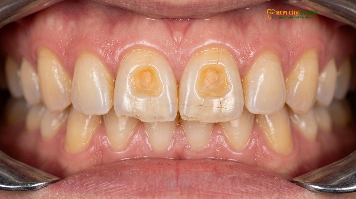
Once the enamel is breached, the underlying dentin is exposed. Dentin is significantly softer than enamel—approximately seven times less resistant to wear. When opposing teeth grind against exposed dentin, the rate of tooth destruction accelerates exponentially. Clinically, this presents as deep, yellow-colored craters on the occlusal (biting) surfaces. Because dentin contains thousands of microscopic tubules that communicate directly with the tooth’s nerve (pulp), patients at this stage often experience severe thermal sensitivity and sharp pain when consuming hot, cold, or sweet foods.
Biomechanics of Abfraction and Micro-Fractures
The damage from severe bruxism is not limited to the biting surfaces. The massive lateral forces generated by grinding cause the teeth to flex microscopically at their necks (the cementoenamel junction, or CEJ), where the enamel is thinnest. This constant flexing causes the enamel rods to break away, creating deep, V-shaped notches at the gumline known as abfraction lesions. These lesions further weaken the structural integrity of the tooth and create highly sensitive areas that are prone to rapid decay.
“The structural compromise seen in severe bruxism is not merely a cosmetic issue; it is a biomechanical crisis. Once the enamel architecture is lost, the tooth enters an accelerated phase of degradation that inevitably leads to catastrophic pulpal exposure or vertical root fracture if interceptive therapy is not initiated.”
— Dr. Nguyen Van Cuong, Lead Prosthodontist
Simultaneously, the immense pressure creates microscopic cracks (craze lines) throughout the remaining enamel. Over time, these micro-fractures propagate deeper into the dentin. When a patient with heavily micro-fractured teeth bites down on a hard piece of food, the weakened tooth structure can suddenly give way, resulting in a catastrophic cusp fracture or a split tooth that may require immediate extraction and replacement with implant-supported restorations.
Loss of Vertical Dimension of Occlusion (VDO)
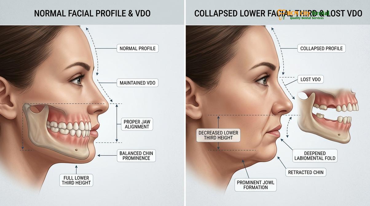
As teeth are progressively ground down, the lower jaw over-closes, resulting in a collapsed bite that alters facial aesthetics and impairs chewing function. Restoring this lost vertical height is one of the most complex challenges in reconstructive dentistry.
The vertical dimension of occlusion (VDO) refers to the specific distance between the upper and lower jaws when the teeth are in maximum contact. This dimension is dictated by the height of the clinical crowns of the teeth. In a healthy mouth, the VDO provides optimal muscle resting length, proper temporomandibular joint positioning, and harmonious facial proportions. However, as severe bruxism damage literally erases the height of the teeth, the VDO inevitably decreases, leading to a condition known as occlusal collapse.
The Pathophysiology of Bite Collapse
When vertical dimension loss occurs, the mandible (lower jaw) must travel further upward to allow the upper and lower teeth to meet. This over-closure drastically alters the biomechanics of the entire stomatognathic system. The muscles of mastication, particularly the masseter and temporalis, are forced to operate at a shortened, inefficient length, which can trigger chronic muscle spasms and tension headaches [2].
Furthermore, the loss of VDO severely impacts the patient’s chewing efficiency. Without the natural cusps and proper vertical height, the teeth can no longer shear and crush food effectively. Patients often find themselves struggling to chew fibrous meats or raw vegetables, leading to dietary compromises and potential gastrointestinal issues due to poorly masticated food.
Aesthetic and Phonetic Consequences
The aesthetic impact of occlusal collapse is profound and often distressing for the patient. Because the lower jaw over-closes, the lower third of the face appears shortened, mimicking the appearance of advanced aging or complete tooth loss (edentulism). This structural collapse causes the soft tissues of the face to lose their underlying support, resulting in:
- Deepening of the nasolabial folds (smile lines).
- Pronounced marionette lines extending from the corners of the mouth to the chin.
- A protruding or “witch-like” appearance of the chin.
- Angular cheilitis, a painful inflammatory condition characterized by cracking and pooling of saliva at the corners of the mouth.
| Stage | Clinical Presentation | Aesthetic Impact | Functional Impact |
|---|---|---|---|
| Mild | Flattened cusps, wear facets confined to enamel. | Minimal; slight shortening of anterior teeth. | Normal chewing function; occasional sensitivity. |
| Moderate | Dentin exposed, loss of 1-2mm of crown height. | Visible wear; teeth appear unusually flat and yellow. | Increased thermal sensitivity; slight muscle fatigue. |
| Severe | Loss of >3mm of crown height, deep dentin craters. | Facial collapse, deep wrinkles, shortened lower face. | Impaired chewing, chronic muscle pain, joint strain. |
| Catastrophic | Teeth worn to the gumline, pulpal exposure. | Severe pseudo-aging, angular cheilitis. | Inability to chew, severe pain, complete bite collapse. |
Phonetically, the loss of tooth height alters the space required for the tongue to articulate properly against the teeth. Patients with severe vertical dimension loss often develop a lisp or struggle to clearly pronounce sibilant sounds (like “s” and “z”) and fricative sounds (like “f” and “v”), which rely heavily on precise tooth positioning.
Periodontal Support Failure: Bone Loss and Loose Teeth
The massive lateral forces generated during severe bruxism overwhelm the periodontal ligament, triggering localized inflammation and accelerating alveolar bone resorption. This traumatic occlusion ultimately causes otherwise healthy teeth to become dangerously mobile.
While the visible destruction of the clinical crowns is the most obvious sign of severe bruxism damage, some of the most insidious harm occurs beneath the gumline. The teeth are anchored into the jawbone (alveolar bone) by a complex hammock of connective tissue fibers known as the periodontal ligament (PDL). The PDL is designed to act as a shock absorber, cushioning the teeth against the vertical forces of normal chewing. However, it is highly vulnerable to the extreme, sustained lateral (side-to-side) forces generated during bruxism.
Primary Occlusal Trauma and Osteoclast Activation
When a tooth is subjected to the relentless lateral grinding forces of severe bruxism, the PDL is violently compressed on one side and stretched on the other. This condition is clinically diagnosed as primary occlusal trauma. The severe compression cuts off the blood supply to localized areas of the PDL, leading to tissue necrosis and acute inflammation [3].
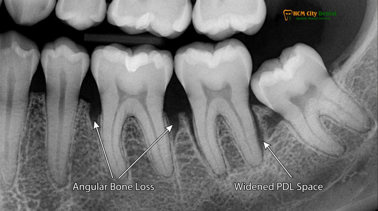
In response to this trauma, the body’s immune system releases inflammatory cytokines that activate osteoclasts—specialized cells responsible for breaking down bone tissue. The osteoclasts begin to resorb the alveolar bone surrounding the traumatized tooth in an attempt to move the bone away from the source of the pressure. On a dental radiograph, this presents as a distinct widening of the periodontal ligament space and angular (vertical) bone defects alongside the tooth roots.
Clinical Warning: Tooth mobility in the absence of heavy plaque or tartar buildup is a major red flag for severe occlusal trauma. If you notice that your teeth feel loose or shift slightly when you bite down (a condition known as fremitus), immediate clinical intervention is required to prevent irreversible bone loss and eventual tooth exfoliation.
The Synergy of Bruxism and Periodontitis
The situation becomes exponentially more dangerous if the patient already suffers from pre-existing gum disease (periodontitis). In a healthy mouth, primary occlusal trauma causes bone loss, but the connective tissue attachment to the tooth remains intact. However, in a mouth with active periodontal disease, the bacterial infection has already compromised the connective tissue attachment. When severe bruxism forces are applied to these already weakened teeth (secondary occlusal trauma), the rate of bone destruction accelerates dramatically. The combination of bacterial inflammation and mechanical trauma creates a highly destructive synergy that can lead to rapid, catastrophic tooth loss.
Clinically, teeth subjected to severe occlusal trauma will exhibit increasing degrees of mobility. Dentists grade this mobility on a scale from Class I (slight movement) to Class III (severe movement in all directions, including vertically). Without intervention, such as occlusal adjustment and the use of custom-fabricated nightguards to distribute the forces, these teeth will eventually require extraction.
Neuromuscular Strain: Permanent Joint Displacement
Relentless clenching overworks the masseter and temporalis muscles, transferring destructive loads directly to the temporomandibular joint. Over time, this chronic strain degrades the articular disc, leading to irreversible joint displacement and chronic facial pain.
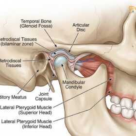
The temporomandibular joint (TMJ) is one of the most complex joints in the human body, functioning as a sliding hinge that connects the mandible to the skull. Between the condyle (the rounded end of the lower jaw) and the glenoid fossa (the socket in the skull) lies a small, fibrous, shock-absorbing cushion called the articular disc. In a healthy system, this disc ensures smooth, frictionless movement of the jaw during speaking and chewing.
Biomechanics of TMJ Degradation
Severe bruxism places an unprecedented and unsustainable load on the TMJ complex. During intense clenching episodes, the powerful masseter and temporalis muscles contract with maximum force, driving the condyle upward and backward into the joint space. This immense pressure compresses the articular disc far beyond its physiological limits. Over months and years of chronic bruxism, the disc begins to thin, fray, and lose its structural integrity [4].
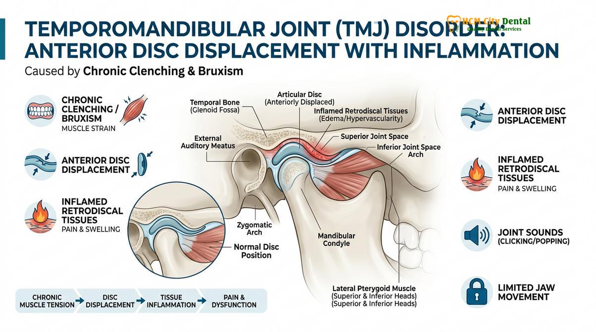
Eventually, the relentless pressure forces the articular disc to slip out of its proper position, usually displacing anteriorly (forward). When the disc is displaced, the condyle no longer rests on the protective cushion but instead impinges directly on the highly innervated and vascularized retrodiscal tissues behind the joint. This impingement causes acute inflammation (retrodiscitis), resulting in severe, radiating pain in front of the ear, which often mimics a severe earache.
“The temporomandibular joint is the silent victim of severe bruxism. While patients often notice the visible wear on their teeth, the internal derangement of the TMJ—characterized by disc displacement and osteoarthritic changes—is frequently the source of their most debilitating chronic pain.”
— International Consensus on Temporomandibular Disorders
Muscle Hypertrophy and Chronic Pain Syndromes
The neuromuscular system also suffers profoundly from severe bruxism. The muscles of mastication are forced into a state of chronic hyperactivity and spasm. Just as lifting heavy weights causes skeletal muscles to grow, the constant clenching causes the masseter muscles to undergo hypertrophy (enlargement). This can visibly alter the patient’s facial appearance, resulting in a widened, highly pronounced, square-shaped jawline.
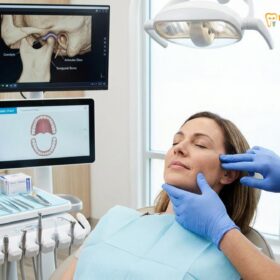
Furthermore, the chronic muscle tension leads to the development of painful trigger points within the muscle fibers. This myofascial pain often radiates throughout the head and neck, manifesting as severe tension headaches, neck stiffness, and even pain behind the eyes. Managing this complex web of neuromuscular and joint pathology requires specialized comprehensive temporomandibular joint therapy to deprogram the muscles and stabilize the joint position.
Comprehensive Treatment Workflows for Advanced Cases
Rehabilitating a bruxism-destroyed dentition requires a phased approach, beginning with acute symptom management and culminating in full-mouth structural restoration. Custom occlusal splints serve as the foundational therapy to deprogram muscles and protect future restorations.
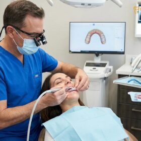
Addressing severe bruxism damage is rarely a simple fix; it requires a meticulously planned, multi-disciplinary approach to restore both form and function. At HCMC Dental Clinic in Ho Chi Minh City, the clinical workflow for full-mouth rehabilitation is divided into distinct, highly controlled phases to ensure predictable, long-lasting outcomes.
Phase 1: Neuromuscular Deprogramming and Stabilization
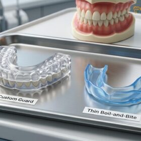
The absolute first step in treating severe bruxism is to halt the ongoing destruction and relieve the acute neuromuscular pain. This is achieved through the fabrication of a hard acrylic stabilization splint (often referred to as a Michigan splint or a flat-plane occlusal guard). Unlike soft, over-the-counter mouthguards—which can actually encourage more chewing and clenching—a rigid, custom-calibrated splint provides a perfectly even biting surface [5].
When the patient wears the splint, the teeth glide smoothly against the acrylic, preventing the interlocking of cusps that triggers muscle spasms. This allows the hyperactive masseter and temporalis muscles to relax and “deprogram,” while simultaneously decompressing the temporomandibular joint. The splint also serves as a diagnostic tool; the dentist can observe the wear patterns on the acrylic to determine the exact vectors of the patient’s bruxism forces.
Phase 2: Provisionalization and VDO Testing
Once the muscles are relaxed and the joint is stable, the next challenge is determining the correct vertical dimension of occlusion to restore the lost tooth height. Dr. Nguyen Van Cuong emphasizes that this new bite position must be tested before any permanent restorations are placed. Using advanced digital smile design and articulated models, the dental team creates a wax-up of the proposed new teeth.
Clinical Case Review: A 45-year-old patient presented to HCMC Dental Clinic in Ho Chi Minh City with severe enamel attrition, chronic TMJ pain, and a collapsed bite resulting in a shortened lower facial profile. Under the care of Dr. Cuong, the patient underwent a 3-month stabilization phase using a custom hard acrylic splint to deprogram the jaw muscles. Following stabilization, the lost vertical dimension was restored using provisional composite overlays. After confirming neuromuscular comfort, the dentition was permanently rehabilitated with high-strength full-coverage dental crowns, completely eliminating the joint pain and restoring a youthful facial aesthetic.
This wax-up is then transferred to the patient’s mouth using temporary composite resin materials (provisionalization). The patient lives with these temporary restorations for several weeks to months. This critical testing phase ensures that the new vertical height is comfortable for the TMJ, allows for proper speech, and meets the patient’s aesthetic expectations.
Phase 3: Definitive Structural Restoration
Once the new VDO is verified as comfortable and functional, the temporary restorations are systematically replaced with definitive, high-strength ceramics. Depending on the extent of the damage to individual teeth, the restorative options may include:
- Porcelain Overlays/Onlays: Used when the core structure of the tooth is intact, but the biting surface needs to be rebuilt to the new vertical height.
- Full-Coverage Crowns: Required for teeth that have suffered catastrophic structural failure, deep micro-fractures, or extensive abfraction lesions.
- Endodontic Therapy: If the bruxism damage has exposed the dental pulp or caused irreversible pulpitis, endodontic therapy is necessary before the tooth can be crowned.
Following the completion of the full-mouth rehabilitation, the patient is fitted with a new, final occlusal guard. Because bruxism is a centrally mediated neurological habit, the grinding will likely continue. The final nightguard ensures that these immense forces are directed against the replaceable acrylic appliance rather than the newly placed ceramic restorations.
When to See a Doctor
Severe bruxism is a progressive condition that becomes exponentially more difficult and expensive to treat as the damage accumulates. You should schedule a specialized clinical evaluation immediately if you experience any of the following red flag symptoms:
- Visible Yellow Craters: The appearance of deep, yellow indentations on the biting surfaces of your teeth indicates that the enamel has been completely destroyed and the dentin is exposed.
- Severe Thermal Sensitivity: A sudden, sharp increase in sensitivity to hot or cold foods suggests that micro-fractures have reached the inner nerve of the tooth.
- Jaw Locking or Popping: If your jaw frequently clicks, pops loudly, or temporarily locks in an open or closed position, the TMJ articular disc is likely displaced.
- Unexplained Tooth Mobility: Teeth that feel loose or shift when you bite down are suffering from severe occlusal trauma and require immediate stabilization.
- Chronic Morning Headaches: Waking up consistently with dull, aching pain in your temples, jaw, or neck is a primary indicator of intense nocturnal muscle clenching.
Early intervention is the key to preserving your natural dentition. If you suspect you are suffering from the effects of chronic grinding, scheduling a specialized consultation with a prosthodontist or TMJ specialist is critical to developing a protective and restorative treatment plan.
Frequently Asked Questions
Can bruxism completely wear down my teeth?
Yes, chronic bruxism can completely wear down the clinical crowns of your teeth to the gum line if left untreated. Once the hard outer enamel is breached, the softer underlying dentin erodes at a significantly accelerated rate. This rapid degradation not only destroys the visible tooth structure but also exposes the sensitive inner pulp, often necessitating complex root canal therapies and full-coverage crowns to salvage the remaining roots.
How does teeth grinding cause bone loss?
Teeth grinding causes bone loss by subjecting the periodontal ligament to excessive lateral forces that it was not designed to withstand. This traumatic occlusion triggers localized inflammation, activating osteoclasts (bone-resorbing cells) that break down the alveolar bone supporting the tooth roots. Over time, this mechanical trauma leads to widened periodontal spaces, angular bone defects, and dangerous tooth mobility, especially if compounded by pre-existing gum disease.
Is worn-down teeth repairable?
Yes, severely worn-down teeth are repairable through comprehensive full-mouth rehabilitation. Depending on the extent of the damage, treatments may include custom occlusal splints, composite build-ups, porcelain crowns, or overlays to restore both the structural integrity and the proper vertical dimension of the bite. The process requires careful neuromuscular planning to ensure the new restorations are in harmony with the jaw joints and muscles.
What is the best way to stop severe bruxism damage at night?
The most effective way to halt nocturnal bruxism damage is the consistent use of a custom-fabricated hard acrylic nightguard. This appliance provides a protective barrier between the upper and lower teeth, evenly distributes occlusal forces, and helps deprogram hyperactive jaw muscles during sleep. Unlike soft over-the-counter guards, a hard, professionally adjusted splint prevents the teeth from locking together, significantly reducing muscle strain and joint compression.
Does severe teeth grinding change your face shape?
Yes, prolonged and severe teeth grinding can significantly alter your facial shape. The constant overworking of the masseter muscles can cause them to hypertrophy (enlarge), resulting in a widened, square-looking jawline. Additionally, as the teeth wear down and lose their vertical height, the lower third of the face can collapse, leading to a shortened facial profile, deepened wrinkles, and a prematurely aged appearance.
References
- Journal of the American Dental Association. Biomechanics of severe enamel attrition and occlusal wear. (2021).
- Journal of Prosthetic Dentistry. Vertical dimension of occlusion: clinical management and restorative protocols. (2020).
- Journal of Periodontology. The relationship between occlusal trauma and alveolar bone loss. (2019).
- Journal of Oral Rehabilitation. Temporomandibular joint disc displacement and neuromuscular strain in bruxism. (2022).
- Clinical Oral Investigations. Efficacy of hard acrylic stabilization splints in managing severe bruxism. (2018).