Platelet-Rich Fibrin (PRF) dental healing is an advanced biological technique that utilizes a concentration of your own blood cells to support tissue regeneration[1]. By applying natural growth factors directly to surgical sites, PRF can significantly reduce discomfort, help protect against infection, and encourage a smoother recovery after extractions or implant placements. In the modern landscape of regenerative dentistry, the shift toward autologous (self-derived) materials represents a profound advancement in how clinicians manage postoperative healing, moving away from synthetic alternatives toward solutions that work synergistically with the human body’s innate repair mechanisms.
Clinical Summary:
PRF therapy represents a paradigm shift in modern oral surgery, utilizing autologous blood concentrates to form a robust fibrin matrix rich in platelets, leukocytes, and mesenchymal stem cells. This bioactive scaffold releases essential growth factors—such as PDGF, TGF-β, and VEGF—continuously over a period of up to 14 days. Clinically, PRF is highly effective for socket preservation, supporting implant osseointegration, and aiding in the healing of complex bony defects. Because the material is entirely autologous, it eliminates the risk of foreign body rejection or allergic reactions, making it a foundational protocol in biological dentistry for achieving optimal, chemical-free healing.
Key Takeaways:
- PRF is derived entirely from the patient’s own blood without the use of chemical additives or artificial anticoagulants.
- The three-dimensional fibrin matrix releases regenerative growth factors continuously for up to two weeks.
- Application of PRF generally lowers the risk of painful dry socket following complex tooth extractions.
- Leukocytes trapped within the PRF matrix provide natural, localized protection against oral pathogens.
- The therapy supports both soft tissue closure and the stabilization of bone grafts in implantology.
What is Platelet-Rich Fibrin (PRF) in Dental Surgery?
Platelet-Rich Fibrin (PRF) is a second-generation autologous blood concentrate that creates a bioactive scaffold to stimulate bone and soft tissue regeneration in oral surgery.
In the evolving landscape of regenerative medicine and biological dentistry, the quest for materials that can naturally and safely support human tissue repair has led to the development of autologous blood concentrates. Platelet-Rich Fibrin, commonly referred to as PRF, stands at the forefront of this clinical evolution. Unlike synthetic grafting materials, allografts (human donor bone), or xenografts (animal-derived collagens), PRF is created entirely from the patient’s own peripheral blood. This fundamental characteristic means it is a highly biocompatible substance, carrying no risk of immune rejection, disease transmission, or allergic response. When utilized in dental surgery, PRF acts as a biological catalyst, fundamentally altering the trajectory of postoperative healing by providing the surgical site with a concentrated dose of the body’s own regenerative cells[2].
To fully understand the clinical advantages of PRF, it is helpful to look at its predecessor: Platelet-Rich Plasma (PRP). First-generation platelet concentrates like PRP required the addition of chemical anticoagulants (such as bovine thrombin or calcium chloride) to prevent the blood from clotting during the centrifugation process. While PRP was effective in delivering a quick burst of growth factors to a surgical site, the chemical additives altered the natural coagulation cascade. This resulted in a weak, unstable fibrin network that dissolved rapidly, usually within a matter of hours, limiting its long-term regenerative potential. Furthermore, the use of bovine thrombin introduced a small but present risk of coagulopathies and immune reactions.
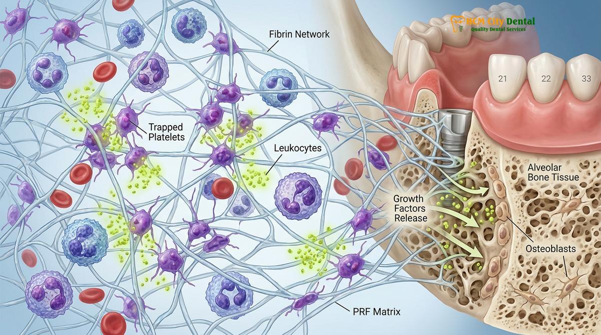
PRF completely eliminates the need for chemical additives, marking it as a true second-generation concentrate. By allowing the blood to coagulate naturally during a specific, carefully timed centrifugation protocol, PRF forms a dense, complex, tri-dimensional fibrin matrix. This robust architectural scaffold physically traps platelets, leukocytes (white blood cells), and circulating stem cells. Because the fibrin network is highly polymerized and structurally sound, it resists rapid degradation. Instead, it allows the trapped cells to slowly and continuously secrete growth factors over an extended period, providing sustained regenerative support exactly when the healing tissues need it most.
The Centrifugation Process: Preparing PRF from Your Blood
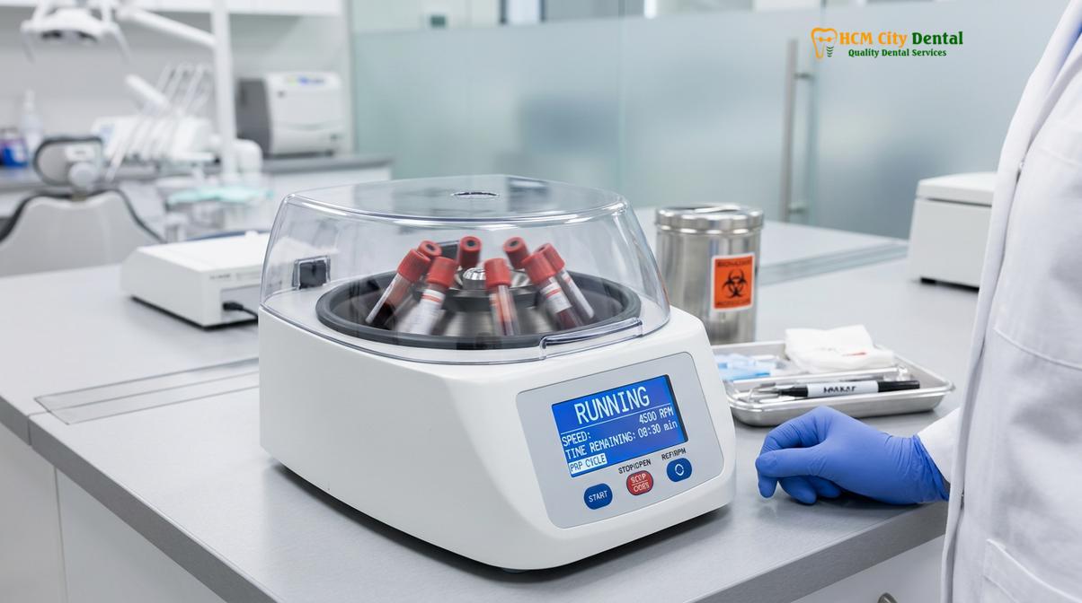
The preparation of PRF involves drawing a small blood sample and spinning it in a specialized centrifuge to separate the regenerative cells from red blood cells without using chemical additives.
The clinical workflow for preparing Platelet-Rich Fibrin is a seamless, highly controlled process that takes place entirely within the dental clinic, immediately prior to or during the surgical procedure. Because the efficacy of PRF relies entirely on the natural coagulation cascade, timing, temperature, and precision are of the utmost importance. The process begins with a standard venipuncture, typically performed on the patient’s arm, similar to a routine medical blood draw. A small volume of peripheral blood—usually between two to four vials, depending on the extent of the surgery—is collected into sterile glass or silica-coated plastic tubes. It is critical that these specific tubes contain absolutely no anticoagulants. The absence of anticoagulants means that the blood will begin to clot the moment it leaves the body and contacts the tube walls, necessitating immediate transfer to the centrifuge.
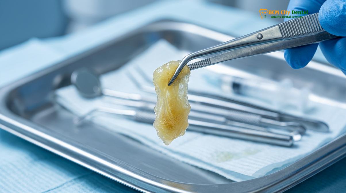
Once the vials are placed into the centrifuge, the machine is set to a specific protocol of revolutions per minute (RPM) and time. The physics of centrifugation apply a relative centrifugal force (RCF) that separates the whole blood components based on their specific gravity and molecular weight. During this spin cycle, the heavier red blood cells (erythrocytes) are forced to the bottom of the vial. The lighter, acellular blood plasma (platelet-poor plasma) rises to the top. Suspended perfectly in the middle is the “buffy coat”—a dense, yellowish, gelatinous clot. This middle layer is the Platelet-Rich Fibrin matrix.
After the centrifugation cycle is complete, the clinician carefully removes the vials. Using sterile surgical tweezers, the PRF clot is extracted from the tube. The red blood cell layer is gently separated and discarded, leaving only the pure, bioactive fibrin matrix. Depending on the surgical requirement, this clot can be used in its natural three-dimensional plug form to fill an extraction socket, or it can be placed into a specialized PRF compression box. The compression box gently flattens the clot into a strong, suturable membrane that can be used to cover bone grafts, protect sinus perforations, or guide soft tissue healing. The fluid expressed during this compression process, known as PRF exudate, is also highly rich in proteins and can be used to hydrate bone grafting materials, further enhancing their regenerative potential.
How PRF Growth Factors Speed Up Bone and Gum Regeneration
The dense fibrin network of PRF acts as a biological reservoir, slowly releasing a concentrated payload of essential growth factors that orchestrate the complex phases of tissue repair.
The remarkable healing capabilities of Platelet-Rich Fibrin are primarily driven by the high concentration of growth factors trapped within its matrix. When tissue is injured during dental surgery, the body initiates a complex cascade of biological events to repair the damage. This process involves hemostasis, inflammation, cellular proliferation, and tissue remodeling. PRF significantly accelerates and optimizes these phases by delivering a concentrated dose of the exact signaling molecules the body uses to direct cellular repair. Because the fibrin network in PRF is naturally polymerized, it degrades slowly, allowing for the sustained release of these growth factors over a period of 7 to 14 days, which perfectly aligns with the critical early stages of wound healing[3].
Several key growth factors are responsible for the regenerative power of PRF. Platelet-Derived Growth Factor (PDGF) is one of the most prominent. PDGF is a powerful mitogen, meaning it stimulates the division and proliferation of mesenchymal stem cells and osteoblasts (bone-forming cells). It also plays a crucial role in angiogenesis, the formation of new blood vessels, which is essential for delivering oxygen and nutrients to the healing site. Transforming Growth Factor Beta (TGF-β) is another critical component. TGF-β regulates the inflammatory response, helping to prevent excessive swelling, and stimulates the synthesis of extracellular matrix proteins like collagen, which form the structural foundation of new gum tissue and bone.
“The sustained release of autologous growth factors from a natural fibrin scaffold represents the pinnacle of biomimetic healing, allowing clinicians to guide tissue regeneration with unprecedented predictability.”
Additionally, PRF contains high levels of Vascular Endothelial Growth Factor (VEGF), which is the primary signaling molecule responsible for initiating angiogenesis. By promoting the rapid development of a robust microvascular network, VEGF ensures that the surgical site receives the necessary biological support to sustain newly forming tissues. Insulin-like Growth Factor (IGF) is also present, further supporting cellular proliferation and differentiation. Beyond growth factors, the PRF matrix is rich in leukocytes (white blood cells). These immune cells play a dual role: they help orchestrate the transition from the inflammatory phase to the proliferative phase of healing, and they provide localized, natural antimicrobial protection, significantly reducing the risk of postoperative infections in the oral cavity.
PRF Applications: Wisdom Teeth, Implants, and Cavitations
PRF is a versatile biomaterial utilized across various oral surgeries, from preserving extraction sockets to enhancing the stability of dental implants and treating jawbone cavitations.
The biological versatility of Platelet-Rich Fibrin allows it to be seamlessly integrated into almost any surgical dental procedure where enhanced healing, reduced inflammation, and accelerated tissue regeneration are desired. One of the most common and impactful applications is during the extraction of teeth, particularly impacted mandibular wisdom teeth. By placing a PRF clot directly into the empty socket immediately after extraction, the surgeon provides an instant biological seal. This seal protects the exposed alveolar bone, stabilizes the underlying blood clot, and significantly reduces postoperative pain and swelling. Furthermore, it aids in socket preservation, minimizing the natural bone resorption that occurs after a tooth is lost, thereby maintaining the structural integrity of the jaw ridge.
In the realm of implantology, PRF has become an indispensable tool for many biological dentists. When placing dental implants, achieving rapid and robust osseointegration—the vital structural and functional fusion between the titanium or zirconia implant and the patient’s living jawbone—is the ultimate goal. PRF is frequently mixed with particulate bone grafting materials (whether autogenous, allogenic, or xenogenic) to create a composite known as “sticky bone.” This preparation not only makes the particulate graft much easier for the surgeon to handle and shape but also infuses the graft with a high concentration of growth factors, accelerating the maturation of the new bone. Additionally, PRF membranes are often draped over the implant site before suturing to enhance soft tissue thickness and improve the final aesthetic outcome of the gums.
Furthermore, PRF is increasingly utilized in the specialized treatment of jawbone cavitations, clinically referred to as Neuralgia-Inducing Cavitational Osteonecrosis (NICO) or Fatty Degenerative Osteonecrosis of the Jawbone (FDOJ). These are areas of necrotic, ischemic (oxygen-starved) bone that often suffer from chronically poor blood supply, making natural healing exceedingly difficult even after thorough surgical debridement. The application of PRF into these cleaned cavitational defects introduces a concentrated dose of VEGF, which powerfully stimulates angiogenesis. This revitalizes the previously ischemic tissue, promoting the regeneration of healthy, dense, well-vascularized bone and aiding in the resolution of chronic localized inflammation.
| Dental Procedure | Traditional Healing Challenges | PRF Healing Benefits |
|---|---|---|
| Wisdom Tooth Extraction | High risk of dry socket, prolonged swelling, and delayed soft tissue closure. | Instantly seals the socket, protects exposed bone, and minimizes postoperative pain. |
| Dental Implants | Slower osseointegration, potential graft failure, and longer waiting periods. | Accelerates bone fusion, stabilizes grafting materials, and enhances soft tissue aesthetics. |
| Cavitation Surgery | Poor blood supply in necrotic bone areas leading to incomplete healing. | Stimulates angiogenesis and new blood vessel formation to revitalize ischemic tissue. |
| Sinus Lift Procedures | Risk of Schneiderian membrane perforation and delayed graft consolidation. | Repairs minor membrane tears naturally and accelerates the maturation of the sinus bone graft. |
Safe, Chemical-Free Healing Protocols at HCMC Dental
Our clinical protocols emphasize the use of 100% autologous materials, ensuring that your surgical recovery is supported by your body’s own natural regenerative capabilities.
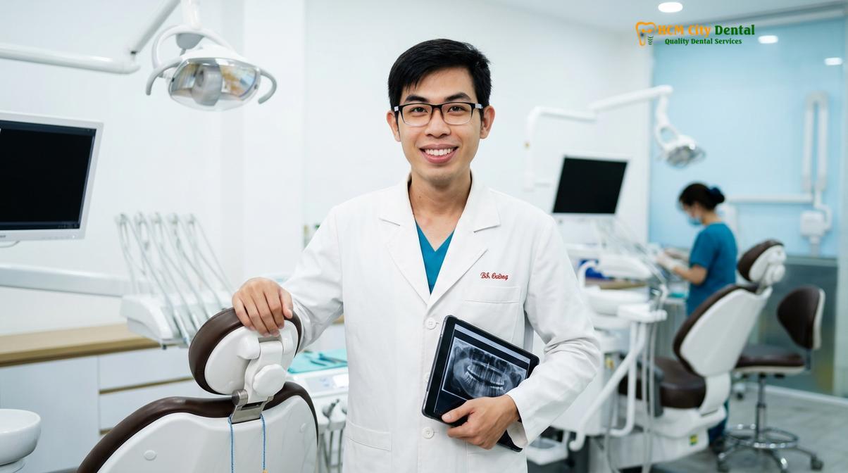
At HCMC Dental Clinic in Ho Chi Minh City, our biological approach prioritizes patient safety and optimal clinical outcomes through advanced, autologous treatments. Dr. Nguyen Van Cuong, a leading specialist at the clinic, meticulously oversees the PRF preparation process to ensure optimal cellular viability and strict adherence to sterile protocols. By utilizing the patient’s own blood without any synthetic additives, Dr. Cuong and his dedicated surgical team provide a highly biocompatible healing matrix that perfectly aligns with the body’s natural regenerative pathways. This unwavering dedication to chemical-free dentistry minimizes the risk of adverse immune reactions, prevents foreign body rejection, and promotes a healthier, more predictable recovery for every patient.
The clinical workflow at HCMC Dental Clinic is designed to be as minimally invasive and efficient as possible. The blood draw is performed swiftly and comfortably by trained professionals just moments before the surgical procedure begins. Because the PRF is generated on-site within minutes using state-of-the-art centrifugation equipment, the bioactive matrix is applied to the surgical site while the growth factors and living cells are at their absolute peak concentration. This immediate application is a cornerstone of our safe healing protocols, ensuring that the delicate cellular architecture is preserved and utilized to its maximum potential before any degradation can occur.
Furthermore, the integration of PRF into our standard surgical protocols reflects a broader commitment to holistic and biological dental principles. We recognize that the mouth is not an isolated system, but a vital gateway to the rest of the body. By choosing materials that actively support the immune system and facilitate rapid, natural healing, we reduce the systemic burden of inflammation and minimize the need for extended courses of postoperative pharmaceuticals, such as heavy analgesics or prophylactic antibiotics, whenever clinically appropriate.
Preventing Dry Socket and Post-Extraction Pain Naturally
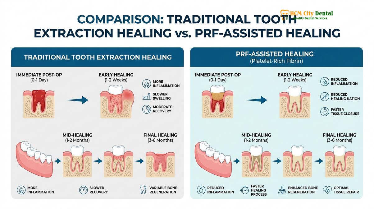
Placing a PRF clot directly into an extraction socket protects exposed bone, stabilizes the blood clot, and drastically reduces the incidence of painful dry socket and postoperative inflammation.
One of the most common and distressing complications following routine dental extractions, particularly the surgical removal of impacted mandibular wisdom teeth, is alveolar osteitis, universally known among patients as “dry socket.” This highly painful condition occurs when the physiological blood clot that normally forms in the extraction socket fails to develop properly, is dislodged prematurely by mechanical forces (such as aggressive rinsing or using a straw), or dissolves prematurely due to localized fibrinolysis. When this protective clot is lost, the underlying alveolar bone and delicate nerve endings are left completely exposed to the harsh oral environment, including food debris, bacteria, and digestive enzymes. This results in severe, radiating pain that typically begins three to five days after surgery and can last for weeks. Traditional management of dry socket is largely reactive, involving the repeated placement of medicated, palliative dressings. PRF, however, offers a highly effective, proactive method for helping to prevent this complication from occurring in the first place[4].
When a tooth is extracted, the resulting bony defect is essentially a hollow cavern that the body must fill with new tissue. By utilizing PRF, the surgeon effectively bypasses the vulnerable initial phase of natural clot formation. Immediately after the tooth is removed and the socket is thoroughly debrided, a pre-formed, dense PRF clot is physically packed into the bony defect. Because the PRF clot is highly polymerized and structurally resilient, it acts as a robust biological plug. It instantly seals the socket, providing a physical barrier that protects the exposed bone and nerve endings from the oral environment.
Moreover, the PRF matrix is highly resistant to the enzymatic breakdown that typically causes a natural blood clot to fail. The dense fibrin network holds its shape, providing a stable scaffold for the migration of fibroblasts and osteoblasts, which are essential for soft tissue closure and bone regeneration. The continuous release of growth factors also accelerates the epithelialization process, meaning the gums close over the extraction site much faster than they would naturally. This immediate physical protection, combined with accelerated tissue regeneration, is the primary reason why patients treated with PRF consistently report significantly lower pain scores, less swelling, and a drastically reduced incidence of alveolar osteitis in the days following complex oral surgery.
Clinical Case Study: PRF Healing at HCMC Dental Clinic
Real-world clinical applications demonstrate how PRF therapy provides a superior healing experience, minimizing downtime and enhancing the structural integrity of the jawbone.
Patient Profile: A 45-year-old patient visiting HCMC Dental Clinic in Ho Chi Minh City presented with a severely fractured mandibular first molar that required a complex surgical extraction and subsequent dental implant placement. The patient had a documented history of delayed healing from previous dental procedures and expressed high anxiety regarding postoperative pain and the potential for complications.
Clinical Intervention: After a comprehensive evaluation, Dr. Nguyen Van Cuong recommended a biological surgical approach utilizing PRF dental healing to mitigate the patient’s risks. Following the general regenerative guidelines supported by the Vietnam Odonto-Stomatology Association for advanced surgical recovery, a PRF matrix was prepared from the patient’s own peripheral blood. Immediately following the atraumatic extraction of the fractured molar, the PRF clot was applied directly into the socket to provide an instant biological seal and initiate the regenerative cascade.
Outcome: The application of PRF provided a robust biological scaffold that stabilized the surgical site. The patient experienced minimal facial swelling and reported almost no pain during the critical first week of recovery, demonstrating the profound impact of autologous growth factors on optimal tissue regeneration[5]. Follow-up radiographic imaging at the three-month mark revealed excellent bone density and complete socket fill, creating an ideal, structurally sound foundation for the successful placement of the future dental implant.
“Biological dentistry prioritizes materials that work synergistically with the human body. PRF exemplifies this philosophy by utilizing the patient’s own cellular resources to guide and accelerate the healing cascade, transforming a traumatic surgical event into a controlled regenerative process.”
When to Consult a Doctor and Important Clinical Notes
While PRF therapy is highly safe and effective, proper postoperative care and professional monitoring remain essential for a successful and complication-free recovery.
Although Platelet-Rich Fibrin significantly enhances the healing process, accelerates tissue regeneration, and reduces the likelihood of common complications, patients must still remain vigilant and proactive during their recovery period. The success of any surgical procedure relies heavily on patient compliance with postoperative instructions. It is imperative to follow all guidelines provided by your dental team. This typically includes maintaining a soft food diet for the first few days, strictly avoiding smoking or the use of tobacco products (which severely impair blood flow and healing), and refraining from vigorous rinsing, spitting, or using straws, as these actions create negative pressure that could dislodge the healing PRF matrix.
Patients should expect some degree of mild swelling and minor oozing during the first 24 to 48 hours following surgery; this is a normal part of the physiological inflammatory response. However, it is crucial to distinguish between normal healing symptoms and signs of potential complications.
Important Clinical Note: You should consult your dental professional immediately if you experience severe, throbbing pain that worsens after three to four days, excessive bleeding that does not subside with gentle biting pressure, purulent discharge (pus) from the surgical site, or persistent swelling accompanied by a fever or difficulty swallowing. While exceedingly rare when PRF protocols are properly utilized, these symptoms may be indicative of an underlying issue, a dislodged clot, or a secondary infection that requires prompt clinical evaluation and intervention.
References
- Journal of Oral and Maxillofacial Surgery. Platelet-Rich Fibrin: A Second-Generation Platelet Concentrate.
- International Journal of Periodontics & Restorative Dentistry. The use of L-PRF in socket preservation and ridge augmentation.
- Clinical Implant Dentistry and Related Research. Growth factor release kinetics of autologous platelet concentrates.
- Journal of the American Dental Association. Prevention of alveolar osteitis using platelet-rich fibrin.
- Biomaterials Research. Autologous blood concentrates in biological dentistry and maxillofacial surgery.
For more information on how biological approaches and advanced tissue engineering can improve your surgical outcomes, explore our comprehensive General Dentistry services. If you are preparing for a complex extraction, bone grafting procedure, or implant placement, schedule a consultation with our expert team at HCMC Dental Clinic in Ho Chi Minh City to discuss your personalized, chemical-free treatment plan.
For pricing, booking, and a free clinical assessment, visit our Biological Dentistry service page at HCMC Dental Clinic in Ho Chi Minh City.