Platelet-Rich Fibrin (PRF) significantly accelerates wisdom teeth recovery by utilizing a concentrated matrix of your own white blood cells, platelets, and growth factors. Placed directly into the extraction socket, this autologous graft minimizes pain, promotes rapid tissue regeneration, and drastically reduces the risk of developing a painful dry socket.
Clinical Summary:
The integration of Platelet-Rich Fibrin (PRF) into dentoalveolar surgery represents a paradigm shift in postoperative recovery, particularly following the surgical removal of impacted third molars. By harvesting a small volume of the patient’s own blood and centrifuging it without anticoagulants, clinicians create a dense, three-dimensional fibrin scaffold. This biomaterial is rich in leukocytes, platelets, and essential cytokines (such as VEGF and PDGF) that are released gradually over 7 to 14 days. Clinically, the application of PRF within the extraction socket serves as a biological membrane that protects exposed bone, accelerates epithelialization, and significantly lowers the incidence of alveolar osteitis (dry socket). Because it is an entirely autologous product, PRF exhibits unparalleled biocompatibility, making it an optimal adjunctive therapy for enhancing healing trajectories and minimizing postoperative morbidity.
Key Takeaways:
- PRF is created entirely from your own blood, ensuring complete biocompatibility without synthetic additives.
- The dense fibrin matrix acts as a biological bandage, protecting the socket and preventing dry socket.
- Growth factors are released slowly over several weeks, sustaining the cellular repair process.
- The procedure is seamlessly integrated into the extraction appointment, requiring only a simple blood draw.
- Clinical evidence demonstrates significantly reduced postoperative pain, swelling, and reliance on analgesics.
What is PRF (Platelet-Rich Fibrin) in Dentistry?
PRF is an advanced autologous biomaterial derived from a patient’s own blood, creating a dense fibrin scaffold rich in growth factors to stimulate rapid cellular repair in dental surgery.
In the realm of modern oral and maxillofacial surgery, optimizing the body’s natural healing capacity is a primary objective. Platelet-Rich Fibrin (PRF) is a revolutionary, second-generation platelet concentrate that has transformed how clinicians manage postoperative recovery. Originally developed in France by Dr. Joseph Choukroun in the early 2000s, PRF was designed to overcome the limitations of earlier blood concentrates, such as Platelet-Rich Plasma (PRP). Unlike PRP, which requires the addition of chemical anticoagulants (like bovine thrombin or calcium chloride) to prevent the blood from clotting during processing, PRF is completely natural. It relies entirely on the body’s intrinsic coagulation cascade to form a robust, flexible biomaterial[1].
When discussing platelet rich fibrin wisdom teeth procedures, it is essential to understand that PRF is not a medication or a synthetic drug; it is an autologous graft. “Autologous” means that the material is harvested from the same individual who will receive it. This eliminates any risk of allergic reactions, immune rejection, or disease transmission. The resulting PRF clot is a complex, three-dimensional architectural matrix composed of fibrin, fibronectin, and vitronectin. Entrapped within this meshwork are high concentrations of platelets (the cells responsible for clotting and healing) and leukocytes (white blood cells that fight infection and regulate inflammation)[2].
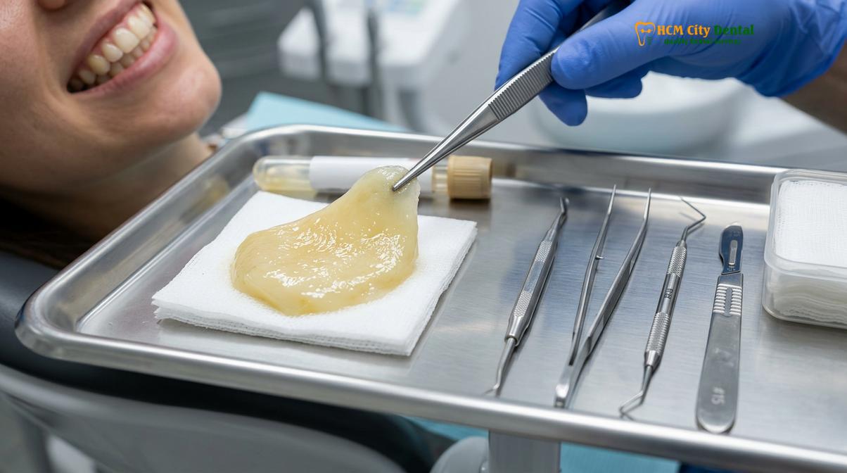
In clinical practice, this fibrin clot can be utilized in two primary forms. It can be left in its natural, gelatinous “plug” state to fill the void left by an extracted tooth, or it can be gently compressed in a specialized sterile container to form a thin, resilient membrane. This membrane can be sutured over bone grafts or used to close soft tissue defects. The versatility of PRF makes it an invaluable tool in a wide array of dental procedures, including implantology, periodontal regeneration, and, most notably, the surgical extraction of impacted third molars.
The transition from traditional extraction methods—where the socket is simply left to fill with a natural, unassisted blood clot—to the proactive placement of a PRF matrix represents a significant advancement in patient care. A natural blood clot is fragile; it can be easily dislodged by rinsing, negative pressure from a straw, or mechanical trauma from food. In contrast, a PRF plug is dense, highly organized, and mechanically stable. It adheres firmly to the bony walls of the extraction socket, providing immediate hemostasis (bleeding control) and establishing an optimal microenvironment for subsequent tissue regeneration.
The Science: How PRF Uses Your Own Blood to Heal
By spinning blood without anticoagulants, PRF traps platelets and leukocytes within a three-dimensional fibrin matrix, ensuring a slow, sustained release of healing cytokines over several weeks.
To truly appreciate the clinical value of PRF, one must delve into the cellular and molecular biology of wound healing. The human body possesses an extraordinary capacity to repair itself, orchestrated by a complex symphony of cellular signals. When tissue is injured—such as during a surgical Tooth Extraction—the body immediately initiates a four-phase healing process: hemostasis, inflammation, proliferation, and remodeling. PRF actively enhances and accelerates each of these phases by delivering a concentrated payload of the very molecules required for repair.
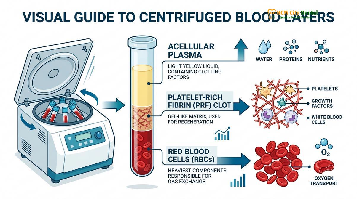
The magic of PRF lies in its unique centrifugation protocol. Because no anticoagulants are used, the blood begins to coagulate naturally the moment it hits the glass or silica-coated walls of the collection tube. The centrifuge spins the blood at a specific, carefully calibrated speed and duration (typically around 2700 to 3000 RPM for 10 to 12 minutes). This centrifugal force separates the blood based on the density of its components. The heavy red blood cells (erythrocytes) are driven to the bottom of the tube. The lighter, acellular plasma rises to the top. In the middle, suspended between these two layers, a dense fibrin clot forms. This middle layer is the PRF[3].
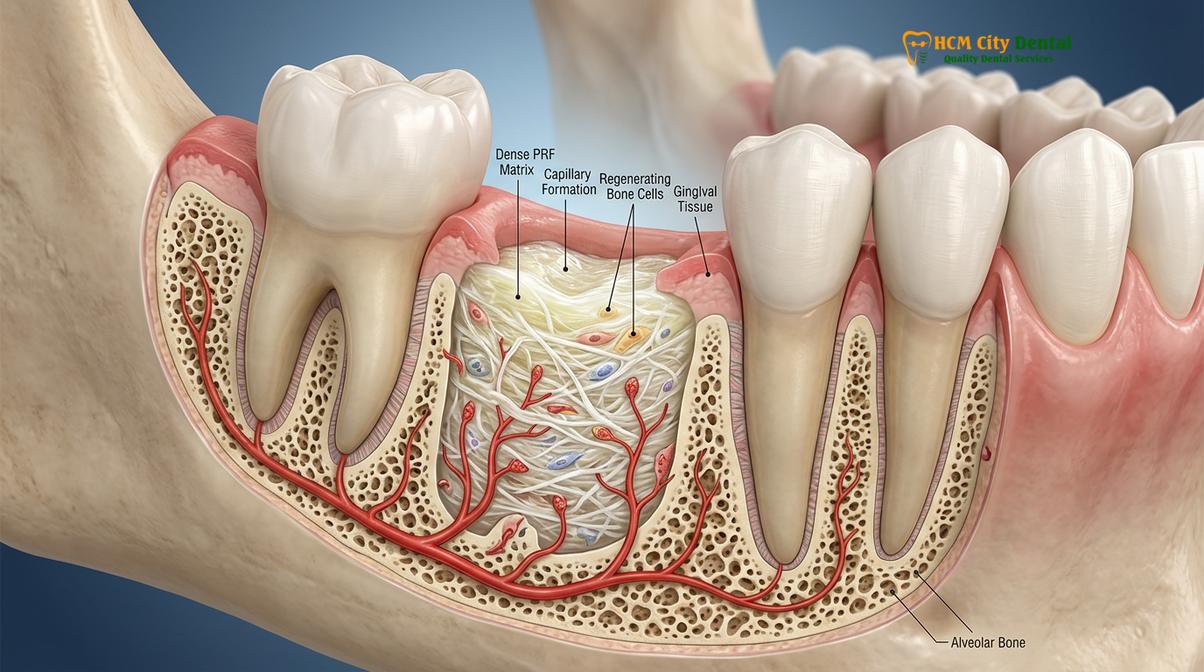
During this slow polymerization process, the fibrin network forms a fine, flexible, and highly porous structure. This specific architecture is crucial because it acts as a biological trap. It captures nearly 100% of the platelets and over 50% of the leukocytes present in the original blood sample. More importantly, it traps the cytokines and growth factors secreted by these cells. The key growth factors involved include:
- Platelet-Derived Growth Factor (PDGF): Stimulates the replication of connective tissue cells and promotes angiogenesis (the formation of new blood vessels).
- Transforming Growth Factor-Beta (TGF-β): Regulates inflammation and stimulates the production of collagen, the primary structural protein in gums and bone.
- Vascular Endothelial Growth Factor (VEGF): The master regulator of blood vessel formation, ensuring the healing tissue receives an adequate supply of oxygen and nutrients.
- Insulin-like Growth Factor (IGF): Promotes cell survival and supports the differentiation of osteoblasts (bone-forming cells).
“The true clinical advantage of PRF is not just the concentration of growth factors, but the kinetics of their release. Unlike liquid PRP, which releases its cytokines in a rapid burst over a few hours, the dense fibrin matrix of PRF degrades slowly, providing a sustained, steady release of healing factors directly into the surgical site for up to 14 days.”
— Clinical Perspective on Biomaterials in Oral Surgery
This sustained release is the cornerstone of prf dental socket healing. In a standard extraction without PRF, the natural blood clot may shrink or break down prematurely, leaving the socket vulnerable. The PRF matrix, however, maintains its structural integrity, acting as a scaffold for migrating fibroblasts and endothelial cells. Furthermore, the high concentration of leukocytes within the PRF provides a localized immune defense. These white blood cells actively phagocytize (engulf and destroy) bacteria and necrotic debris, significantly reducing the risk of postoperative infection in the highly contaminated environment of the oral cavity.
Step-by-Step PRF Extraction Socket Grafting Process
The PRF workflow involves drawing a small blood sample during sedation, centrifuging it to isolate the fibrin clot, and immediately placing this biological membrane into the fresh extraction site.
The integration of PRF therapy into a wisdom tooth extraction procedure is highly streamlined and adds very little time to the overall surgical appointment. Because the entire process is performed chairside, the biomaterial is fresh, highly active, and perfectly matched to the patient’s immediate surgical needs. The workflow requires precise timing and coordination by the dental team to ensure the fibrin clot is processed and placed before it fully polymerizes outside the body.
The process begins with venipuncture. If the patient is undergoing intravenous (IV) sedation for their wisdom teeth removal, the blood sample is conveniently drawn from the existing IV line, meaning no additional needle sticks are required. If the procedure is being performed under local anesthesia, a standard blood draw is performed from a vein in the antecubital fossa (the bend of the arm). Typically, two to four sterile, 10mL glass or silica-coated tubes are collected, depending on the number of teeth being extracted and the size of the anticipated bony defects.
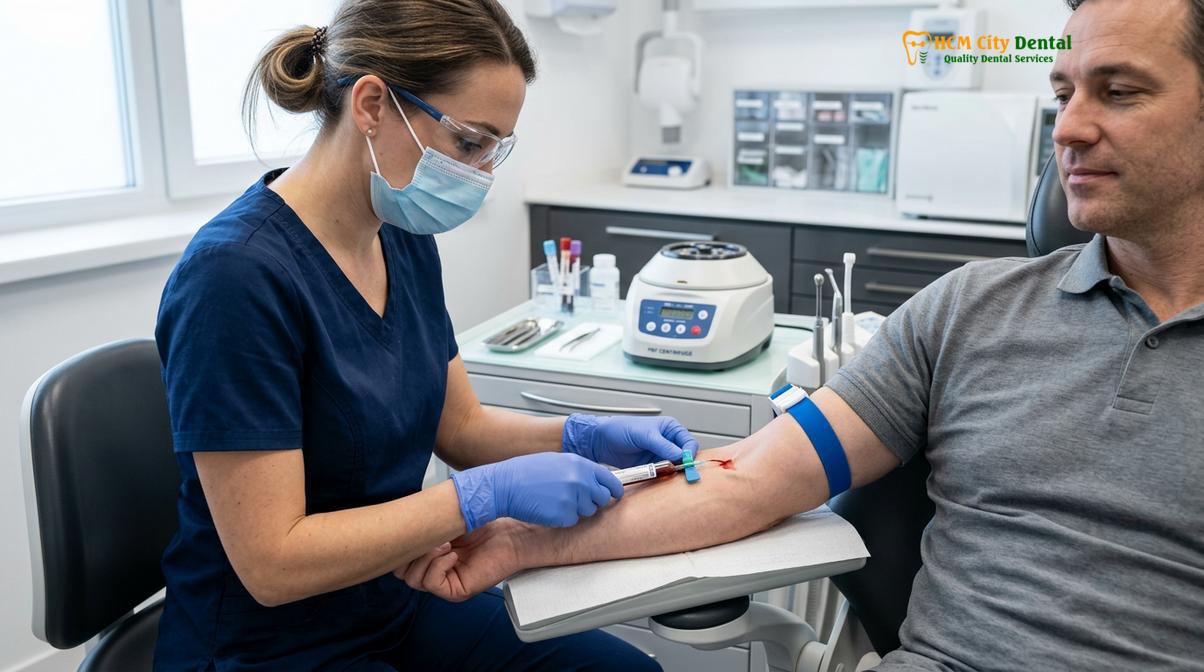
Once the blood is drawn, time is of the essence. The tubes must be placed into the centrifuge immediately—usually within 60 to 90 seconds. If the blood sits for too long before spinning, it will begin to clot diffusely throughout the tube, preventing the formation of a distinct, usable PRF layer. The centrifuge is then activated, spinning the blood at the precise parameters required to separate the components without destroying the delicate cellular structures.
While the blood is spinning (a process that takes about 10 to 12 minutes), the oral surgeon proceeds with the surgical extraction of the wisdom teeth. This simultaneous workflow ensures maximum efficiency. Once the extraction is complete and the socket has been thoroughly debrided (cleaned of any infected tissue or debris), the centrifuge cycle will finish. The dental assistant carefully removes the tubes. Using sterile forceps, the distinct, yellowish PRF clot is extracted from the tube. The red blood cell layer attached to the bottom of the clot is gently scraped away and discarded.
| Procedural Aspect | Standard Wisdom Tooth Extraction | Extraction with PRF Therapy |
|---|---|---|
| Socket Preparation | Socket is cleaned and left to fill with natural blood. | Socket is cleaned; PRF plug is actively packed into the void. |
| Clot Stability | Fragile; susceptible to dislodgement (dry socket risk). | Highly stable; dense fibrin matrix adheres to bone walls. |
| Growth Factor Release | Minimal, rapid depletion within 24-48 hours. | Sustained, slow release over 7 to 14 days. |
| Soft Tissue Closure | Slower epithelial migration over the open socket. | Accelerated closure; PRF acts as a biological bridge. |
| Postoperative Pain | Moderate to severe, often requiring prescription analgesics. | Significantly reduced due to protected nerve endings. |
The prepared PRF can then be customized for the specific surgical site. For a deep wisdom tooth socket, the PRF is often left as a cylindrical plug. The surgeon gently packs this plug directly into the alveolar defect, ensuring it contacts the bony walls. If a larger area of bone is exposed, or if a bone graft is being placed simultaneously, the PRF clot can be placed into a specialized metal compression box. This box gently squeezes out the excess serum, flattening the clot into a strong, suturable membrane. The surgeon then sutures the gingival tissue over the PRF, securing it in place. This biological dressing immediately begins its work, sealing the wound and initiating the regenerative cascade.
Major Benefits: Accelerating Healing & Preventing Dry Socket
PRF acts as a biological bandage that protects exposed bone, accelerates soft tissue closure, and provides immune-boosting leukocytes that virtually eliminate the incidence of alveolar osteitis.
The clinical benefits of utilizing PRF in dentoalveolar surgery are extensively documented in contemporary medical literature. For patients undergoing the removal of impacted third molars, the postoperative period is often associated with significant morbidity, including pain, facial swelling (edema), trismus (restricted mouth opening), and the dreaded complication of alveolar osteitis, commonly known as dry socket. The application of PRF addresses each of these challenges directly, transforming a notoriously uncomfortable recovery into a much more manageable experience.
Alveolar osteitis occurs when the primary blood clot that forms in the extraction socket is either dislodged prematurely or fails to form adequately. This leaves the underlying alveolar bone and highly sensitive nerve endings exposed to the harsh environment of the oral cavity, including air, food debris, and bacteria. The resulting pain is often described as a severe, throbbing ache that radiates to the ear or neck, typically peaking three to five days after surgery. Standard treatments involve packing the socket with medicated dressings, which requires additional clinical visits and prolongs the overall healing timeline.
This is where prf dry socket prevention becomes a game-changer. Because the PRF matrix is physically robust and densely cross-linked, it is highly resistant to mechanical disruption. It acts as a secure, biological bandage that physically shields the bone and nerves. Furthermore, the high concentration of leukocytes within the PRF creates an acidic microenvironment that is hostile to the specific anaerobic bacteria associated with the breakdown of blood clots (fibrinolysis). Clinical studies have consistently shown that the prophylactic placement of PRF in lower third molar extraction sites reduces the incidence of dry socket to near zero, even in high-risk populations[4].
Clinical Case Example: HCMC Dental Clinic
A 26-year-old patient presented to HCMC Dental Clinic in Ho Chi Minh City with bilaterally impacted lower wisdom teeth. Due to a history of delayed healing and high anxiety regarding dry socket, Dr. Nguyen Van Cuong recommended integrating PRF therapy into the surgical plan. Following the atraumatic extraction of both molars, autologous PRF plugs were placed into the sockets and secured with resorbable sutures. At the 7-day follow-up, the patient reported minimal discomfort, required no prescription pain medication after the first 24 hours, and exhibited excellent soft tissue closure with zero signs of alveolar osteitis.
Beyond preventing complications, PRF actively accelerates the regeneration of both soft and hard tissues. The sustained release of Vascular Endothelial Growth Factor (VEGF) promotes rapid angiogenesis. New blood vessels quickly infiltrate the PRF matrix, bringing oxygen and nutrients essential for tissue repair. This enhanced vascularization leads to faster epithelialization—the process where gum tissue grows over the wound to seal it. Patients treated with PRF typically exhibit soft tissue closure days or even weeks faster than those healing by secondary intention alone.
Additionally, the osteogenic potential of PRF cannot be overstated. The extraction of a deeply impacted wisdom tooth often leaves a significant bony defect, particularly on the distal aspect of the adjacent second molar. If this bone does not regenerate properly, it can lead to chronic periodontal pockets and eventual tooth loss. The growth factors in PRF, particularly TGF-β and IGF, stimulate the proliferation and differentiation of osteoblasts, encouraging the body to fill the defect with dense, healthy bone rather than fibrous scar tissue.
Is PRF Safe and Who Should Choose It?
Because PRF is created entirely from your own blood without synthetic additives, it is highly biocompatible, exceptionally safe, and recommended for patients with impacted molars or compromised healing.
When evaluating any adjunctive medical therapy, safety is the paramount concern. The safety profile of Platelet-Rich Fibrin is virtually unmatched in the field of biomaterials. Because PRF is a strictly autologous product—meaning the donor and the recipient are the exact same person—there is absolutely zero risk of foreign body rejection, allergic reaction, or the transmission of blood-borne pathogens (such as HIV or Hepatitis) that can be associated with allogeneic (donor) or xenogeneic (animal-derived) grafts. The material is completely natural, utilizing only the intrinsic healing components already circulating within your vascular system.
Dr. Nguyen Van Cuong frequently emphasizes during patient consultations that while PRF is an extraordinary tool, it is part of a comprehensive surgical approach. “The application of PRF provides a profound biological advantage, significantly enhancing the body’s natural regenerative capacity. However, it requires precise surgical technique and strict adherence to postoperative care protocols to achieve optimal outcomes,” notes Dr. Cuong. The procedure is exceptionally safe, with no published instances of adverse systemic reactions directly attributable to the PRF matrix itself[5].
Important Clinical Consideration
While PRF drastically reduces the risk of complications like dry socket and accelerates healing, it does not replace the need for proper postoperative care. Patients must still strictly follow all instructions regarding oral hygiene, diet modifications (soft foods), and the avoidance of smoking or using straws during the initial recovery phase to ensure the stability of the graft.
While almost any patient undergoing a surgical extraction can benefit from the accelerated healing and reduced pain associated with PRF, it is particularly highly recommended for specific clinical demographics. Patients who are at an elevated risk for postoperative complications should strongly consider PRF therapy. This includes:
- Smokers: Nicotine causes vasoconstriction, significantly reducing blood flow to the surgical site and drastically increasing the risk of dry socket and delayed healing. PRF helps bridge this vascular deficit by promoting localized angiogenesis.
- Older Adults: As we age, cellular metabolism and regenerative capacity naturally decline. PRF provides a concentrated boost of growth factors to stimulate sluggish cellular responses.
- Diabetic Patients: Systemic conditions like diabetes impair microcirculation and immune function, making patients more susceptible to postoperative infections. The high leukocyte concentration in PRF provides vital localized immune support.
- Complex Impactions: Teeth that are deeply embedded in the jawbone require more extensive surgical intervention, resulting in larger bony defects. PRF is crucial for promoting adequate bone fill and protecting the adjacent second molar.
Contraindications for PRF are exceedingly rare but do exist. Patients with severe bleeding disorders, profound thrombocytopenia (abnormally low platelet count), or those currently undergoing intensive anticoagulant therapy may not be ideal candidates, as their blood may not yield a stable, usable fibrin clot. A thorough preoperative medical history and clinical evaluation are always required to determine individual candidacy.
PRF Therapy Options and Costs at HCMC Dental
HCMC Dental Clinic offers integrated PRF therapy during wisdom tooth removal, providing a cost-effective investment in faster recovery and reduced postoperative complications.
At HCMC Dental Clinic, the integration of advanced regenerative techniques is a core component of our surgical philosophy. We believe that minimizing patient discomfort and maximizing clinical outcomes should be the standard of care, not an exception. When planning a wisdom tooth extraction, our clinical team will discuss the potential benefits of incorporating PRF therapy into your personalized treatment plan. The process is seamlessly integrated into your surgical appointment, ensuring a smooth and efficient experience.
Understanding the prf dental cost saigon is an important aspect of treatment planning. While there is an additional fee associated with the specialized equipment, sterile collection kits, and clinical time required to process the blood and prepare the PRF matrix, this cost is generally quite modest compared to the overall surgical fee. More importantly, it is crucial to view PRF not merely as an added expense, but as a strategic investment in your recovery.
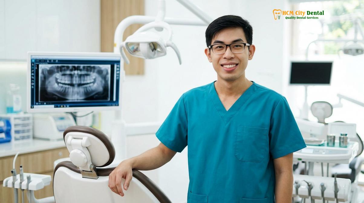
Patients who opt for PRF therapy frequently experience a faster return to their normal daily activities, work, and social obligations. By drastically reducing the incidence of complications like dry socket, PRF often eliminates the need for unexpected, emergency follow-up visits, additional localized treatments, and extended courses of prescription pain medications. In many cases, the upfront investment in PRF ultimately saves the patient both time and money, while sparing them significant physical discomfort.
“Investing in PRF therapy is essentially investing in a smoother, more predictable recovery. By harnessing the patient’s own biology, we can proactively manage inflammation and pain, rather than simply reacting to complications after they occur.”
— Advanced Dentoalveolar Surgery Guidelines
During your initial consultation at our Ho Chi Minh City facility, our specialists will conduct a comprehensive clinical and radiographic examination (often utilizing 3D CBCT imaging) to assess the position of your wisdom teeth and evaluate your specific risk factors. Based on this diagnostic data, we will provide a transparent, itemized treatment plan that clearly outlines the recommended procedures, including the option for PRF enhancement. Our goal is to empower you with the knowledge needed to make informed decisions about your oral health and surgical care.
When to See a Doctor (Post-Extraction Complications)
While PRF significantly enhances healing, patients must remain vigilant for signs of infection or abnormal bleeding and contact their clinician if severe symptoms arise.
Even with the advanced biological support of Platelet-Rich Fibrin, surgical extractions are significant medical procedures, and the body requires time to heal. It is normal to experience mild to moderate swelling, localized discomfort, and minor blood-tinged saliva during the first 24 to 48 hours. However, it is critical to distinguish between normal postoperative healing and signs of potential complications that require immediate clinical evaluation.
You should contact your dental provider promptly if you experience any of the following symptoms:
- Excessive or Uncontrolled Bleeding: While slight oozing is normal, active bleeding that rapidly fills the mouth or does not stop after applying firm, continuous pressure with a gauze pad for 45 minutes is a medical concern.
- Severe, Worsening Pain: Pain that progressively worsens after the third day, or pain that is not manageable with prescribed analgesics, may indicate an infection or the rare failure of the PRF graft.
- Fever or Systemic Illness: A persistent elevated temperature, chills, or a general feeling of malaise can be indicative of a spreading systemic infection.
- Difficulty Breathing or Swallowing: Rapidly expanding swelling that compromises the airway or makes swallowing difficult is a medical emergency requiring immediate attention.
- Pus or Foul Odor: The presence of purulent discharge (pus) from the extraction site, accompanied by a persistently foul taste or odor, strongly suggests a localized infection requiring professional debridement and antibiotic therapy.
Timely intervention is crucial for managing postoperative complications effectively. Always err on the side of caution and reach out to your clinical team if you have any concerns regarding your recovery trajectory.
Frequently Asked Questions
What does PRF stand for in wisdom teeth extraction?
PRF stands for Platelet-Rich Fibrin, an advanced autologous biomaterial derived entirely from your own blood. It is created by centrifuging a small blood sample to concentrate platelets, white blood cells, and growth factors into a dense fibrin matrix. When placed into a surgical site, it acts as a biological scaffold that significantly accelerates tissue regeneration and reduces postoperative complications.
How does PRF prevent dry socket?
PRF prevents dry socket by acting as a secure, biological bandage that protects the exposed alveolar bone and nerve endings. Unlike a standard blood clot that can easily dislodge, the dense fibrin matrix of PRF adheres strongly to the socket walls. Furthermore, it continuously releases growth factors that stimulate rapid soft tissue closure over the extraction site.
Is PRF worth the extra cost for wisdom teeth?
For most patients, PRF is highly worthwhile as it drastically reduces pain, swelling, and the risk of complications. The minor additional fee for processing the blood is often offset by the reduced need for pain medication, fewer follow-up visits for delayed healing, and a much faster return to normal daily activities.
How is PRF blood collected and processed?
A small sample of blood is drawn from your arm and immediately spun in a specialized centrifuge without any chemical additives. This rapid spinning separates the red blood cells from the plasma, forming a concentrated fibrin clot in the middle of the vial. This clot is then compressed into a membrane for surgical use.
Can PRF speed up bone healing after wisdom tooth surgery?
Yes, PRF significantly accelerates both soft tissue and early bone healing by releasing osteogenic growth factors directly into the socket. The concentrated cytokines and cellular components stimulate osteoblasts (bone-forming cells) and promote angiogenesis (new blood vessel formation), which are critical steps in regenerating the jawbone after a complex extraction.
References
- Journal of Oral and Maxillofacial Surgery. The effects of platelet-rich fibrin on post-surgical complications following removal of impacted wisdom teeth. (2021).
- International Journal of Oral and Maxillofacial Surgery. Autologous platelet concentrates in dentoalveolar surgery: A clinical review. (2019).
- British Dental Journal. Alveolar osteitis incidence and prevention utilizing second-generation platelet-rich fibrin. (2020).
- Journal of the American Dental Association. Mandibular third molar impaction classifications and healing trajectories. (2022).
- Clinical Oral Investigations. Cytokine release kinetics from advanced platelet-rich fibrin matrices. (2018).