Impacted wisdom tooth symptoms typically include localized jaw pain, red or swollen gums behind the last molar, difficulty opening the mouth, and persistent bad breath. These signs indicate that the third molar lacks sufficient space to erupt normally, often requiring clinical evaluation to prevent further dental complications.
Clinical Summary:
An impacted wisdom tooth occurs when the third molar fails to fully emerge through the gums due to anatomical space limitations within the jaw. Common symptoms range from mild tenderness and localized swelling to severe radiating jaw pain, trismus (restricted mouth opening), and pericoronitis (acute gum infection). Diagnosis relies on comprehensive clinical examination and 3D CBCT imaging to assess the tooth’s exact position relative to critical anatomical structures like the inferior alveolar nerve. Treatment typically involves surgical extraction, utilizing modern techniques such as ultrasonic Piezotome devices to minimize bone trauma, preserve surrounding tissues, and significantly accelerate the patient’s post-operative recovery.
Key Takeaways:
- Pain and swelling at the back of the jaw are the most frequent early indicators of third molar impaction.
- Partial impaction often leads to pericoronitis, a painful bacterial infection of the overlying gum tissue flap.
- Untreated impactions can cause irreversible damage to the adjacent second molars through decay or root resorption.
- 3D CBCT scans are the clinical gold standard for diagnosing the exact angle and nerve proximity of the trapped tooth.
- Modern surgical removal using ultrasonic technology significantly reduces post-operative discomfort, swelling, and recovery time.
What is an Impacted Wisdom Tooth?
An impacted wisdom tooth is a third molar that remains partially or completely trapped within the jawbone or gum tissue because there is not enough room for it to erupt normally.
Wisdom teeth, clinically referred to as third molars, are the final set of adult teeth to develop and emerge in the human dentition. They typically begin to erupt between the late teenage years and the early twenties. In a perfectly developed jaw with adequate arch length, these four teeth—two in the upper maxilla and two in the lower mandible—will emerge smoothly and align properly with the rest of the dentition, functioning effectively for mastication.
However, human evolution and changes in dietary habits over millennia have led to a gradual reduction in the size of the human jaw. Consequently, a significant percentage of the modern population lacks the necessary anatomical space at the posterior aspect of the dental arch to accommodate these late-arriving molars. When a wisdom tooth is physically blocked from reaching its functional position in the oral cavity, it is classified as “impacted.”[1]
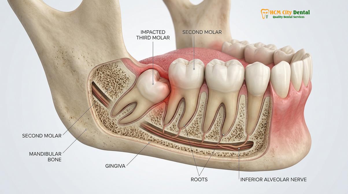
Impaction is not merely a cosmetic issue; it is a complex anatomical anomaly. The tooth may be obstructed by dense cortical bone, thick fibrous gingival tissue, or the physical barrier of the adjacent second molar. Because the tooth continues to develop its roots and eruptive force despite the blockage, it exerts continuous pressure on surrounding structures. This biological mechanism is the primary driver behind the various impacted wisdom tooth symptoms that patients eventually experience. The severity of the impaction dictates the clinical approach required, ranging from careful monitoring to complex surgical intervention.
Common Symptoms of Wisdom Tooth Impaction
The most frequent symptoms include throbbing jaw pain, tender and bleeding gums, difficulty chewing, and a persistent bad taste in the mouth caused by trapped bacteria.
Recognizing the early impacted wisdom teeth signs is crucial for preventing severe oral health complications. Because the third molars are located at the very back of the mouth, early developmental issues often go unnoticed until significant inflammation or structural pressure occurs. The symptomatology of an impacted wisdom tooth can be highly variable, ranging from asymptomatic (silent) to acute, debilitating pain.
One of the primary indicators is localized discomfort. Wisdom tooth impaction pain often begins as a dull, intermittent ache in the posterior jaw. As the tooth attempts to push through the bone and gum tissue, this ache can escalate into a sharp, throbbing sensation. Because of the complex network of the trigeminal nerve, which supplies sensation to the face and jaw, this pain frequently radiates. Patients may experience referred pain manifesting as tension headaches, earaches, or discomfort extending down into the neck and shoulder region.
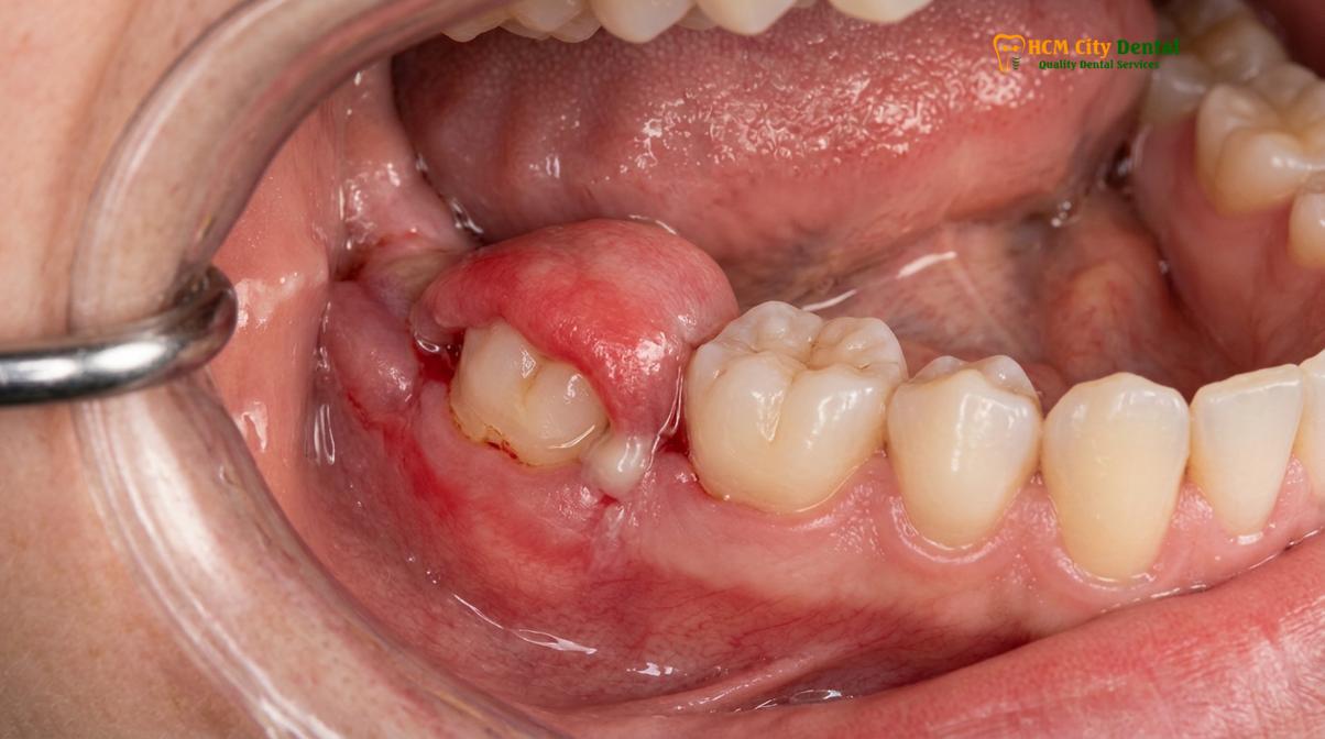
Another hallmark symptom is impacted tooth swelling. When a wisdom tooth is partially erupted, it creates a small opening in the gums. This opening acts as a trap for food particles, dental plaque, and oral bacteria. Because this area is notoriously difficult to clean with a standard toothbrush, the trapped debris rapidly breeds anaerobic bacteria. This leads to an inflammatory condition known as pericoronitis. The gum tissue overlying the tooth (the operculum) becomes red, highly swollen, and exquisitely tender to the touch. In severe cases, the swelling can extend to the cheek and submandibular lymph nodes.
“Many patients dismiss early signs of third molar impaction as temporary discomfort. However, persistent localized swelling and a foul taste are clear indicators of bacterial entrapment that require immediate clinical intervention to prevent systemic spread,” explains Dr. Nguyen Van Cuong.
Furthermore, patients often report a persistent bad breath (halitosis) or an unpleasant, metallic, or salty taste in their mouth. This is a direct result of the purulent exudate (pus) produced by the localized bacterial infection. As the inflammation worsens, patients may also experience trismus—a clinical condition characterized by spasms in the masticatory muscles, making it difficult or painful to open the mouth fully. Difficulty chewing, especially on the affected side, and bleeding gums during routine brushing are also strong indicators that a clinical evaluation is necessary.
Types of Wisdom Tooth Impaction (Partial vs Full)
Wisdom teeth can be partially impacted, where a portion breaks through the gum, or fully impacted, remaining entirely hidden beneath the tissue and jawbone.
In oral and maxillofacial surgery, impacted wisdom teeth are categorized based on their depth of penetration and their anatomical angulation. Understanding these classifications is vital for the dental surgeon to assess the surgical difficulty and predict potential complications. The primary distinction lies between soft tissue and hard tissue impactions.
A soft tissue impaction occurs when the crown of the wisdom tooth has successfully penetrated through the jawbone but remains covered, either partially or completely, by the gingival (gum) tissue. Conversely, a hard tissue impaction (or bony impaction) means the tooth is still partially or entirely encased within the jawbone. Hard tissue impactions are further divided into partial bony (part of the crown is exposed from the bone) and full bony (the tooth is completely submerged in bone).[2]
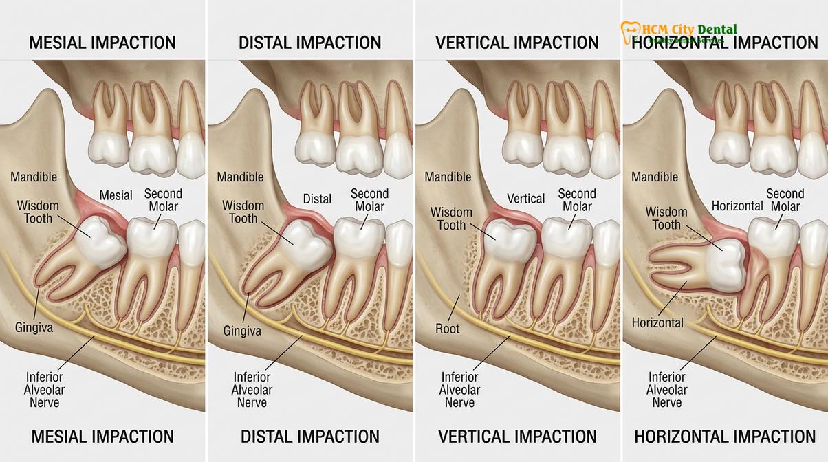
Beyond depth, dentists classify impactions based on the tooth’s angulation relative to the adjacent second molar. The Winter’s classification system is the standard clinical framework used worldwide:
| Type of Impaction | Anatomical Description | Clinical Frequency & Challenge |
|---|---|---|
| Mesial Impaction | The wisdom tooth is angled forward, pushing directly against the back of the adjacent second molar. | Most common type (approx. 44%). High risk of causing decay on the second molar. Moderate surgical difficulty. |
| Distal Impaction | The tooth is angled backward, pointing toward the rear of the jawbone (ramus). | Rarest type. Often requires significant bone removal during surgery. High surgical difficulty. |
| Vertical Impaction | The tooth is oriented straight up and down but remains trapped beneath the gums or bone due to lack of space. | Common. May erupt normally if space allows, but often requires extraction if it causes crowding or pericoronitis. |
| Horizontal Impaction | The tooth is lying completely on its side, perpendicular to the adjacent second molar. | Highly problematic. Exerts severe pressure on adjacent roots. Often fully encased in bone. Highest surgical difficulty. |
The Pell and Gregory classification system is also utilized to evaluate the depth of the impacted tooth relative to the occlusal plane (the biting surface of adjacent teeth) and the amount of space available between the second molar and the ascending ramus of the mandible. A tooth that is deeply embedded in the bone and located far back in the ramus presents a significantly more complex surgical scenario than a superficially impacted tooth. Accurate classification dictates the surgical instruments required, the flap design, and the method of tooth sectioning.
Complications of Leaving Impacted Teeth Untreated
Ignoring an impacted wisdom tooth can lead to severe complications, including chronic gum infections, cyst formation, and irreversible decay or structural damage to the adjacent second molar.
A common misconception among patients is that if an impacted wisdom tooth is not currently causing acute pain, it can be safely ignored. However, asymptomatic does not mean disease-free. The anatomical positioning of an impacted tooth creates a highly compromised environment that makes the area susceptible to a cascade of progressive, and often irreversible, dental pathologies.[3]
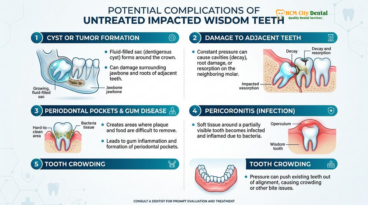
The most immediate and frequent complication is pericoronitis. As previously mentioned, a partially erupted tooth creates a deep periodontal pocket. This pocket is an ideal incubator for pathogenic bacteria. If left untreated, acute pericoronitis can evolve into a chronic condition, characterized by recurrent flare-ups of intense pain, swelling, and purulent discharge. In severe cases, the infection can spread into the fascial spaces of the head and neck, leading to life-threatening conditions such as Ludwig’s angina, which compromises the airway.
Another critical complication is the damage inflicted on the adjacent second molar. When a wisdom tooth is mesially or horizontally impacted, it exerts continuous mechanical pressure against the root and crown of the second molar. This pressure can cause root resorption, where the body begins to dissolve the root structure of the healthy tooth. Furthermore, the inability to clean the tight space between the impacted wisdom tooth and the second molar almost inevitably leads to deep dental caries (cavities). Because these cavities form below the gum line, they are difficult to detect early and often result in the loss of both the wisdom tooth and the otherwise healthy second molar.
Clinical Warning: A partially erupted wisdom tooth creates a deep periodontal pocket that is impossible to clean with a standard toothbrush. This environment rapidly breeds anaerobic bacteria, leading to acute pericoronitis, which can spread infection to the neck and facial spaces if left untreated.
In addition to infection and decay, impacted wisdom teeth are associated with the development of odontogenic cysts and tumors. Wisdom teeth develop within a sac of tissue inside the jawbone. If the tooth remains impacted, this sac can fill with fluid, forming a dentigerous cyst. As the cyst expands, it hollows out the jawbone, weakening its structural integrity and potentially leading to pathological jaw fractures. It can also damage the roots of neighboring teeth and compress the inferior alveolar nerve. While rare, benign tumors (such as ameloblastomas) can develop from the walls of these cysts, requiring extensive maxillofacial surgery to remove the lesion and reconstruct the jaw.
Finally, there is the issue of dental crowding. While the theory that erupting wisdom teeth exert enough forward pressure to cause anterior crowding (shifting of the front teeth) is debated in orthodontic literature, it is widely accepted that the eruptive forces can disrupt the stability of the dental arch, particularly in patients who have previously undergone orthodontic treatment. Removing impacted third molars is often recommended as a preventative measure to maintain orthodontic alignment.
How Impacted Wisdom Teeth Are Diagnosed (3D CBCT)
Dentists diagnose impacted wisdom teeth through a comprehensive oral examination combined with advanced imaging, specifically 3D Cone Beam Computed Tomography (CBCT), to map the tooth’s exact position.
Determining how to tell if wisdom tooth is impacted requires a professional clinical assessment. The diagnostic workflow begins with a thorough review of the patient’s medical and dental history, followed by a detailed physical examination of the oral cavity. The dentist will palpate the posterior gingival tissues, checking for swelling, tenderness, or the presence of a partially erupted crown. Periodontal probing may be used to assess the depth of the gingival pockets and check for signs of exudate or bleeding.
However, because the majority of an impacted tooth’s structure is hidden beneath the gums and bone, radiographic imaging is absolutely essential for a definitive diagnosis. Historically, the standard imaging modality has been the panoramic radiograph (OPG). An OPG provides a broad, two-dimensional overview of the entire upper and lower jaw, allowing the dentist to see the presence, number, and general angulation of the wisdom teeth. It helps identify gross pathologies such as large cysts or obvious decay on adjacent teeth.
Despite its utility, a 2D panoramic X-ray has significant limitations, particularly when planning surgical extraction. It cannot accurately depict the three-dimensional relationship between the roots of the lower wisdom tooth and the inferior alveolar nerve—a major sensory nerve that runs through the lower jaw. Damage to this nerve during extraction can result in temporary or permanent numbness (paresthesia) of the lower lip, chin, and tongue.
Clinical Case Study: A 23-year-old patient presented at HCMC Dental Clinic in Ho Chi Minh City complaining of intermittent headaches and jaw stiffness. While the oral cavity appeared normal upon visual inspection, a 3D CBCT scan revealed a horizontally impacted lower right third molar pressing directly against the root of the second molar. The precise 3D imaging allowed the surgical team to map the exact location of the inferior alveolar nerve and plan a safe extraction trajectory, successfully resolving the patient’s chronic pain without neurological complications.
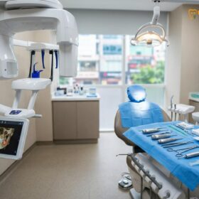
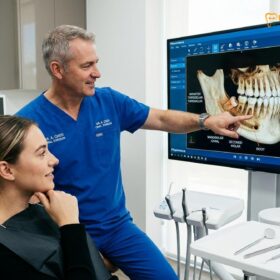
To overcome the limitations of 2D imaging, modern dental practices utilize 3D Cone Beam Computed Tomography (CBCT). A CBCT scan captures hundreds of high-resolution images from different angles, reconstructing them into a highly detailed 3D model of the patient’s maxillofacial anatomy. This allows the oral surgeon to view the impacted tooth in axial, coronal, and sagittal planes.[4]
With a 3D CBCT scan, the surgeon can precisely measure the distance between the tooth roots and the nerve canal, identify complex root curvatures (dilacerations), assess the exact thickness of the surrounding cortical bone, and determine the spatial relationship with the maxillary sinus for upper wisdom teeth. This level of diagnostic precision is paramount for minimizing surgical risks, optimizing the surgical approach, and ensuring the highest standard of patient safety.
Modern Surgical Removal Options in Vietnam
Modern extraction techniques utilize minimally invasive ultrasonic instruments and advanced sedation protocols to safely remove impacted teeth while maximizing patient comfort and accelerating recovery.
The surgical management of impacted wisdom teeth has evolved significantly. Traditional methods often relied heavily on rotary drills and manual elevators, which, while effective, could result in considerable bone trauma, post-operative swelling, and extended recovery periods. Today, leading facilities like HCMC Dental Clinic employ advanced, minimally invasive protocols that prioritize tissue preservation and patient comfort.
One of the most significant advancements in oral surgery is the use of piezoelectric technology (Piezotome). Unlike conventional rotary burs that cut both bone and soft tissue indiscriminately, a Piezotome utilizes high-frequency ultrasonic micro-vibrations to cut bone with extreme precision. The unique physical properties of ultrasonic surgery mean that it only cuts mineralized tissue (bone and tooth) while leaving soft tissues—such as nerves, blood vessels, and mucosal membranes—completely unharmed, even in the event of accidental contact.[5]
When performing professional wisdom tooth extraction services, the surgeon uses the Piezotome to gently gutter the bone surrounding the impacted tooth and section the tooth into smaller, easily removable pieces. This technique drastically reduces surgical trauma, minimizes bleeding, and significantly lowers the incidence of post-operative edema (swelling) and pain.
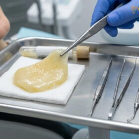
In addition to advanced surgical instrumentation, modern extraction protocols heavily incorporate regenerative medicine, specifically the use of Platelet-Rich Fibrin (PRF). Following the removal of the impacted tooth, the surgeon draws a small amount of the patient’s own blood and centrifuges it to isolate a concentrated matrix of platelets, white blood cells, and growth factors. This PRF membrane is then placed directly into the empty extraction socket before suturing.
The application of PRF serves multiple critical functions. It acts as a biological bandage, stabilizing the blood clot and virtually eliminating the risk of developing a painful “dry socket” (alveolar osteitis). Furthermore, the slow release of growth factors from the PRF membrane accelerates the regeneration of both soft gingival tissue and hard alveolar bone, cutting the overall healing time significantly.
Patient comfort during the procedure is also a primary focus. Depending on the complexity of the impaction and the patient’s anxiety levels, procedures can be performed under profound local anesthesia, nitrous oxide (laughing gas), or intravenous (IV) sedation. A comprehensive pre-operative consultation is essential to determine the most appropriate surgical and anesthetic approach tailored to the individual’s specific clinical presentation.
When to See a Doctor
While mild discomfort during the normal eruption of wisdom teeth can sometimes be managed with over-the-counter pain relievers and warm saltwater rinses, certain symptoms indicate a severe underlying issue that requires immediate professional medical attention. Delaying treatment in the presence of these red flag symptoms can lead to systemic infections and permanent structural damage to the jaw and adjacent teeth.
You should seek an urgent clinical evaluation if you experience any of the following:
- Severe, unmanageable pain: Pain that is not relieved by standard doses of ibuprofen or acetaminophen, or pain that radiates intensely to the ear, head, or neck.
- Significant facial swelling: Swelling that extends beyond the gums into the cheek, jawline, or under the chin, indicating a spreading infection.
- Pus discharge: The presence of a foul-tasting, yellowish or greenish fluid oozing from the gums around the back teeth.
- Difficulty swallowing or breathing: This is a critical medical emergency. It suggests that the infection has spread into the deep fascial spaces of the neck, potentially compromising the airway.
- Fever and chills: Systemic signs of infection indicating that the localized bacteria have entered the bloodstream.
“If you experience sudden trismus—an inability to open your mouth fully—accompanied by facial swelling, this is no longer a simple toothache. It is a dental emergency indicating that the infection has penetrated deeper fascial spaces,” warns Dr. Cuong.
Regular dental check-ups, typically every six months, are the most effective way to monitor the development of wisdom teeth. Through routine clinical examinations and periodic radiographic imaging, a dentist can track the trajectory of the erupting molars and intervene proactively before impaction causes acute symptoms or irreversible damage. If you are experiencing any signs of impaction, scheduling a diagnostic consultation at a reputable facility in Ho Chi Minh City is the safest course of action.
Frequently Asked Questions
What does impacted wisdom tooth pain feel like?
Impacted wisdom tooth pain typically feels like a dull, continuous ache at the back of the jaw that can escalate into sharp, throbbing pain. This discomfort often radiates toward the ear, temple, or neck. Patients frequently experience increased pain when chewing food or applying pressure to the affected side, and the surrounding gum tissue may feel highly sensitive and tender to the touch.
Can an impacted wisdom tooth cause headaches?
Yes, an impacted wisdom tooth can frequently cause tension headaches and migraines. Because the third molars are located near the temporomandibular joint (TMJ) and share complex neural pathways via the trigeminal nerve, the pressure and inflammation from an impacted tooth can radiate upward. This referred pain often manifests as a persistent headache, earache, or generalized facial discomfort on the affected side.
What happens if you don’t remove an impacted wisdom tooth?
Leaving an impacted wisdom tooth untreated can lead to severe, irreversible dental complications. The most common consequence is chronic pericoronitis, a painful bacterial infection of the gums. Additionally, the trapped tooth can cause structural damage, root resorption, or deep decay in the adjacent second molar. In rare cases, fluid-filled cysts or benign tumors can form around the impacted tooth, destroying surrounding jawbone.
How do dentists diagnose an impacted wisdom tooth?
Dentists diagnose an impacted wisdom tooth through a combination of visual clinical examination and advanced radiographic imaging. While a standard panoramic X-ray (OPG) provides a general overview of the jaw, a 3D Cone Beam Computed Tomography (CBCT) scan is the gold standard. The 3D scan allows the dentist to precisely map the tooth’s angulation, root morphology, and proximity to the inferior alveolar nerve.
Is removal of an impacted wisdom tooth painful?
The surgical removal of an impacted wisdom tooth is not painful during the procedure, as it is performed under profound local anesthesia or intravenous sedation. While patients may feel pressure, there is no sharp pain. Post-operative discomfort is normal but is effectively managed with prescribed analgesics, anti-inflammatory medications, and cold compresses. Modern techniques like ultrasonic surgery further minimize trauma and reduce recovery pain.
References
- Journal of Oral and Maxillofacial Surgery. Pathophysiology and management of impacted third molars. (2021).
- International Journal of Oral and Maxillofacial Surgery. Efficacy of 3D CBCT in assessing inferior alveolar nerve proximity. (2020).
- British Dental Journal. Clinical complications of asymptomatic impacted wisdom teeth. (2019).
- Journal of the American Dental Association. Pell and Gregory classification in predicting surgical difficulty. (2022).
- Clinical Oral Investigations. Piezoelectric surgery versus conventional rotary instruments in third molar extraction. (2023).