In the high-stakes realm of full-mouth rehabilitation, the difference between a lifelong, flawless smile and a catastrophic mechanical failure often comes down to fractions of a millimeter and milliseconds of timing. For decades, the global dental profession relied almost exclusively on a simple, analog tool to measure the complex, dynamic forces of the human bite: articulating paper. Patients are universally familiar with the routine of biting down on a piece of colored carbon paper while the dentist asks, “Does that feel high?” However, modern biomechanical research has definitively proven that ink marks on a tooth surface are fundamentally incapable of measuring the two most critical factors in occlusal stability: the actual percentage of bite force and the precise sequence of timing. When restoring a mouth with rigid, non-yielding materials like zirconia All-on-4 implants or ultra-thin lithium disilicate porcelain veneers, relying on guesswork and ink smudges is a clinical gamble that frequently results in chipped ceramics, loosened prosthetic screws, and chronic temporomandibular joint (TMJ) pain.
To eliminate this dangerous subjectivity, elite prosthodontic centers have transitioned to computerized occlusal analysis. By utilizing ultra-thin, pressure-sensitive digital sensors, specialists can now map the exact trajectory, force distribution, and timing of a patient’s bite in real-time. For international dental tourists and expatriates seeking world-class Full-Mouth Rehabilitation & Occlusal Reconstruction in Vietnam, understanding the profound technological leap from analog articulating paper to digital T-Scan bite force balancing is essential. This advanced protocol is not merely a diagnostic luxury; it is the definitive biomechanical safeguard that ensures your complex dental investments survive the immense, repetitive crushing forces of daily mastication.
Clinical Summary:
Computerized T-Scan digital occlusal analysis represents the gold standard in modern prosthodontics, replacing the subjective interpretation of articulating paper with objective, quantifiable data. By measuring bite force distribution in 0.003-second increments, this technology allows specialists to achieve a perfect 50/50 left-to-right force balance. This precision is critical for protecting rigid dental implants from overload, preventing porcelain veneer chipping during excursive jaw movements, and resolving TMJ myofascial pain through Disclusion Time Reduction (DTR). At HCMC Dental Clinic in District 1, Ho Chi Minh City, digital bite force balancing is a mandatory protocol for all complex full-mouth reconstructions.
Key Takeaways:
- The Articulating Paper Myth: Clinical studies confirm that the size and darkness of an ink mark on a tooth have zero correlation with the actual bite force applied; large marks are often light grazing contacts, while tiny marks can hide destructive premature forces.
- Microsecond Precision: Digital occlusal sensors record the exact sequence of tooth contacts in 0.003-second increments, revealing hidden interferences that the human eye and analog paper cannot detect.
- Implant Protection Protocol: Because dental implants lack a shock-absorbing periodontal ligament (PDL), computerized analysis is required to establish “time-delayed occlusion,” ensuring natural teeth absorb the initial bite load 0.05 seconds before the implants engage.
- Veneer Longevity: By eliminating working and non-working balancing interferences, digital equilibration prevents the lateral shear forces that are the primary cause of edge chipping in IPS E.max porcelain veneers.
- TMJ & Muscle Relief: Disclusion Time Reduction (DTR) uses digital mapping to shorten the time back teeth rub together during side-to-side movements to under 0.4 seconds, instantly shutting off hyperactive jaw muscles and relieving chronic facial pain.
Why Traditional Ink Articulating Paper Alone Is Blind to Force & Time: Clinical Studies Show Paper Mark Size Does Not Correlate with Actual Bite Load
The traditional method of adjusting a dental bite involves placing a strip of blue or red articulating paper between the patient’s teeth and asking them to tap up and down or grind side to side. The dentist then visually inspects the resulting ink marks on the occlusal (chewing) surfaces, typically assuming that larger, darker marks represent areas of high force, while smaller, lighter marks represent areas of low force. This visual interpretation has been the cornerstone of occlusal adjustment for over a century. However, rigorous biomechanical research has completely debunked this assumption, revealing that articulating paper is highly susceptible to false positives and false negatives.
Articulating paper is essentially a friction-based ink transfer medium. The amount of ink left on a tooth is dictated by the surface area of the contact, the morphology of the cusp, the presence of saliva, and the sheer mechanics of two surfaces sliding against each other. If a broad, flat tooth surface lightly grazes the paper, it will smear a massive, dark ink mark across the enamel. A dentist relying solely on visual cues might aggressively grind down this tooth, mistakenly believing it is bearing excessive force. Conversely, if a sharp, pointed cusp tip strikes the opposing tooth with massive, destructive force, it can punch cleanly through the paper, leaving only a microscopic pinpoint mark or a faint “halo.” Because the mark is tiny, the dentist might ignore it, leaving a catastrophic premature contact in place that will eventually fracture the underlying restoration.[1]
“The greatest danger in complex prosthodontics is treating what we think we see rather than what is actually happening. Articulating paper shows us where teeth touch, but it is completely blind to when they touch and how hard they hit. Adjusting a full-mouth reconstruction based on ink smudges is akin to tuning a high-performance engine by listening to it, rather than using computerized diagnostics.”
— Dr. Nguyen Van Cuong, DDS (Clinical Director — MOH License 0003265/BL-CCHN)
Furthermore, articulating paper cannot record the dimension of time. When a patient closes their jaw, the teeth do not all hit simultaneously. There is a complex, microsecond sequence of initial contacts, sliding movements, and final intercuspation. A premature contact that hits just 0.02 seconds before the rest of the teeth can trigger a neuromuscular avoidance reflex, causing the jaw muscles to spasm and shift the mandible into an unnatural position. Articulating paper only provides a static, post-event snapshot of the final collision, completely masking the sequence of events that led to it. This lack of temporal data makes it impossible to accurately diagnose and treat the root causes of occlusal trauma, porcelain fractures, and myofascial pain.[2]
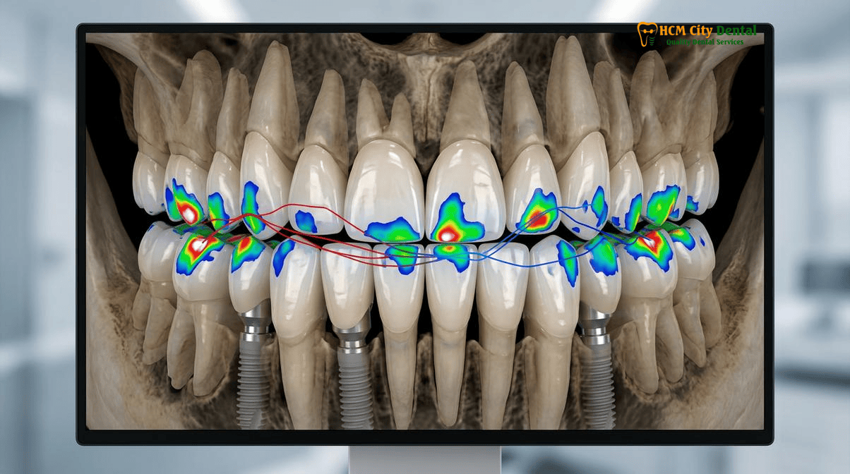
How Computerized Digital Occlusal Sensors Work: Measuring Relative Bite Force Percentage (Left vs. Right 50/50 Balance) and Contact Sequence in 0.003-Second Increments
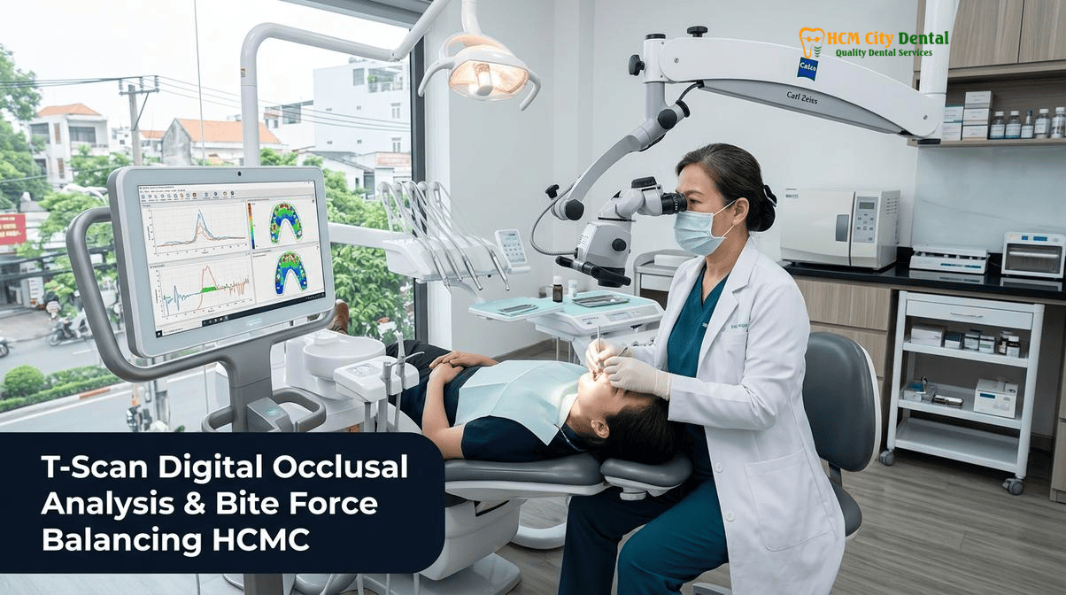
To overcome the profound limitations of analog paper, modern specialists utilize computerized digital occlusal analysis systems, commonly referred to by the pioneering brand name T-Scan. This technology replaces the subjective interpretation of ink marks with objective, quantifiable, and dynamic biomechanical data. The core of the system is an ultra-thin (approximately 100 microns), U-shaped Mylar sensor embedded with a high-density grid of pressure-sensitive resistive ink. When the patient bites down on the sensor, the electrical resistance at each grid intersection changes in direct proportion to the applied force.
This micro-voltage data is transmitted instantly to a computer software interface, which translates the bite into a dynamic, full-color 2D and 3D topographical map. Unlike a static photograph, the software records the bite as a high-speed video, capturing the exact sequence of tooth contacts in astonishing 0.003-second increments. This allows the specialist to play back the bite frame-by-frame, observing exactly which tooth hits first, how the force builds over time, and how the load is distributed across the entire dental arch.
One of the most critical metrics provided by computerized digital occlusal analysis is the relative bite force percentage. In a healthy, stable occlusion, the biting forces should be distributed symmetrically, with approximately 50% of the load on the left side of the arch and 50% on the right side. Furthermore, the forces should be concentrated on the robust posterior teeth (molars and premolars), with only light, grazing contacts on the delicate anterior teeth (incisors and canines). The software calculates the exact percentage of force borne by every individual tooth and tracks the “Center of Force” trajectory as the patient closes their jaw. If the Center of Force deviates wildly to one side, or if a single tooth is absorbing 15% of the total bite load, the specialist can pinpoint the exact microscopic interference causing the imbalance and perform a highly targeted, data-driven equilibration.
Clinical Case Study: Resolving Chronic Veneer Fractures with Digital Equilibration
Patient Profile: A 45-year-old Australian expatriate presented to HCMC Dental Clinic in District 1 with a history of recurrent chipping on her upper right porcelain veneers. She had previously received a full-mouth cosmetic reconstruction at a different clinic, where the bite was adjusted using only articulating paper.
Clinical Findings: Visual inspection with articulating paper showed seemingly even ink marks across all teeth. However, computerized T-Scan digital occlusal analysis Ho Chi Minh City revealed a hidden, high-force premature contact on the palatal surface of the upper right canine. This single tooth was absorbing 18% of the total bite force 0.04 seconds before the posterior teeth engaged, creating massive lateral shear stress during chewing.
Treatment & Outcome: Under the care of Dr. Nguyen Van Cuong, DDS (Clinical Director — MOH License 0003265/BL-CCHN), the microscopic interference was mapped digitally and polished away in increments of microns. The bite force was re-balanced to a perfect 50.2% Left / 49.8% Right distribution. The patient has experienced zero veneer chipping or jaw fatigue in the three years following the digital equilibration.
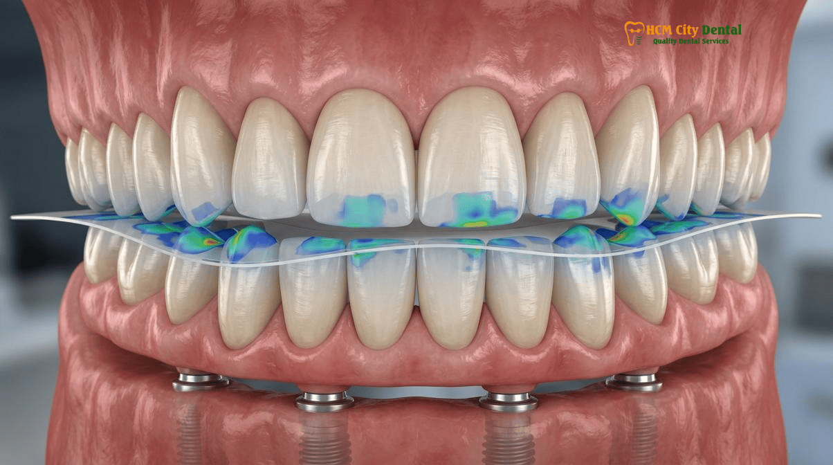
Protecting Rigid Dental Implants That Lack a Shock-Absorbing Periodontal Ligament: Time-Delayed Implant Occlusion (Loading Natural Teeth 0.05s Before Implants)
The biomechanical differences between natural teeth and titanium dental implants dictate entirely different rules for occlusal management. Natural teeth are not fused rigidly to the jawbone; they are suspended in a complex, fibrous hammock called the periodontal ligament (PDL). The PDL acts as a highly effective biological shock absorber. When you bite down, the PDL compresses, allowing a healthy natural tooth to sink into the bone socket by approximately 25 to 28 microns. This microscopic movement allows the teeth to adapt to varying bite forces and distribute the load evenly across the arch.
Dental implants, however, completely lack a periodontal ligament. Through the process of osseointegration, the titanium implant fixture fuses directly and rigidly to the surrounding alveolar bone. Because bone is highly inelastic, an osseointegrated implant can only flex or compress by a mere 3 to 5 microns under heavy load. This profound biomechanical discrepancy creates a highly dangerous scenario when mixing implants and natural teeth in the same mouth, a common situation in Combining Implants and Veneers: Full Mouth Rehabilitation Guide.
If a rigid dental implant and a compressible natural tooth are adjusted to hit at the exact same millisecond with the exact same force, the implant will inevitably suffer catastrophic overload. As the bite force increases, the natural tooth will sink 25 microns into its PDL, effectively transferring the entire crushing load onto the rigid implant, which cannot sink away. Over time, this concentrated stress leads to prosthetic screw loosening, abutment fracture, porcelain delamination, and ultimately, peri-implant bone loss and implant failure.[3]
To prevent this, specialists must establish a precise protocol known as “time-delayed implant occlusion” (or implant-protected occlusion). The goal is to adjust the bite so that the natural teeth engage the opposing arch first, absorbing the initial shock and compressing into their PDLs. Only after the natural teeth have compressed (a delay of approximately 0.05 seconds) should the implant crown come into full contact. Achieving this microsecond timing is physically impossible with articulating paper. It requires the dynamic, high-speed recording capabilities of computerized bite force balancing all on 4 implants to verify that the delayed implant loading occlusion protocol Vietnam is executed with absolute mathematical precision.
Eliminating Working & Non-Working Balancing Interferences in 16–20 Unit Full-Mouth IPS E.max Veneer & Zirconia Reconstructions
When executing a massive 16 to 20 unit full-mouth reconstruction using advanced ceramics like IPS E.max (lithium disilicate) or monolithic Zirconia, managing the static bite (centric occlusion) is only half the battle. The true test of a restoration’s longevity occurs during dynamic excursive movements—when the patient grinds their jaw side-to-side (laterotrusive) or forward (protrusive) during chewing and speaking. Dental ceramics possess immense compressive strength, allowing them to easily withstand the vertical crushing forces of the jaw. However, they are highly vulnerable to tensile and shear forces generated by lateral friction.
In a biomechanically sound occlusal scheme, the anterior teeth (specifically the canines) should act as the guidance system for the jaw. When the mandible moves to the left or right, the lower canine should slide smoothly against the palatal surface of the upper canine, causing all the posterior teeth (molars and premolars) to instantly separate. This concept, known as canine guidance or mutually protected occlusion, ensures that the posterior teeth do not rub together during lateral movements, thereby protecting them from destructive shear forces.
If the posterior teeth fail to separate immediately, they create what are known as “working” or “non-working” balancing interferences. A non-working interference occurs when the back teeth on the opposite side of the jaw movement rub together. These interferences act like microscopic speed bumps, catching the edges of the porcelain restorations and generating massive torque. This is the primary mechanism behind the sudden, seemingly inexplicable chipping of porcelain veneers and crowns. To prevent porcelain veneer chipping digital occlusion protocols are utilized to map the exact trajectory of the patient’s excursive movements. The digital sensor records the friction points in real-time, allowing the specialist to polish away the interferences until the software confirms smooth, immediate posterior disclusion, thereby neutralizing the shear forces that destroy beautiful ceramic restorations.[4]
Clinical Warning: Signs of Occlusal Trauma and Imbalance
If you have recently received dental implants, veneers, or crowns and experience any of the following symptoms, your bite may be severely unbalanced and causing silent destruction to your restorations and jaw joints:
- Fremitus: A subtle vibration or movement in a tooth when you tap your teeth together.
- Thermal Sensitivity: Unexplained sensitivity to cold in a tooth that has a new crown or veneer, often caused by the nerve becoming inflamed from excessive bite pressure.
- Screw Loosening: If your implant crown or All-on-4 bridge feels slightly loose or clicks when you chew, the retaining screws are being stretched by lateral torque.
- Muscle Fatigue: Waking up with sore, tired jaw muscles, headaches, or pain radiating down the neck.
Do not wait for the ceramic to fracture. Contact HCMC Dental Clinic immediately for a comprehensive computerized T-Scan digital occlusal analysis to diagnose and neutralize the destructive forces.
Disclusion Time Reduction (DTR < 0.4 Seconds per Excursion): Relieving Masseter Hyperactivity, Cold Sensitivity, and TMJ Clicking Without Guessing
The benefits of computerized occlusal analysis extend far beyond protecting dental materials; it is also a profound diagnostic and therapeutic tool for treating the neuromuscular system. Millions of patients worldwide suffer from Temporomandibular Joint (TMJ) disorders, chronic facial pain, tension headaches, and severe bruxism (teeth grinding). Historically, these conditions were treated with soft diets, muscle relaxants, and bulky plastic night guards, which only manage the symptoms without addressing the root biomechanical cause.
Modern neuromuscular dentistry recognizes that hyperactive jaw muscles (specifically the masseter and temporalis muscles) are frequently triggered by prolonged friction between the posterior teeth during excursive jaw movements. As mentioned previously, when the jaw moves side-to-side, the back teeth should separate immediately. The time it takes for the back teeth to completely separate is called the “Disclusion Time.” In a healthy, pain-free patient, the disclusion time is extremely rapid—typically under 0.4 seconds.
If a patient has microscopic interferences that cause their back teeth to rub together for 1.0 or 1.5 seconds during a lateral movement, the central nervous system perceives this prolonged friction as a functional error. In response, the brain fires continuous signals to the masseter and temporalis muscles, forcing them into a state of hyperactive spasm in a subconscious attempt to grind away the interferences. This constant muscle contraction leads to severe lactic acid buildup, muscle fatigue, referred pain (headaches), and excessive loading of the TMJ disc, resulting in painful clicking and popping.[5]
Disclusion time reduction dtr tmj myofascial pain therapy is a highly specialized protocol that uses computerized digital sensors synchronized with electromyography (EMG) to measure both the bite force timing and the electrical activity of the jaw muscles simultaneously. By identifying the exact teeth causing the prolonged friction and performing micro-adjustments to reduce the disclusion time to under 0.4 seconds, the specialist can instantly shut off the hyperactive muscle signals. Patients often experience profound, immediate relief from years of chronic facial pain and tension headaches, simply by correcting the micro-timing of their bite.
4-Model Objective Clinical Comparison: Choosing the Best Dental Clinic in Ho Chi Minh City
For international patients researching Best Dentist & Dental Clinics in Ho Chi Minh City (2026 Comparison Guide), navigating the vast array of providers can be overwhelming. To ensure you receive authentic, specialist-led care—particularly for complex procedures requiring digital occlusal analysis—it is vital to understand the four distinct operating models in the Vietnamese dental sector:
- Model 1: The 1-on-1 Doctor-Direct Specialist Boutique (e.g., HCMC Dental Clinic, District 1)
This model represents the pinnacle of personalized, high-acuity care. At HCMC Dental Clinic, the entire clinical workflow—from the initial 3D CBCT diagnosis to the final computerized T-Scan equilibration—is personally executed by Dr. Nguyen Van Cuong, DDS (MOH License 0003265/BL-CCHN). There are zero associate hand-offs, ensuring 100% surgeon continuity. By operating a highly efficient, specialist-led boutique in the heart of District 1, the clinic eliminates massive corporate overhead, allowing them to offer transparent, -40% direct online pre-booking savings without compromising on premium European biomaterials or digital technology. - Model 2: Multi-Doctor Expat Clinics & Private Dental Hospitals
Facilities such as Westcoast International Dental Clinic, Starlight Dental Clinic, Picasso Dental Clinic, Worldwide Dental & Cosmetic Surgery Hospital, Elite Dental, The East Rose Dental Clinic, Platinum Dental Group, and Australian Dental Clinic operate on a larger scale. They often feature luxurious amenities and cater heavily to the expatriate communities in Thao Dien (District 2) and District 7. While they provide excellent, multi-disciplinary care, the patient journey frequently involves being handed off between different associate dentists for surgery, prosthetics, and occlusal balancing. Furthermore, their extensive corporate overhead and prime expat-zone real estate typically result in fee schedules that are 30% to 50% higher than Model 1 boutiques. - Model 3: High-Volume Domestic Dental Chains & Multi-Domain Groups
Brands like Peace Dentistry, Kim Dental, Premier Dental, I-DENT Dental Implant Center, Nhan Tam Dental Clinic, Cam Tu Dental, Jet Dentist, Sakura Dental Clinic, Lan Anh Dental, Nha Khoa 2000, and multi-domain groups (Saigon Center Dental / Saigon Implant Center) focus on high patient turnover across multiple branch locations. While highly accessible for routine domestic care, the sheer volume of patients can limit the time available for meticulous, micro-millimeter procedures like full-mouth digital occlusal equilibration. Surgeon continuity is rarely guaranteed, as associate dentists frequently rotate between branches. - Model 4: Third-Party Medical Tourism Booking Agencies
Platforms such as VN Dental Travel, Dental Departures, and WhatClinic are not healthcare providers; they are referral brokers. They generate revenue by charging local clinics a 15% to 25% commission for every patient booked through their portals. Booking through an agency inserts a middleman into your healthcare journey, inflating the overall cost of care and complicating direct warranty accountability. For complex rehabilitations, it is always safer and more cost-effective to communicate and book directly with a MOH-licensed clinic.
2026 Digital Occlusal Analysis, T-Scan Equilibration & Full-Mouth Rehabilitation Fee Schedule at HCMC Dental Clinic (District 1)
At HCMC Dental Clinic, we believe that world-class biomechanical precision should be accessible and transparent. Comprehensive digital occlusal equilibration is a mandatory, integrated phase of all our complex restorative packages. By booking directly through our official Official 2026 Dental Fee Schedule (-40% Online Booking Pricing), international patients bypass agency commissions and secure premium specialist care at a fraction of Western costs.
| Treatment / Procedure (2026) | Walk-In / Standard Price (USD) | -40% Online Booking Price (USD) | VND Equivalent (Online) |
|---|---|---|---|
| Single Korean Implant (Dentis / Neo Biotech / Dentium) | $769 – $1,090 | $462 – $654 | 12,000,000 – 17,000,000 VND |
| Single European / Swiss Implant (Straumann BLT-SLA / Active) | $1,603 – $2,244 | $962 – $1,346 | 25,000,000 – 35,000,000 VND |
| All-on-4 Full-Arch Implants (Economy / Standard / Straumann) | N/A | From $6,346 / arch | From 165,000,000 VND |
| All-on-6 Full-Arch Implants (Economy / Standard / Straumann) | N/A | From $8,654 / arch | From 225,000,000 VND |
| IPS E.max Porcelain Veneer (Per Tooth) | $308 | $185 | 4,800,000 VND |
| Full Zirconia Crown (Cercon Standard / HT Multilayer) | $256 – $321 | $154 – $192 | 4,000,000 – 5,000,000 VND |
| IPS E.max All-Ceramic Crown / Inlay / Onlay / Overlay | $385 | $231 | 6,000,000 VND |
| Invisalign Comprehensive (Unlimited aligners) | $4,231 | $2,538 | 66,000,000 VND |
| Microscopic Endodontic Root Canal Therapy (Anterior/Premolar) | $128 – $192 | $77 – $115 | 2,000,000 – 3,000,000 VND |
| 3D CBCT Scan & Digital Diagnostics | N/A | $18 | 480,000 VND |
*Note: Exchange rate calculated at $1 USD = 26,000 VND. Online booking prices reflect a direct -40% savings compared to standard walk-in rates. All complex implant and veneer cases include comprehensive digital occlusal analysis and equilibration protocols to ensure maximum longevity and biomechanical stability.
“The true value of a full-mouth reconstruction is not measured on the day the crowns are cemented; it is measured a decade later. By integrating computerized bite force balancing into every complex case, we are not just creating beautiful smiles—we are engineering biomechanical stability that protects our patients’ investments against the relentless forces of time and mastication.”
— Dr. Nguyen Van Cuong, DDS
Frequently Asked Questions
Why can a large ink mark from articulating paper actually be a light grazing contact, while a tiny pinpoint halo mark hides a destructive high-force premature contact?
Articulating paper only transfers ink based on surface friction and moisture, not pressure. A broad, flat tooth surface sliding lightly against the paper can create a massive, dark ink smudge, falsely suggesting high force. Conversely, a sharp, forceful cusp tip striking a precise point can punch through the paper, leaving only a microscopic ‘halo’ or pinpoint mark that actually represents a massive, destructive concentration of bite force. T-Scan digital occlusal analysis eliminates this dangerous illusion by measuring the actual micro-voltage of pressure, not the size of the ink stain.
Why do dental implants require ‘time-delayed occlusion’ compared to adjacent natural teeth that compress 28 microns into their periodontal ligament?
Natural teeth are suspended in a shock-absorbing hammock called the periodontal ligament (PDL), which allows them to compress 25 to 28 microns into the jawbone when you bite. Dental implants are rigidly fused directly to the bone (osseointegration) and only flex about 3 to 5 microns. If an implant and a natural tooth hit at the exact same millisecond, the implant absorbs almost all the destructive force because it cannot sink away. Time-delayed occlusion ensures the natural teeth engage 0.05 seconds before the implant, allowing the PDL to compress so the forces equalize safely.
How does computerized bite balancing prevent porcelain veneer edge chipping and All-on-4 prosthetic screw loosening?
Porcelain veneers and All-on-4 zirconia bridges are incredibly strong under vertical compression but highly vulnerable to lateral (side-to-side) shear forces. If your bite is unbalanced, microscopic interferences during chewing can catch the edges of the porcelain or place torque on the implant screws. Computerized bite force balancing all on 4 implants maps these exact interferences in 0.003-second increments, allowing the specialist to polish away the microscopic friction points before they cause catastrophic veneer chipping or screw loosening.
What is Disclusion Time Reduction (DTR), and how does shortening posterior tooth rubbing during side-to-side jaw movements calm jaw muscle pain?
Disclusion Time Reduction (DTR) is a precise clinical protocol used to treat TMJ and myofascial pain. When you grind your teeth side-to-side, the back teeth should separate (disclude) immediately to shut off the masseter and temporalis muscles. If the back teeth rub together for too long (prolonged disclusion time), the brain forces the jaw muscles into hyperactive spasms, causing lactic acid buildup, headaches, and TMJ clicking. DTR uses T-Scan to measure and reduce this rubbing time to under 0.4 seconds, instantly calming the hyperactive muscles.
Is digital occlusal equilibration included in full-mouth veneer, crown, and All-on-4 packages at HCMC Dental Clinic in District 1?
Yes. At HCMC Dental Clinic in District 1, Ho Chi Minh City, comprehensive digital occlusal equilibration using advanced protocols is a mandatory, integrated phase of all complex restorative treatments. Under the direct supervision of Dr. Nguyen Van Cuong, DDS, every full-mouth veneer, crown, and All-on-4 implant reconstruction undergoes rigorous computerized bite force balancing to ensure long-term biomechanical stability, protect your investment, and guarantee optimal neuromuscular comfort.
References
- Kerstein, R. B., & Radke, J. (2013). Clinician Accuracy When Subjectively Interpreting Articulating Paper Markings. The Journal of Craniomandibular & Sleep Practice, 32(1), 13-23.
- Qadeer, S., Kerstein, R., Kim, R. J., Huh, J. B., & Shin, S. W. (2012). Relationship between articulation paper mark size and percentage of force measured with computerized occlusal analysis. Journal of Dentistry, 40(4), 282-290.
- Luo, Q., & Ding, Q. (2020). Biomechanical analysis of implant-supported prostheses with different occlusal schemes: A 3D finite element study. International Journal of Oral & Maxillofacial Implants, 35(5), 921-929.
- Thumati, P., & Kerstein, R. B. (2014). Disclusion Time Reduction Therapy (DTR) for treating TMJ/Myofascial Pain Dysfunction Syndrome. Journal of Indian Prosthodontic Society, 14(1), 126-132.
- Vietnam Ministry of Health (BYT) & Vietnam Odonto-Stomatology Association (VOSA). National Clinical Guidelines on Specialist Dental Care, Biomaterial Traceability & EN 13060 Class B Infection Control. (2024).