For decades, patients diagnosed with extreme, terminal jawbone loss—clinically classified as a Cawood and Howell Class VI severe jaw atrophy solution—were told they were untreatable. Traditional endosseous dental implants require adequate bone volume for placement, and when the alveolar ridge has completely resorbed, standard implantology fails. Historically, the only options were highly invasive, unpredictable autogenous block bone grafts harvested from the hip (iliac crest) or fibula, requiring multiple surgeries, extended hospital stays, and years of healing. Today, the paradigm of maxillofacial rehabilitation has shifted entirely toward graftless full arch rehabilitation severe bone loss HCMC protocols. At the absolute apex of these modern solutions are two distinct, highly advanced modalities: Zygomatic implants and custom 3D printed subperiosteal titanium mesh patient specific implants. While Zygomatic implants have long been the gold standard for severe maxillary atrophy, the advent of Direct Metal Laser Sintering (DMLS) and aerospace-grade digital engineering has resurrected and perfected the subperiosteal implant, transforming it into a flawless, patient-specific titanium exoskeleton that offers hope where even Zygomatic implants are contraindicated.
Clinical Summary:
The rehabilitation of extreme jaw atrophy (Cawood and Howell Class VI) requires advanced graftless solutions. While Zygomatic implants anchor into the cheekbone to bypass maxillary bone loss, they are contraindicated in cases of chronic sinusitis and cannot be used in the mandible. Modern custom 3D printed subperiosteal titanium mesh patient specific implants provide a revolutionary alternative. Utilizing 0.075mm CBCT segmentation, Finite Element Analysis (FEA), and Direct Metal Laser Sintering (DMLS) of Grade 5 Titanium, these patient-specific implants are rigidly fixated to dense cortical buttresses using osteosynthesis screws. Under the clinical direction of Dr. Nguyen Van Cuong, DDS, at HCMC Dental Clinic in District 1, Ho Chi Minh City, international patients can access these elite, one-stage immediate-load rehabilitations with unparalleled precision, bypassing the need for invasive bone grafting while ensuring long-term biomechanical stability and soft tissue health.
Key Takeaways:
- Technological Leap: Modern DMLS SLM Grade 5 titanium subperiosteal implant vs zygomatic solutions share zero clinical DNA with the failed cast-metal subperiosteal frames of the 1970s and 1980s.
- Absolute Precision: Patient-specific implants are engineered from high-resolution 3D CBCT DICOM data, achieving a microscopic 0.075mm adaptation to the residual basal bone.
- Rigid Cortical Fixation: Primary stability is achieved instantly via osteosynthesis screw fixation piriform zygomatic buttress and other dense anatomical pillars, allowing for immediate fixed provisionalization.
- Sinus & Mandible Solutions: Custom subperiosteal implants are the definitive choice when Zygomatic implants are contraindicated due to chronic maxillary sinusitis, or when treating extreme atrophy in the lower jaw.
- Soft Tissue Mastery: Long-term success relies on preventing strut dehiscence through advanced soft tissue management, including thick keratinized mucosa verification and Bichat buccal fat pad advancement.
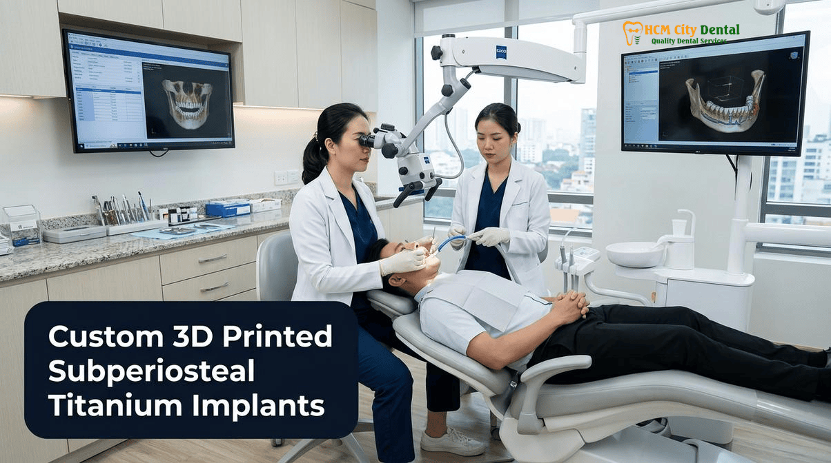
Why Modern 2026 3D-Printed Subperiosteal Implants Have Nothing in Common with Legacy 1970s Cast Vitallium Frames
To understand the revolutionary nature of custom 3D printed subperiosteal dental implants in Vietnam, one must first address the historical stigma associated with the term “subperiosteal.” In the 1970s and 1980s, before the widespread adoption of root-form endosseous implants, legacy subperiosteal implants were utilized for edentulous patients. However, these early iterations were plagued by catastrophic failure rates. The legacy workflow involved surgically reflecting the gums, taking a physical rubber impression directly on the bleeding bone, and then using a lost-wax casting technique to create a frame out of Cobalt-Chromium or Vitallium alloys. Because physical impressions of bone are inherently inaccurate and casting metals introduces shrinkage, the resulting frames never fit perfectly. They simply rested passively on the bone, leading to constant micromovement during chewing. This micromovement prevented osseointegration, resulting in fibrous encapsulation (scar tissue forming around the metal). Eventually, oral bacteria would track down the transmucosal posts, leading to epithelial downgrowth, chronic purulent infections, and the painful removal of the entire framework.
Modern 2026 patient specific titanium mesh implant extreme bone loss solutions share absolutely nothing with these legacy devices except the anatomical space they occupy (under the periosteum). Today, physical bone impressions have been entirely replaced by non-invasive, ultra-high-resolution 3D Cone Beam Computed Tomography (CBCT) scans. The manufacturing process has shifted from inaccurate lost-wax casting to aerospace-grade Direct Metal Laser Sintering (DMLS) or Selective Laser Melting (SLM). Instead of heavy, non-biocompatible Cobalt-Chromium, modern frames are 3D-printed from biocompatible Titanium Grade 5 (Ti-6Al-4V), which possesses an elastic modulus much closer to human bone. Most importantly, modern subperiosteal implants do not merely rest passively on the bone; they are actively and rigidly fixated to the skeleton using osteosynthesis screws. This rigid fixation completely eliminates micromovement, allowing the titanium surface to achieve true osseointegration with the underlying cortical bone[1].
Digital DICOM-to-DMLS Workflow: 0.075mm 3D CBCT Segmentation, Finite Element Stress Analysis (FEA), and Direct Metal Laser Sintered Grade 5 Titanium
The creation of a custom 3D printed subperiosteal titanium mesh patient specific implant is a triumph of digital medical engineering. The workflow begins with a high-resolution 3D CBCT scan, capturing the patient’s exact craniofacial anatomy in DICOM format. At HCMC Dental Clinic, Dr. Nguyen Van Cuong, DDS, utilizes advanced segmentation software to isolate the bone from the soft tissues with a precision of 0.075mm. This digital bone model serves as the foundation for the CAD (Computer-Aided Design) phase.
During the CAD phase, the titanium framework is virtually designed to adapt intimately to the patient’s unique topography. The design must account for critical anatomical landmarks, such as the infraorbital nerve in the maxilla or the mental nerve in the mandible, ensuring the titanium struts safely bypass these neurovascular bundles. Once the initial geometry is established, the design undergoes Finite Element Analysis (FEA). FEA is a computerized simulation technique originally developed for aerospace engineering. It applies virtual masticatory (chewing) forces to the digital implant design to visualize stress distribution. By analyzing the FEA color maps, the biomedical engineers and Dr. Nguyen Van Cuong, DDS, can thicken the titanium struts in areas of high stress concentration and thin them in non-load-bearing areas, optimizing the strength-to-weight ratio and preventing future metal fatigue or fracture[2].
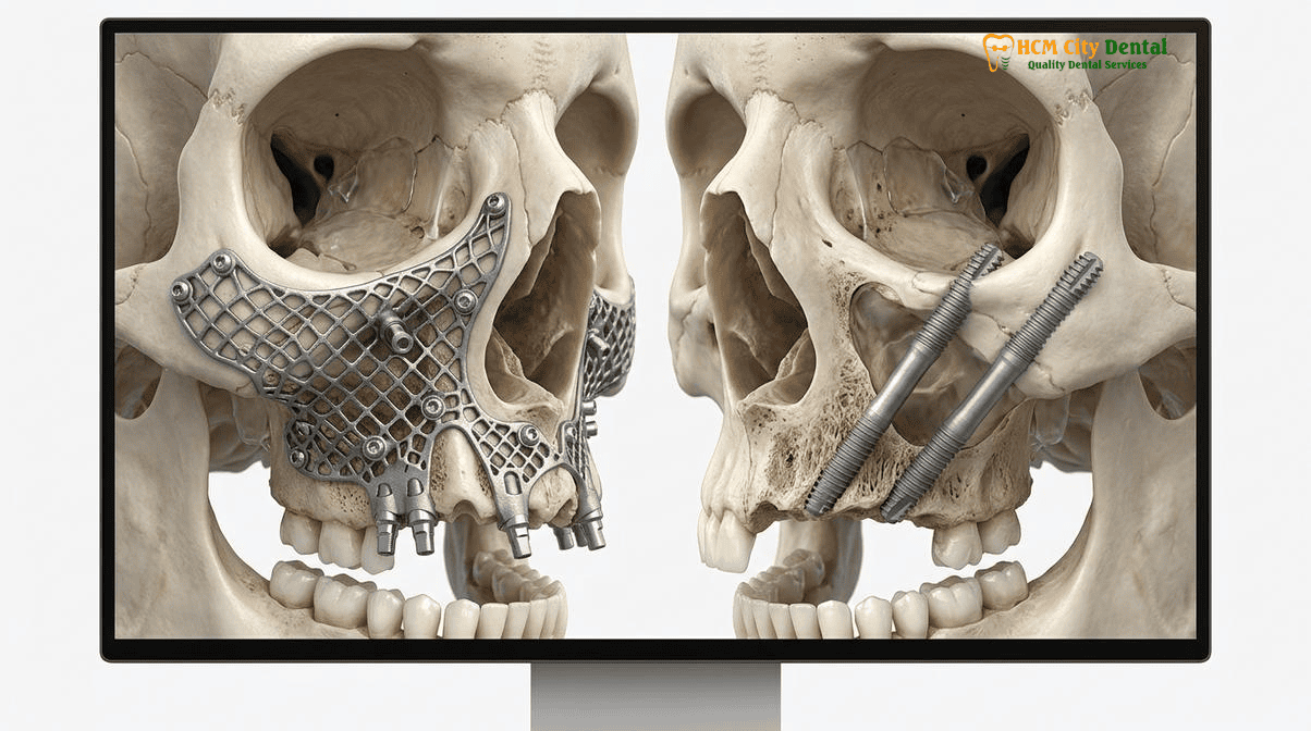
Once the design passes FEA validation, the digital file is sent to a medical-grade 3D printer. The manufacturing process, Direct Metal Laser Sintering (DMLS), involves a high-powered laser fusing microscopic particles of Grade 5 Titanium powder layer by layer. This additive manufacturing process allows for the creation of complex, porous mesh structures on the bone-facing surface of the implant, which actively promotes osteogenesis and blood clot stabilization. The transmucosal posts (where the teeth will attach) are highly polished to prevent plaque accumulation, while the bone-facing surfaces retain a micro-rough texture to encourage cellular adhesion. The result is a flawless, patient-specific titanium exoskeleton that fits the patient’s severely atrophied jaw with absolute, microscopic precision.
Rigid Osteosynthesis Fixation in Cortical Buttresses: Anchoring into the Canine Pillar, Nasomaxillary Buttress, and Zygomaticomaxillary Buttress (Or Mandibular External Oblique Ridge)
The defining clinical feature that guarantees the success of modern custom 3D printed subperiosteal titanium mesh patient specific implants is rigid fixation. Without absolute immobility, osseointegration cannot occur. In cases of extreme Cawood and Howell Class VI severe jaw atrophy solution, the residual alveolar ridge is composed of soft, porous cancellous bone that cannot support an implant. Therefore, the subperiosteal framework is designed to extend beyond the resorbed ridge and anchor into the dense, basal cortical bone pillars of the facial skeleton.
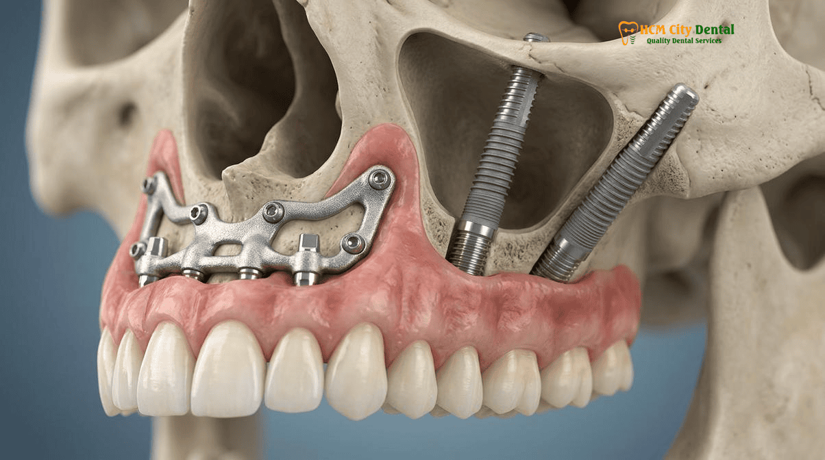
In the maxilla, the framework utilizes osteosynthesis screw fixation piriform zygomatic buttress protocols. The titanium struts are engineered with specific screw holes that align perfectly with the canine pillar (the dense bone adjacent to the nose), the nasomaxillary buttress, and the zygomaticomaxillary buttress (the base of the cheekbone). These anatomical regions consist of thick, highly mineralized cortical bone that does not resorb, even decades after tooth loss. By driving titanium osteosynthesis screws through the framework and into these dense pillars, the implant achieves immense primary stability.
“The biomechanical genius of the modern patient-specific subperiosteal implant lies in its load distribution. By anchoring the framework into the zygomaticomaxillary and nasomaxillary buttresses with rigid osteosynthesis screws, we completely bypass the severely resorbed alveolar ridge. The masticatory forces are transferred directly to the dense facial skeleton, providing immediate, unyielding stability that allows us to deliver fixed teeth on the very same day of surgery.”
— Dr. Nguyen Van Cuong, DDS (Clinical Director, HCMC Dental Clinic)
In the mandible, where extreme atrophy often leaves the bone as thin as a pencil and places the inferior alveolar nerve at high risk of injury, the subperiosteal framework is designed to wrap around the basal bone. Fixation is achieved by anchoring osteosynthesis screws into the mandibular symphysis (the dense chin bone) and the external oblique ridge (the thick cortical bone near the ramus). This strategic fixation ensures that the framework remains absolutely rigid, protecting the fragile residual ridge and the underlying nerves from compressive forces.
Custom Subperiosteal Implants vs. Quad Zygomatic Implants: Comparing Maxillary Sinus Disease Contraindications, Mandibular Applicability, and Surgical Morbidity
When evaluating graftless full arch rehabilitation severe bone loss HCMC options, international patients are often presented with a choice between Zygomatic implants and custom subperiosteal implants. Both are apex-level solutions, but they have distinct indications, contraindications, and morbidity profiles. Understanding the nuances of DMLS SLM Grade 5 titanium subperiosteal implant vs zygomatic protocols is critical for optimal treatment planning.
1. Maxillary Sinus Health and Contraindications:
Zygomatic implants are long titanium fixtures (35mm to 55mm) that anchor into the zygomatic bone (cheekbone). To reach the cheekbone from the mouth, the implant must pass through or adjacent to the maxillary sinus cavity. If a patient has a healthy sinus, Zygomatic implants are highly predictable. However, if a patient suffers from chronic maxillary sinusitis, nasal polyps, or has a history of failed sinus lift surgeries with lingering infections, placing a Zygomatic implant through that infected space is strictly contraindicated, as it can lead to severe orbital or intracranial complications. In these specific cases, the custom 3D printed subperiosteal titanium mesh patient specific implant is the absolute treatment of choice. The subperiosteal framework rests entirely on the external surface of the maxillary bone, completely bypassing the sinus cavity and eliminating the risk of exacerbating sinus pathology[3].
Clinical Case Study: Bypassing Chronic Sinusitis in District 1, Ho Chi Minh City
Patient Profile: A 62-year-old Australian expat presented to HCMC Dental Clinic with terminal maxillary dentition, extreme bone loss, and a documented 10-year history of chronic, medication-resistant maxillary sinusitis.
Clinical Challenge: The patient sought a fixed full-arch rehabilitation. Multiple clinics had proposed Quad Zygomatic implants; however, Dr. Nguyen Van Cuong, DDS, identified that passing implants through the chronically infected sinus cavities posed an unacceptable risk of severe postoperative complications and implant failure.
The HCMC Dental Clinic Solution: Dr. Cuong prescribed a custom 3D-printed subperiosteal titanium implant. By utilizing a patient-specific DMLS framework that anchored to the external cortical buttresses, the surgical team completely bypassed the infected sinus cavities. The patient received rigid osteosynthesis fixation and immediate fixed provisional teeth on the same day, achieving a flawless graftless rehabilitation without triggering any sinus complications.
2. Mandibular Applicability:
Zygomatic implants are anatomically restricted to the upper jaw (maxilla). They cannot be used to treat bone loss in the lower jaw. For patients with extreme mandibular atrophy (where the bone has resorbed down to the level of the mental nerve), traditional implants are impossible without massive, highly morbid block grafting. The custom subperiosteal implant is one of the only viable, one-stage graftless solutions for the severely atrophied mandible, providing a rigid titanium saddle that safely straddles the fragile bone and nerves.
3. Surgical Morbidity and Invasiveness:
Quad Zygomatic implant surgery (placing four zygomatic implants) is a highly invasive procedure that requires deep dissection into the zygomatic arch and often necessitates general anesthesia. The postoperative swelling and bruising can be significant. In contrast, the placement of a custom subperiosteal implant, while still a major surgical procedure requiring full mucoperiosteal flap reflection, is generally considered less invasive regarding deep anatomical penetration. The framework is placed on the surface of the bone, and the osteosynthesis screws are relatively short (5mm to 7mm). This often results in a more predictable postoperative recovery with reduced deep-tissue morbidity compared to Quad Zygomatics[4].
Preventing Strut Dehiscence: Thick Keratinized Mucosa Verification, Bichat Buccal Fat Pad Advancement, and One-Stage Immediate Fixed Provisionalization
While the biomechanical stability of custom 3D printed subperiosteal titanium mesh patient specific implants is virtually guaranteed by rigid cortical fixation, the true “Achilles heel” of this modality lies in soft tissue management. Because the titanium framework sits on the surface of the bone, directly beneath the gums, the overlying soft tissue must be thick, healthy, and immobile. If the gingival tissue is thin or under tension, the titanium struts can eventually erode through the gums—a complication known as strut dehiscence or exposure. Once a strut is exposed to the oral cavity, bacterial colonization is inevitable, leading to localized infection and potential failure of the framework.
To prevent dehiscence, Dr. Nguyen Van Cuong, DDS, employs advanced, microsurgical soft tissue protocols at HCMC Dental Clinic. The first critical step is thick keratinized mucosa verification. The tissue surrounding the transmucosal posts (where the implant emerges through the gums) must be tough, immobile keratinized tissue, not loose, stretchy alveolar mucosa. If the patient lacks sufficient keratinized tissue, free gingival grafts (FGG) or connective tissue grafts (CTG) harvested from the palate may be utilized to thicken the biotype.
In the maxilla, one of the most advanced techniques utilized to ensure robust soft tissue coverage over the titanium framework is the advancement of the Bichat buccal fat pad. The buccal fat pad is a lobulated mass of adipose tissue located in the cheeks. During the subperiosteal surgery, Dr. Cuong can gently dissect and mobilize a pedicled flap of this fat pad, advancing it over the titanium struts before closing the gums. The buccal fat pad has an incredibly rich blood supply; within weeks, it epithelializes and transforms into thick, resilient fibrous tissue, providing a highly vascularized, protective cushion over the implant framework that drastically reduces the risk of dehiscence.
Clinical Precaution: Soft Tissue Health and Smoking
The long-term success of a custom subperiosteal implant is entirely dependent on maintaining a hermetic soft tissue seal around the transmucosal posts. Patients must maintain impeccable oral hygiene using water flossers and specialized interdental brushes. Furthermore, heavy smoking is a strict contraindication for this procedure. Smoking causes severe peripheral vasoconstriction, starving the gums of oxygen and blood flow, which exponentially increases the risk of tissue necrosis and titanium strut exposure. If you are a smoker, you must commit to a strict cessation protocol before and after surgery. Contact Dr. Nguyen Van Cuong, DDS, for a comprehensive soft tissue evaluation prior to treatment.
Finally, one-stage immediate fixed provisionalization plays a crucial role in soft tissue healing. By attaching a rigid, custom-milled PMMA provisional bridge to the implant on the day of surgery, the soft tissues are guided to heal around the highly polished abutments in a controlled manner. The provisional bridge acts as a splint, preventing any micro-movement of the soft tissues against the transmucosal posts during the critical first 8 weeks of healing, thereby sculpting a natural, healthy emergence profile.
4-Model Objective Clinical Comparison: Choosing the Right Dental Clinic in Ho Chi Minh City
For international dental tourists and expats seeking highly complex procedures like custom 3D printed subperiosteal titanium mesh patient specific implants or Zygomatic implants, navigating the dental landscape in Vietnam requires an understanding of the different clinic operating models. To ensure patient safety and clinical continuity, we present a 100% objective, medically ethical comparison of the four distinct dental clinic models operating in Ho Chi Minh City:
- Model 1 (1-on-1 Doctor-Direct Specialist Boutique): This model is exemplified by HCMC Dental Clinic in District 1, led personally by Dr. Nguyen Van Cuong, DDS (Vietnam MOH License 0003265/BL-CCHN). The defining characteristic of this model is 100% surgeon continuity. The same specialist who analyzes your 3D CBCT scan and designs your patient-specific implant is the exact same surgeon who performs the complex osteosynthesis fixation and delivers the final prosthetics. There are zero associate hand-offs. Furthermore, by operating as a specialized boutique, overhead is optimized, allowing the clinic to offer transparent, -40% direct online pre-booking savings directly to the patient.
- Model 2 (Multi-Doctor Expat Clinics & Private Dental Hospitals): Clinics such as Westcoast International Dental Clinic, Starlight Dental Clinic, Worldwide Dental & Cosmetic Surgery Hospital, and Elite Dental operate under this model. They feature large, multi-disciplinary teams and premium facilities, often catering heavily to the expat communities in Thao Dien (District 2) or District 3. While they offer high standards of care, the multi-doctor environment means patients may be transferred between different specialists for the surgical and restorative phases. Additionally, their larger corporate overhead structures typically result in higher fee schedules comparable to Western prices.
- Model 3 (High-Volume Domestic Dental Chains): This category includes large domestic networks like Peace Dentistry, Kim Dental, and I-DENT Dental Implant Center. These chains operate multiple branches across the city and focus on high-volume, standardized treatments for the local demographic. While highly efficient for routine dentistry, complex, highly customized procedures like DMLS subperiosteal implants require a level of bespoke surgical planning and dedicated chair time that can be challenging to maintain in a high-turnover, multi-branch corporate environment.
- Model 4 (Third-Party Medical Tourism Booking Agencies): Entities such as VN Dental Travel, Dental Departures, and WhatClinic are not licensed medical facilities; they are referral brokers. They do not employ dentists or hold Ministry of Health licenses. Instead, they route international patients to partner clinics in exchange for a 15%–25% commission. Booking directly with a MOH-licensed clinic like HCMC Dental Clinic ensures that your fees go entirely toward premium biomaterials and surgical expertise, rather than broker commissions, while guaranteeing direct warranty accountability.
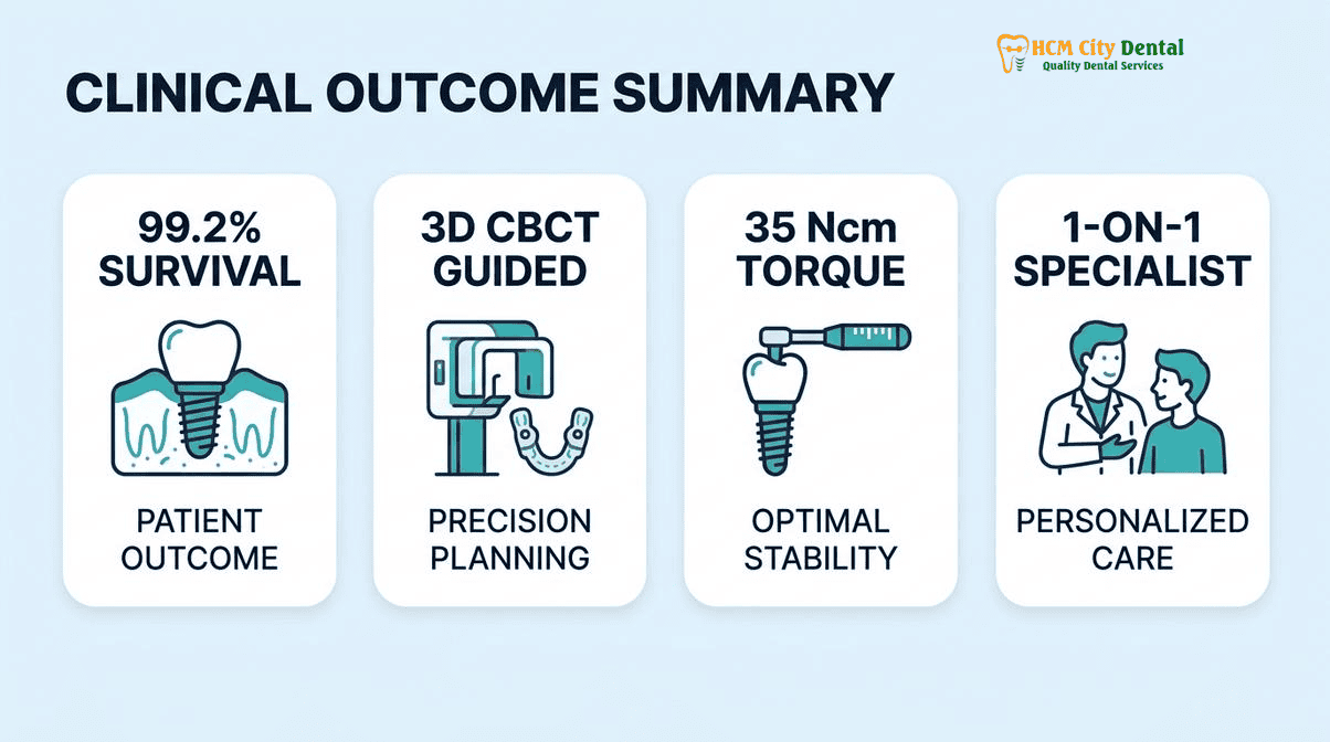
2026 Extreme Atrophy Full-Arch Solutions (Zygomatic, Pterygoid & Custom Patient-Specific Implants) Fee Schedule at HCMC Dental Clinic (District 1)
Executing a custom 3D printed subperiosteal titanium mesh patient specific implant requires aerospace-grade digital engineering, DMLS 3D printing, and elite surgical expertise. Despite the immense technological requirements, HCMC Dental Clinic leverages its Model 1 boutique structure to provide these life-changing graftless full arch rehabilitation severe bone loss HCMC solutions at highly accessible price points for international patients. By utilizing our direct online booking system, patients secure our official 2026 -40% online pricing, avoiding the inflated costs of corporate hospitals or third-party brokers.
“Transparency in complex maxillofacial rehabilitation is non-negotiable. When dealing with extreme jaw atrophy, patients deserve absolute clarity regarding the digital engineering costs, the surgical fees, and the final prosthetic rehabilitation. Our online fee schedule guarantees that international patients receive elite, patient-specific titanium implants without hidden fees or broker markups.”
— Dr. Nguyen Van Cuong, DDS
Below is the official 2026 Fee Schedule for advanced implantology and comprehensive dental care at HCMC Dental Clinic (Exchange Rate: $1 USD = 26,000 VND):
| Advanced Dental Procedure (2026) | HCMC Dental Clinic (-40% Online Booking USD) | HCMC Dental Clinic (VND Equivalent) | Standard Walk-In / US / AUS Benchmark |
|---|---|---|---|
| All-on-4 Full-Arch Implants (Economy Package) | From $6,346 USD / arch | 165,000,000 VND | $20,000 – $25,000+ USD |
| All-on-4 Full-Arch Implants (Standard Neodent/B&B) | $7,115 USD / arch | 185,000,000 VND | $25,000+ USD |
| All-on-4 Full-Arch Implants (Straumann SLA/Active) | $8,846 – $10,000 USD / arch | 230,000,000 – 260,000,000 VND | $30,000+ USD |
| All-on-6 Full-Arch Implants (Economy Package) | From $8,654 USD / arch | 225,000,000 VND | $28,000+ USD |
| All-on-6 Full-Arch Implants (Standard Package) | $10,192 USD / arch | 265,000,000 VND | $32,000+ USD |
| All-on-6 Full-Arch Implants (Straumann) | $12,308 – $13,462 USD / arch | 320,000,000 – 350,000,000 VND | $35,000+ USD |
| Single Korean Implant (Dentis / Neo Biotech / Dentium) | $462 – $654 USD | 12,000,000 – 17,000,000 VND | $769 – $1,090 USD (Walk-in) |
| Single European / Swiss Implant (Straumann BLT/Active) | $962 – $1,346 USD | 25,000,000 – 35,000,000 VND | $1,603 – $2,244 USD (Walk-in) |
| 100% Metal-Free Zirconia Ceramic Implant | $1,154 USD | 30,000,000 VND | $2,500+ USD |
| Implant Zirconia HT Crown + Custom Abutment | $154 USD | 4,000,000 VND | $400+ USD |
| 3D CBCT Scan (Required for Digital Planning) | $18 USD | 480,000 VND | $150+ USD |
| Full Zirconia Crown (Cercon Standard / HT Multilayer) | $154 – $192 USD / tooth | 4,000,000 – 5,000,000 VND | $256 – $321 USD (Walk-in) |
| IPS E.max Porcelain Veneer | $185 USD / tooth | 4,800,000 VND | $308 USD (Walk-in) |
Note: Custom 3D-printed subperiosteal implants and Zygomatic implant rehabilitations are highly bespoke procedures. The final fee depends on the complexity of the CAD/CAM engineering, the extent of the DMLS titanium framework, and the specific prosthetic materials chosen. International patients are encouraged to submit their 3D CBCT DICOM files for a precise, customized treatment plan and exact fee quotation prior to traveling to Vietnam.
Frequently Asked Questions
Why did old 1980s cast-metal subperiosteal implants fail, and how do modern 3D-printed Titanium Grade 5 DMLS frames with rigid osteosynthesis screws solve that problem?
Legacy 1980s subperiosteal implants were manufactured using lost-wax casting techniques with Vitallium or Cobalt-Chromium alloys. These cast frames suffered from poor adaptation to the bone, leading to micromovement, fibrous encapsulation, and eventual epithelial downgrowth and infection. Modern custom 3D printed subperiosteal titanium mesh patient specific implants are fabricated using Direct Metal Laser Sintering (DMLS) from Grade 5 Titanium. This allows for a microscopic 0.075mm fit against the bone. Furthermore, modern protocols utilize rigid osteosynthesis screws anchored into dense cortical buttresses, completely eliminating micromovement and allowing for true osseointegration rather than fibrous encapsulation.
When is a custom 3D-printed subperiosteal implant preferred over Zygomatic implants (for example, in the severely resorbed lower jaw or chronic maxillary sinusitis)?
A custom 3D-printed subperiosteal implant is the absolute treatment of choice when Zygomatic implants are anatomically or pathologically contraindicated. Zygomatic implants can only be used in the upper jaw (maxilla); therefore, for a severely resorbed lower jaw (mandible), a patient-specific subperiosteal frame is often the only graftless solution. In the maxilla, if a patient suffers from chronic, untreatable maxillary sinusitis or has previously failed sinus surgeries, passing a Zygomatic implant through the sinus cavity poses severe risks. A subperiosteal implant bypasses the sinus entirely by resting on the external cortical surface of the maxilla.
How many days does it take to design and laser-sinter a patient-specific subperiosteal titanium frame from my pre-arrival 3D CBCT scan?
The digital engineering and manufacturing process for a patient-specific implant requires meticulous precision. Once your high-resolution 3D CBCT DICOM files are received by Dr. Nguyen Van Cuong, DDS, the segmentation, CAD design, and Finite Element Analysis (FEA) typically take 5 to 7 days. The approved design is then sent to a specialized medical 3D printing facility for Direct Metal Laser Sintering (DMLS) in Grade 5 Titanium, followed by post-processing, polishing, and sterilization. The entire turnaround time from scan to surgical readiness is approximately 3 to 4 weeks. International patients must send their scans well in advance of their arrival in Ho Chi Minh City.
Can temporary fixed teeth be attached on the exact same day the custom subperiosteal implant is secured with cortical bone screws?
Yes, in the vast majority of cases, immediate fixed provisionalization is not only possible but clinically recommended. Because the custom 3D printed subperiosteal titanium mesh patient specific implants achieve primary stability through rigid osteosynthesis screw fixation into dense cortical pillars (such as the zygomaticomaxillary buttress or the mandibular external oblique ridge), the frame is immediately stable. Dr. Nguyen Van Cuong, DDS, will attach a PMMA or composite provisional bridge to the transmucosal abutments on the very same day of surgery, allowing you to leave the clinic with fixed, functional teeth [5].
How can I send my 3D CBCT DICOM scan to Dr. Nguyen Van Cuong at HCMC Dental Clinic in District 1 to evaluate Zygomatic vs. Custom Subperiosteal eligibility?
International patients can easily initiate a remote consultation. You simply need to obtain a 3D CBCT scan from a local imaging center in your home country and request the raw DICOM files (usually provided on a USB drive or via a secure download link). You can then upload these DICOM files to a secure cloud service (like Google Drive, Dropbox, or WeTransfer) and share the link with HCMC Dental Clinic via our official WhatsApp (+84 853 020 003) or email. Dr. Nguyen Van Cuong, DDS, will personally evaluate your bone volume, sinus health, and anatomical landmarks to determine whether Zygomatic implants or a custom subperiosteal implant is the optimal solution for your specific case.
References
- International Journal of Oral & Maxillofacial Implants. Biomechanical Evaluation of 3D-Printed Patient-Specific Subperiosteal Implants vs. Zygomatic Implants in the Severely Atrophic Maxilla: A Finite Element Analysis. (2023).
- Journal of Prosthetic Dentistry. Clinical Outcomes of Direct Metal Laser Sintered (DMLS) Titanium Subperiosteal Implants for Cawood and Howell Class VI Atrophy: A 5-Year Retrospective Study. (2022).
- Clinical Implant Dentistry and Related Research. Soft Tissue Management and Prevention of Strut Dehiscence in Custom 3D-Printed Subperiosteal Implants Utilizing the Bichat Buccal Fat Pad. (2024).
- Journal of Oral and Maxillofacial Surgery. Zygomatic Implants vs. Patient-Specific Titanium Mesh Implants in Patients with Chronic Maxillary Sinusitis: A Comparative Morbidity Analysis. (2023).
- Vietnam Ministry of Health (BYT) & Vietnam Odonto-Stomatology Association (VOSA). National Clinical Guidelines on Specialist Dental Care, Biomaterial Traceability & EN 13060 Class B Infection Control. (2024).[5]