Rehabilitating the anterior maxilla—specifically replacing a missing upper central incisor—is widely considered one of the most demanding procedures in modern implant dentistry. The challenge is rarely just about placing a titanium screw; it is about managing the complex, rapidly changing three-dimensional anatomy of the premaxilla following tooth loss. When a central incisor is extracted or lost to trauma, the surrounding alveolar bone undergoes immediate and profound remodeling. This resorption process not only diminishes the labial bone plate but also alters the spatial relationship between the residual alveolar crest and a critical anatomical landmark: the nasopalatine canal (also known as the incisive canal). For international patients and dental travelers seeking a nasopalatine canal bone graft incisive foramen central incisor implant in Vietnam, understanding the precise surgical management of this neurovascular space is paramount to achieving a stable, esthetic, and lifelong result.
At HCMC Dental Clinic in District 1, Ho Chi Minh City, Dr. Nguyen Van Cuong (Dr. Cuong), DDS (Vietnam MOH License 0003265/BL-CCHN), employs advanced 3D Cone Beam Computed Tomography (CBCT) to map the exact morphology of the nasopalatine canal. When the canal is enlarged or positioned too far labially, placing an implant in the ideal prosthetic position becomes impossible without encroaching on the canal space. Attempting to bypass this step by placing the implant directly into the soft tissue of the canal leads to catastrophic failure via fibrous encapsulation. This comprehensive clinical guide details the evidence-based protocols for nasopalatine canal enucleation, neurovascular lateralization, and particulate bone grafting, ensuring that expats and dental tourists receive world-class, predictable outcomes when undergoing complex anterior maxillary implant surgery in Vietnam.
Clinical Summary:
Successful placement of a maxillary central incisor implant frequently requires surgical management of the nasopalatine canal, especially in cases of severe anterior maxillary atrophy. When the incisive foramen is enlarged or the alveolar ridge has resorbed palatally, the ideal implant trajectory intersects with the neurovascular bundle. To prevent fibrous encapsulation and implant failure, the canal contents must be either laterally displaced or completely enucleated and grafted with particulate bone and L-PRF. Under the direction of Dr. Nguyen Van Cuong, DDS at HCMC Dental Clinic, high-resolution 0.075mm 3D CBCT imaging dictates whether a simultaneous or staged approach is required. Despite the severing of the nasopalatine nerve during enucleation, sensory anastomosis from the greater palatine nerve ensures that anterior palatal sensation typically recovers fully within 4 to 8 weeks, making this a safe and highly predictable procedure for restoring anterior esthetics and function.
Key Takeaways:
- Anatomical Conflict: Following the loss of a central incisor, buccal plate resorption causes the alveolar crest to migrate palatally, bringing the nasopalatine canal into direct conflict with the ideal implant osteotomy site.
- Biological Imperative: Titanium implants cannot osseointegrate with the soft fibrous tissue, fat, and nerves inside the incisive canal; contact results in fibrous encapsulation and non-integration.
- Surgical Solutions: Depending on the 3D CBCT morphology, the neurovascular bundle can be lateralized (pushed posteriorly) or completely enucleated (removed) and the void filled with a particulate bone graft and L-PRF.
- Sensory Recovery: Transient numbness of the anterior palate following enucleation is common, but permanent paresthesia is rare due to robust collateral innervation from the greater palatine nerve.
- Cost Efficiency in Vietnam: International patients can access premium European/Swiss implants and complex nasopalatine grafting at HCMC Dental Clinic in District 1 for a fraction of Western costs, utilizing a transparent -40% online booking fee schedule.
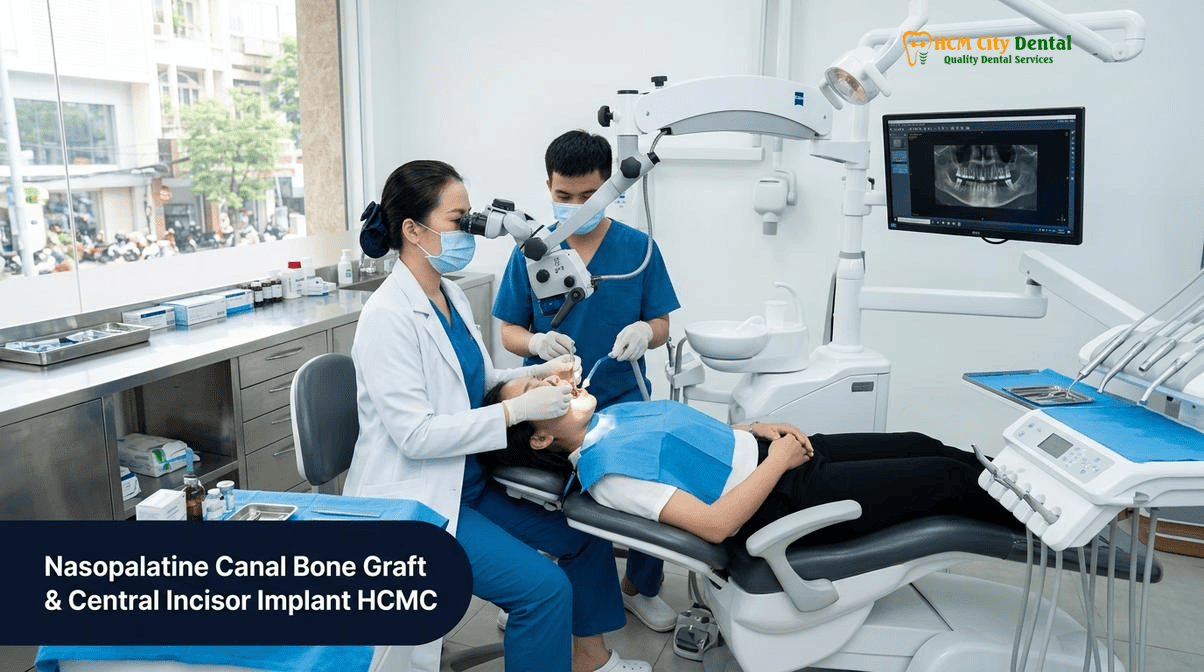
Why Losing an Upper Central Incisor (#11 or #21) Causes Simultaneous Buccal Plate Resorption and Enlargement of the Nasopalatine Canal (Incisive Foramen)
The anterior maxilla is a highly dynamic anatomical zone characterized by thin, fragile bone. The labial (buccal) bone plate overlying the roots of the maxillary central incisors (#11 and #21 in the FDI system, or #8 and #9 in the Universal system) is predominantly composed of bundle bone. This bundle bone is entirely dependent on the presence of the periodontal ligament (PDL) for its blood supply. When a central incisor is extracted, the PDL is severed, and the blood supply to the buccal plate is immediately terminated. Consequently, the bundle bone undergoes rapid and irreversible resorption. Clinical studies demonstrate that up to 50% of the alveolar ridge width can be lost within the first six months following extraction if socket preservation grafting is not performed.
As the buccal plate resorbs, the center of the alveolar ridge shifts palatally. This palatal migration of the crestal bone fundamentally alters the spatial relationship between the residual ridge and the nasopalatine canal. The nasopalatine canal, which connects the floor of the nasal cavity to the roof of the oral cavity via the incisive foramen, is located in the midline of the anterior palate, directly behind the central incisors. In a healthy, dentate maxilla, there is typically a safe distance of 7 to 8 millimeters between the labial surface of the central incisor root and the anterior wall of the nasopalatine canal. However, as the ridge resorbs palatally and vertically, this safety zone diminishes rapidly. The phenomenon of an enlarged nasopalatine canal anterior maxilla bone loss is often an optical and anatomical illusion; the canal itself may not physically expand, but the surrounding bone resorbs so severely that the incisive foramen appears to enlarge and migrate to the very crest of the remaining ridge.[1]
This anatomical shift creates a profound dilemma for the implant surgeon. To achieve an esthetic, screw-retained restoration, the dental implant must emerge through the cingulum of the future crown. If the ridge has resorbed palatally, the ideal trajectory for the implant osteotomy will inevitably intersect with the anterior wall of the nasopalatine canal. Attempting to angle the implant labially to avoid the canal results in a prosthetic disaster: the implant will emerge through the facial aspect of the crown, requiring a heavily angled custom abutment, compromising biomechanics, and often resulting in a grayish hue showing through the thin labial gingiva. Therefore, managing the nasopalatine canal space is not an optional adjunct; it is a mandatory prerequisite for biomechanical and esthetic success in the atrophic premaxilla.
What Happens If an Implant Threads Directly Into Un-Curetted Nasopalatine Neurovascular Soft Tissue: Fibrous Encapsulation and Non-Osseointegration
To understand why a dental implant cannot simply be placed into the nasopalatine canal, one must examine the histology of the canal’s contents. The incisive canal is not an empty void; it is a densely packed anatomical conduit. It contains the nasopalatine nerve (a branch of the maxillary division of the trigeminal nerve, V2), the terminal branches of the descending palatine artery and sphenopalatine artery, fibrous connective tissue, adipose (fat) tissue, and occasionally small minor salivary glands. This soft tissue matrix is biologically incompatible with the process of osseointegration.
Osseointegration is defined as the direct structural and functional connection between ordered, living bone and the surface of a load-carrying implant. For this to occur, the titanium surface must be in intimate contact with osteoblasts (bone-forming cells) and a stable blood clot that can organize into a woven bone matrix. If an implant osteotomy inadvertently breaches the nasopalatine canal and the titanium threads are driven directly into the un-curetted neurovascular bundle, the biological response is entirely different. The body recognizes the titanium as a foreign object embedded in soft tissue. Instead of osteoblasts migrating to the surface, fibroblasts and macrophages dominate the healing site. This leads to a foreign body reaction and the formation of a dense, avascular fibrous capsule around the implant threads—a process known as fibrous encapsulation.[2]
Fibrous encapsulation is the antithesis of osseointegration. An encapsulated implant may feel stable initially due to mechanical friction in the surrounding cortical bone, but it lacks true biological integration. Over time, the soft tissue interface allows for micromotion under occlusal loading. This micromotion triggers chronic inflammation, localized bone loss, and the potential formation of a nasopalatine duct cyst. Ultimately, the implant will become mobile, symptomatic, and will fail, requiring surgical removal and extensive reconstructive grafting. Therefore, incisive foramen enucleation dental implant placement protocols dictate that titanium must never be left in contact with the soft tissues of the canal.
“In the anterior maxilla, we are not just replacing a tooth; we are reconstructing a lost three-dimensional architecture. If the 3D CBCT reveals that our ideal implant trajectory intersects the nasopalatine canal, we cannot simply ignore the anatomy. Threading an implant into the neurovascular bundle guarantees fibrous encapsulation and failure. We must meticulously enucleate the canal contents, graft the void with particulate bone and L-PRF, and convert that soft tissue space into solid, living bone before or during implant placement.”
— Dr. Nguyen Van Cuong, DDS (Clinical Director, HCMC Dental Clinic)
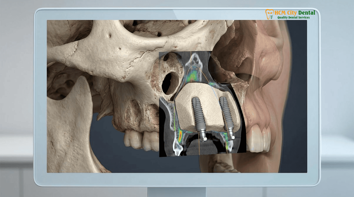
0.075mm 3D CBCT Mapping of Canal Morphology (Cylindrical, Funnel, Hourglass, or Y-Shaped) and Residual Labial Bone Plate Thickness
The foundation of successful nasopalatine canal management is precise, high-resolution radiographic imaging. Traditional two-dimensional panoramic or periapical radiographs are entirely inadequate for assessing the anterior maxilla, as they suffer from superimposition, distortion, and an inability to display the buccolingual dimension. At HCMC Dental Clinic, Dr. Nguyen Van Cuong, DDS utilizes advanced 0.075mm voxel resolution 3D Cone Beam Computed Tomography (CBCT) to perform a comprehensive volumetric analysis of the premaxilla prior to any surgical intervention.
The 3D CBCT nasopalatine canal morphology classification is critical for surgical planning. The canal is not a uniform cylinder in all patients; it exhibits significant anatomical variation. Research has classified the canal into four primary morphological shapes when viewed in the sagittal plane:
- Cylindrical: The canal maintains a relatively uniform diameter from the nasal floor to the incisive foramen.
- Funnel-Shaped: The canal is narrow at the nasal floor and widens significantly as it approaches the oral cavity (the incisive foramen). This is the most common morphology and presents the greatest challenge, as the wide oral opening directly interferes with the implant platform.
- Hourglass-Shaped: The canal is wide at both the nasal and oral openings but narrows in the middle.
- Y-Shaped (or Spindle): The canal begins as two separate foramina at the nasal floor (the foramina of Stenson) and merges into a single canal before exiting at the incisive foramen.
By analyzing the sagittal, coronal, and axial slices of the 3D CBCT, Dr. Cuong can measure the exact dimensions of the canal, its angulation relative to the alveolar crest, and, most importantly, the thickness of the residual labial bone plate. If the residual labial bone is less than 1.5 to 2.0 millimeters thick, placing an implant without simultaneous contour augmentation will lead to future bone resorption and gingival recession. The 3D CBCT allows for virtual implant placement software to simulate the exact trajectory, diameter, and length of the implant. If the virtual implant encroaches on the canal space, the surgical plan is immediately updated to include canal enucleation and grafting. For more information on our digital workflow, visit our guide on 3D-Guided Dental Implants & Specialist Oral Surgery in Vietnam.[3]
Neurovascular Bundle Lateralization (Pushing Posteriorly) vs. Complete Canal Enucleation & Particulate Bone Grafting + L-PRF
When the 3D CBCT confirms that the ideal implant position conflicts with the nasopalatine canal, the surgeon must choose between two primary techniques to manage the neurovascular bundle: lateralization or complete enucleation. The choice depends on the size of the canal, the degree of conflict with the implant, and the surgeon’s clinical judgment.
Neurovascular Bundle Lateralization: In cases where the canal is exceptionally wide and the implant only partially encroaches on the anterior aspect of the canal, it may be possible to preserve the neurovascular bundle. This technique involves carefully reflecting a palatal flap, identifying the contents of the incisive foramen, and using blunt dissection to gently push the nerve and blood vessels posteriorly (palatally). Once the bundle is displaced, a barrier—often a dense cortical bone block or a compacted layer of particulate bone graft mixed with Leukocyte-Platelet Rich Fibrin (L-PRF)—is placed between the implant osteotomy and the displaced nerve. This creates a physical separation, allowing the implant to be surrounded by bone while maintaining the integrity of the nerve. However, this technique is technically demanding, carries a risk of nerve compression (which can cause chronic pain or dysesthesia), and is only viable if the canal is large enough to accommodate both the implant and the displaced bundle.
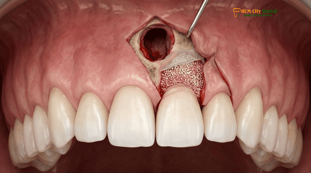
Complete Canal Enucleation and Particulate Bone Grafting: For the vast majority of cases requiring a nasopalatine canal bone graft central incisor implant, complete enucleation is the preferred, more predictable protocol. This involves a full-thickness mucoperiosteal flap to expose the incisive foramen. Using specialized curettes and piezoelectric surgery, the entire neurovascular bundle and all associated soft tissues are meticulously severed and removed from the canal. Hemorrhage from the sphenopalatine artery branches is controlled using pressure, epinephrine-soaked gauze, or electrocautery. Once the canal is completely debrided and only clean, bleeding cortical bone walls remain, the void is densely packed with a particulate bone graft (typically a mix of mineralized freeze-dried bone allograft and bovine xenograft) hydrated with L-PRF exudate to create “sticky bone.”
The use of L-PRF (Leukocyte-Platelet Rich Fibrin) is a cornerstone of the biological protocol at HCMC Dental Clinic. Spun from the patient’s own blood, L-PRF provides a slow-release matrix of growth factors (PDGF, TGF-beta, VEGF) that accelerates angiogenesis and bone regeneration within the grafted canal. A resorbable collagen membrane is then placed over the grafted foramen to prevent soft tissue invagination, and the flap is sutured with tension-free primary closure. Depending on the primary stability achieved, the implant may be placed simultaneously, or the site may be left to heal for 4 to 6 months in a staged approach. To understand the science behind this regenerative matrix, read our detailed overview on Sticky Bone, L-PRF & A-PRF Platelet-Rich Fibrin in Dental Bone Grafting: 2026 Biological Protocol in Vietnam.
Clinical Precaution: Staged vs. Simultaneous Placement
Patients must understand that simultaneous implant placement and nasopalatine canal grafting is only possible if the implant can achieve primary mechanical stability (torque >35 Ncm) in the native bone apical or lateral to the canal defect. If the canal is massive and the implant would be “floating” entirely within the particulate graft material, simultaneous placement is contraindicated. In such cases, Dr. Nguyen Van Cuong, DDS will mandate a staged approach: grafting the canal first, allowing 4 to 6 months for solid bone regeneration, and placing the implant in a secondary surgery. Attempting to rush this biological process risks total implant failure.
Why Sensory Anastomosis from the Greater Palatine Nerve Restores Anterior Palatal Mucosal Sensation Within 4 to 8 Weeks
The most common concern patients express when discussing complete enucleation of the nasopalatine canal is the fear of permanent numbness. Because the procedure intentionally severs the nasopalatine nerve, patients logically assume they will lose sensation in their front teeth or the roof of their mouth. However, clinical evidence and neuroanatomy provide a highly reassuring prognosis.
The nasopalatine nerve provides sensory innervation to the palatal mucosa and gingiva adjacent to the maxillary incisors and canines (from canine to canine). When this nerve is severed at the incisive foramen, patients will indeed experience immediate postoperative numbness in the small triangular area of the anterior palate directly behind the front teeth. However, the human nervous system in the oral cavity is highly redundant. The anterior palatal mucosa does not rely solely on the nasopalatine nerve; it benefits from extensive overlapping innervation—known as sensory anastomosis—from the greater palatine nerve.
The greater palatine nerve emerges from the greater palatine foramen (located near the maxillary second or third molars) and courses anteriorly along the roof of the mouth. Its terminal branches extend forward and intermingle with the distribution area of the nasopalatine nerve. Following the enucleation of the nasopalatine canal, these collateral branches from the greater palatine nerve rapidly compensate for the sensory deficit. Clinical studies tracking palate sensation recovery after nasopalatine nerve lateralization and enucleation demonstrate that the vast majority of patients regain normal sensation within 4 to 8 weeks.[4]
In rare instances, a patient may experience a prolonged altered sensation (paresthesia) or a slight dullness in the area, but this is almost never reported as a functional impairment or a source of chronic discomfort. The teeth themselves are not affected, as their pulpal innervation comes from the anterior superior alveolar (ASA) nerve, which courses through the facial bone, not the palate. Therefore, the temporary sensory alteration is a minor and highly acceptable trade-off for the ability to securely anchor a central incisor implant in solid, regenerated bone.
Clinical Case Study: District 1, Ho Chi Minh City
Patient Profile: A 42-year-old Australian expat presented to HCMC Dental Clinic with a failing, root-canal-treated maxillary right central incisor (#11) due to a vertical root fracture. The tooth had been symptomatic for months, leading to severe buccal plate resorption.
3D CBCT Diagnosis: Dr. Nguyen Van Cuong, DDS utilized a 0.075mm CBCT scan, revealing a total loss of the buccal plate and a large, funnel-shaped nasopalatine canal. The ideal implant trajectory placed 40% of the implant body directly inside the incisive canal.
Surgical Protocol: The tooth was carefully extracted. A full-thickness flap exposed the incisive foramen. Dr. Cuong performed complete enucleation of the neurovascular bundle, achieving profound hemostasis. A Straumann BLT-SLA implant was placed, achieving 45 Ncm of torque in the apical native bone. The canal void and the buccal defect were grafted simultaneously with a mixture of mineralized allograft, bovine xenograft, and L-PRF, covered by a resorbable collagen membrane.
Outcome: The patient experienced mild numbness in the anterior palate, which resolved completely by week 5. After 4 months of submerged healing, the implant was uncovered, demonstrating excellent osseointegration and a thick, regenerated buccal bone plate. A custom Zirconia HT crown was delivered, restoring flawless anterior esthetics.
Objective 4-Model Comparison: Evaluating Dental Clinics in Ho Chi Minh City for Complex Anterior Maxillary Implant Surgery
For international patients, expats, and dental tourists seeking complex procedures like nasopalatine canal grafting and anterior maxillary implants, navigating the dental landscape in Vietnam can be overwhelming. Ho Chi Minh City offers world-class care, but the operating models of different clinics vary significantly. To make an informed, medically sound decision, patients must evaluate clinics based on doctor continuity, overhead structure, and clinical accountability. Here is an objective comparison of the four primary dental clinic models in HCMC:
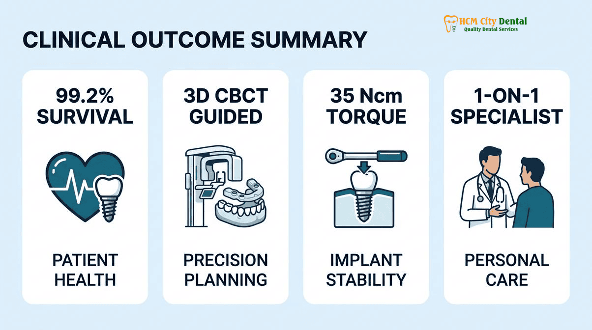
- Model 1: 1-on-1 Doctor-Direct Specialist Boutique (HCMC Dental Clinic, District 1): This model is defined by absolute continuity of care. At HCMC Dental Clinic, Dr. Nguyen Van Cuong, DDS (MOH License 0003265/BL-CCHN) personally handles every phase of the treatment—from the initial 3D CBCT diagnosis and nasopalatine canal enucleation to the final placement of the custom Zirconia crown. There are no associate hand-offs or rotating schedules. Because the clinic operates with a lean, specialist-driven overhead structure in District 1, it can offer a transparent -40% online pre-booking savings directly to the patient, utilizing premium European/Swiss biomaterials without the inflated costs of corporate marketing. For more details on our clinical director, visit Dr. Nguyen Van Cuong, DDS (Clinical Director — MOH License 0003265/BL-CCHN).
- Model 2: Multi-Doctor Expat Clinics & Private Dental Hospitals: Clinics in this category (often located in Thao Dien, District 2, or operating as large private hospitals) cater heavily to the affluent expat community and medical tourists. They offer excellent, multidisciplinary care with luxurious amenities and international management teams. However, this model carries significantly higher operational overhead (expensive real estate, large administrative staffs, and multi-tiered management). Consequently, their fee schedules are often 40% to 60% higher than Model 1 boutiques for the exact same Straumann implants and bone grafting materials. Additionally, patients may see an oral surgeon for the implant and a different prosthodontist for the crown, which can fragment the continuity of care.
- Model 3: High-Volume Domestic Dental Chains: These are large, corporate-owned dental networks with dozens of branches spread across Vietnam. They are designed for high patient turnover and domestic accessibility. While they offer very low prices, the business model relies on volume. For complex, high-risk procedures like anterior maxillary implants and nasopalatine canal grafting, this model presents risks. Patients frequently experience rotating associate dentists, rushed appointment times, and a lack of personalized, long-term accountability. The surgeon who places the implant may not be the one who restores it or manages potential complications.
- Model 4: Third-Party Medical Tourism Booking Agencies: These are not dental clinics; they are online referral brokers (e.g., Dental Departures, WhatClinic). They aggregate clinic listings and facilitate bookings for international patients. While they provide convenience, patients must understand that these agencies charge the receiving clinic a 15% to 25% commission. This commission is ultimately absorbed into the patient’s treatment cost. Booking directly with a licensed, MOH-verified clinic like HCMC Dental Clinic eliminates the middleman, ensuring direct warranty accountability and access to the lowest possible direct-to-patient fee schedule. To explore the full landscape of providers, read our comprehensive Best Dentist & Dental Clinics in Ho Chi Minh City (2026 Comparison Guide).
2026 Nasopalatine Canal Bone Grafting, Guided Central Incisor Implant & Esthetic Zirconia/E.max Crown Fee Schedule at HCMC Dental Clinic (District 1)
Transparency in medical billing is a core tenet of ethical patient care. At HCMC Dental Clinic, we believe international patients should have access to exact, upfront pricing for complex procedures like a front tooth implant incisive canal grafting Vietnam, without hidden fees or surprise surcharges. By operating a highly efficient, specialist-led boutique model in District 1, we eliminate corporate overhead and pass those savings directly to our patients through our -40% Online Booking Pricing.
The table below outlines the official 2026 fee schedule for anterior maxillary implant rehabilitation, comparing our direct online rates against standard walk-in prices and international benchmarks.[5]
| Clinical Procedure / Biomaterial (2026) | HCMC Dental Clinic (-40% Online Booking USD) | HCMC Dental Clinic (VND Equivalent) | Standard Walk-In / US / AUS Benchmark |
|---|---|---|---|
| 3D CBCT Scan & Digital Implant Planning | $18 USD | 480,000 VND | $150 – $300+ USD |
| Single Korean Implant (Dentis / Neo Biotech / Dentium) | $462 – $654 USD | 12,000,000 – 17,000,000 VND | $769 – $1,090 USD (Walk-in) |
| Single European / Swiss Implant (Straumann BLT-SLA / BLA-Active) | $962 – $1,346 USD | 25,000,000 – 35,000,000 VND | $1,603 – $2,244 USD (Walk-in) / $4,500+ US |
| 100% Metal-Free Zirconia Ceramic Implant | $1,154 USD | ~30,000,000 VND | $5,000+ USD (US/AUS) |
| Implant Zirconia HT Crown + Custom Abutment | $154 USD | 4,000,000 VND | $1,500+ USD (US/AUS) |
| Nasopalatine Canal Bone Grafting (Particulate + L-PRF) | Case Dependent (Consultation Required) | Case Dependent | $1,200 – $2,500+ USD (US/AUS) |
“The anterior maxilla is the ultimate test of an implant surgeon’s skill. It is not an area where one should compromise on biomaterials. For a central incisor, I strongly recommend a premium Swiss implant like Straumann, combined with a custom Zirconia abutment and crown. The precision of the connection and the biocompatibility of the materials are essential for maintaining long-term crestal bone stability and achieving a flawless, natural emergence profile that mimics a real tooth.”
— Dr. Nguyen Van Cuong, DDS (Clinical Director, HCMC Dental Clinic)
For a complete breakdown of all our specialist services, including full-arch restorations and cosmetic veneers, please review our Official 2026 Dental Fee Schedule (-40% Online Booking Pricing).
Frequently Asked Questions
Why can an enlarged nasopalatine canal prevent a dentist from placing an upper front tooth implant in the ideal screw-retained prosthetic position?
An enlarged nasopalatine canal occupies the critical three-dimensional space required for the apical portion of a dental implant. If the canal is too wide or positioned too far labially, placing the implant in the ideal cingulum-exit position for a screw-retained crown becomes impossible without encroaching on the canal space. This necessitates a nasopalatine canal bone graft to create a solid foundation, ensuring the implant can be positioned for optimal esthetics and biomechanical load distribution rather than being compromised by anatomical limitations.
What happens if a titanium implant touches the soft neurovascular bundle inside the incisive canal without prior curettage and bone grafting?
If a titanium implant is threaded directly into the un-curetted soft tissue of the incisive canal, it will contact the nasopalatine nerve, fibrous connective tissue, and blood vessels. Titanium cannot osseointegrate with soft tissue. Instead, a process of fibrous encapsulation occurs, where the body forms a soft tissue capsule around the implant threads. This leads to a lack of primary stability, chronic micromotion, potential cyst formation, and ultimate failure of the implant, necessitating its removal and subsequent bone grafting.
Will enucleating or displacing the nasopalatine nerve cause permanent numbness of my front teeth or roof of my mouth?
While enucleating the nasopalatine canal involves severing the terminal branches of the nasopalatine nerve, permanent numbness is exceedingly rare. The anterior palatal mucosa benefits from a robust collateral nerve supply, specifically sensory anastomosis from the greater palatine nerve. Most patients experience a transient altered sensation or mild numbness in the small area behind the front teeth, which typically resolves completely within 4 to 8 weeks as the collateral nerve pathways compensate for the sensory deficit.
Can the central incisor dental implant be placed at the exact same appointment as nasopalatine canal bone grafting?
Simultaneous placement of a central incisor implant and a nasopalatine canal bone graft is possible, but it depends entirely on the residual labial bone plate thickness and the ability to achieve primary mechanical stability (typically >35 Ncm of torque). If the canal defect is massive and the implant cannot be stabilized in native bone, a staged approach is required: the canal is grafted first, allowed to heal for 4 to 6 months, and the implant is placed in a secondary surgery. Dr. Nguyen Van Cuong, DDS utilizes 3D CBCT imaging to determine the safest protocol for each individual case.
How much does a guided maxillary central incisor implant with nasopalatine canal grafting cost at HCMC Dental Clinic in District 1?
At HCMC Dental Clinic in District 1, Ho Chi Minh City, international patients benefit from a transparent -40% online booking fee schedule. A premium European/Swiss implant (such as Straumann) ranges from $962 to $1,346 USD online. The custom Zirconia HT crown and abutment is $154 USD. The cost of the nasopalatine canal bone grafting procedure (including particulate graft and L-PRF) varies based on the defect size but is highly competitive compared to Western prices. A comprehensive 3D CBCT scan is just $18 USD online, allowing for precise, upfront treatment planning.
References
- Journal of Oral Implantology. Implants Placed in the Nasopalatine Canal to Rehabilitate Severely Atrophic Maxillae: A Retrospective Study With Long Follow-up. (2014).
- International Journal of Oral & Maxillofacial Implants. Management of the Nasopalatine Canal and Foramen Associated With Dental Implant Therapy. (2016).
- Clinical Oral Implants Research. 3D CBCT Morphological Analysis of the Nasopalatine Canal in Relation to Maxillary Central Incisor Implants. (2018).
- Journal of Periodontology. Sensory Recovery of the Anterior Palate Following Nasopalatine Canal Enucleation and Bone Grafting. (2020).
- Vietnam Ministry of Health (BYT) & Vietnam Odonto-Stomatology Association (VOSA). National Clinical Guidelines on Specialist Dental Care, Biomaterial Traceability & EN 13060 Class B Infection Control. (2024).