The mandibular first molar is statistically the most frequently endodontically treated tooth in the human dentition, yet it remains one of the most anatomically unpredictable. For decades, traditional Western dental textbooks have taught that the lower first molar typically possesses two roots (one mesial and one distal) housing three to four root canals. However, this generalized morphological assumption fails to account for a critical anatomical variant highly prevalent in Asian populations: the Radix Entomolaris (RE). This supernumerary (extra) distolingual third root harbors a hidden canal that, if missed during initial root canal therapy, inevitably leads to persistent apical periodontitis, chronic pain, and eventual tooth loss. For the successful management of the radix entomolaris extra distolingual root, molar endodontics CBCT imaging has evolved from an optional diagnostic aid into an absolute clinical necessity.
At HCMC Dental Clinic in District 1, Ho Chi Minh City, our endodontic specialty team routinely encounters international patients, expatriates, and dental travelers suffering from failing root canals performed elsewhere. In many of these cases, the root cause of failure is an untreated Radix Entomolaris that was invisible on conventional 2D X-rays. By integrating 0.075mm high-resolution 3D Cone Beam Computed Tomography (CBCT) with 25x Carl Zeiss Extaro 300 microscopic magnification, our clinical director, Dr. Nguyen Van Cuong, DDS, ensures that these complex, severely curved extra roots are accurately mapped, safely negotiated, and hermetically sealed.
Clinical Summary:
The Radix Entomolaris is a supernumerary third root located on the distolingual aspect of mandibular first molars, occurring in 15% to 30% of Asian populations compared to less than 5% in Caucasians. Because it superimposes behind the main distal root on standard 2D X-rays and frequently exhibits a severe 60°–90° buccolingual apical hook (De Moor Type III), it is highly prone to being missed or improperly instrumented by clinicians lacking advanced imaging. Successful treatment requires 0.075mm 3D CBCT diagnostics, a modified trapezoidal access cavity, 25x microscopic visualization, and heat-treated controlled-memory NiTi files to navigate the curvature without instrument separation. At HCMC Dental Clinic, this advanced microscopic protocol ensures the eradication of hidden biofilms and the long-term preservation of the natural tooth.
Key Takeaways:
- High Asian Prevalence: The 3-rooted mandibular first molar (Radix Entomolaris) is present in up to 30% of East and Southeast Asian patients, making it a critical anatomical consideration in Vietnam.
- The 2D Superimposition Trap: Conventional periapical X-rays compress 3D anatomy, hiding the extra distolingual root behind the main distobuccal root. 3D CBCT is mandatory for accurate detection.
- Trapezoidal Access Modification: Standard triangular access cavities will leave the Radix Entomolaris orifice hidden under a dentinal shelf; the access must be extended distolingually into a trapezoid.
- Severe Apical Curvature: De Moor Type III Radix roots feature a sharp 60°–90° buccolingual hook that requires specialized martensitic NiTi files to prevent ledging or file separation.
- Strict Irrigation Safety: Navigating these complex roots requires 100% prevention of Sodium Hypochlorite (NaOCl) extrusion using closed-end side-vented 30G needles and passive ultrasonic agitation.
What Is a ‘Radix Entomolaris’ (Supernumerary Distolingual Third Root) and Why Is It Present in 15%–30% of Asian Patients?
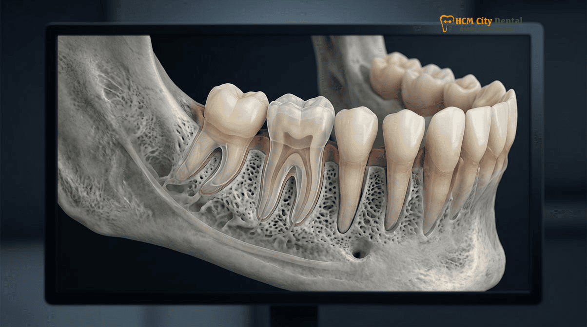
The anatomical complexity of the human root canal system is heavily influenced by genetic and ethnic factors. The supernumerary distolingual root of the mandibular first molar was first documented by Carabelli in 1844 and later formally named the Radix Entomolaris by Bolk in 1915. While the standard mandibular first molar features a mesial root (typically housing two canals) and a distal root (housing one or two canals), the presence of a Radix Entomolaris introduces a distinct, separate third root located on the lingual (tongue) side of the distal aspect.
The clinical significance of the Radix Entomolaris lies in its striking ethnic distribution. In Caucasian, African, and European populations, the prevalence of this extra root is exceptionally low, generally ranging from 3% to 5%. Consequently, many Western-trained dentists are not conditioned to routinely search for a third root in lower molars. However, in Mongoloid populations—including Chinese, Korean, Taiwanese, Native American, and Vietnamese demographics—the prevalence of the 3-rooted mandibular first molar surges dramatically to between 15% and 30%[1]. This high frequency establishes the Radix Entomolaris not merely as a rare anomaly, but as a standard anatomical variant that must be anticipated during every molar endodontic procedure in Asia.
This ethnic predilection for complex molar anatomy in Asian cohorts is not limited to the Radix Entomolaris. It parallels other distinct morphological traits, such as the high prevalence of C-shaped canal systems in mandibular second molars. According to Fan’s C1–C3 classification, C-shaped molars occur in 35% to 44% of East and Southeast Asian patients. Both the Radix Entomolaris and C-shaped canals demand a profound understanding of internal anatomy, as failure to identify and debride these aberrant spaces guarantees endodontic failure. The etiology of the Radix Entomolaris is believed to be linked to the penetrance of an atavistic gene or a polygenetic system that dictates the folding of Hertwig’s epithelial root sheath during odontogenesis.
“When treating international patients and expats in Ho Chi Minh City, we must bridge the gap between Western endodontic training and Asian dental anatomy. A clinician who assumes a lower molar has only two roots will inevitably leave the Radix Entomolaris untouched, condemning the tooth to chronic infection. This is why 3D CBCT mapping is the foundation of our endodontic protocol.”
— Dr. Nguyen Van Cuong, DDS (Clinical Director, HCMC Dental Clinic)
Why Conventional 2D Periapical X-Rays Superimpose the Extra Root Behind the Main Distobuccal Root (And How 0.075mm 3D CBCT Exposes It)
The primary reason the Radix Entomolaris is so frequently missed during initial root canal therapy is the inherent limitation of conventional two-dimensional radiography. A standard periapical X-ray captures a 3D object and compresses it into a flat 2D image. Because the Radix Entomolaris is located on the distolingual aspect of the tooth, it sits in the exact same buccolingual plane as the larger, more dominant distobuccal root. When the X-ray beam is directed orthogonally (straight on), the dense structure of the distobuccal root completely superimposes and eclipses the smaller Radix Entomolaris behind it.
Historically, endodontists attempted to overcome this by applying the SLOB rule (Same Lingual, Opposite Buccal), taking multiple 2D X-rays at 25- to 30-degree mesial or distal horizontal angulations to “separate” the roots visually. While helpful, this technique is highly technique-sensitive, often results in distorted images, and still fails to reveal the true three-dimensional curvature of the extra root[2]. Furthermore, a 2D radiograph provides zero information regarding the exact location of the canal orifice on the pulpal floor.
To eliminate this diagnostic blind spot, HCMC Dental Clinic utilizes high-resolution 3D Cone Beam Computed Tomography (CBCT). By capturing volumetric data at an ultra-fine 0.075mm voxel size, our specialists can digitally slice the tooth in axial, coronal, and sagittal planes. For more information on our imaging technology, visit our guide on the Root Canal CBCT Scan: 3D Endodontic Imaging | HCMC Dental.
The axial slice (viewing the tooth from top to bottom) is particularly revolutionary for diagnosing the Radix Entomolaris. It allows the endodontist to visualize the exact geometry of the pulp chamber floor before the drill ever touches the tooth. Instead of guessing where the extra canal might be, the 3D CBCT provides a precise GPS map, showing the exact distance and angle of the distolingual orifice relative to the main distal canal. This level of diagnostic certainty is what separates modern microscopic endodontics from traditional blind instrumentation.
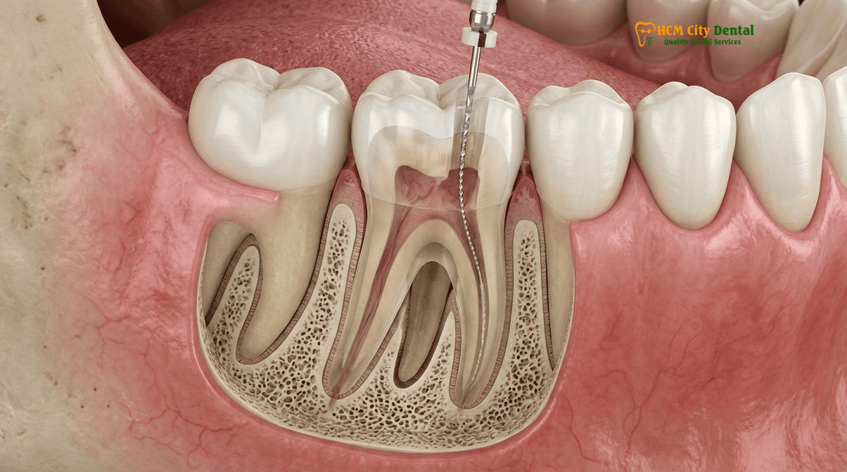
Modifying the Endodontic Access Cavity from Triangular to Trapezoidal Under 25x Carl Zeiss Magnification
Identifying the presence of a Radix Entomolaris on a 3D CBCT scan is only the first step; physically locating and accessing the canal orifice inside the mouth presents a significant clinical challenge. In a standard two-rooted mandibular first molar, the access cavity is traditionally prepared in a triangular shape, with the base of the triangle facing mesially (toward the front) to encompass the mesiobuccal and mesiolingual canals, and the apex pointing distally to uncover the single distal canal.
However, if a clinician applies this standard triangular access to a tooth with a Radix Entomolaris, the distolingual orifice will remain completely hidden beneath a shelf of dentin. The orifice of the RE is typically located far to the distolingual corner, often sharply divergent from the main distal canal. To achieve straight-line access—which is critical to prevent file binding and breakage—the access cavity must be modified. The traditional triangle must be extended distolingually, transforming the access outline into a trapezoidal or rectangular shape[3].
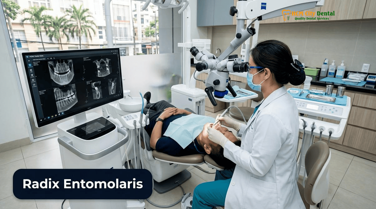
This delicate modification requires extreme precision. Over-preparation of the access cavity can weaken the pericervical dentin, increasing the risk of vertical root fractures, while under-preparation leaves the canal hidden. At HCMC Dental Clinic, this procedure is performed exclusively under a 25x Carl Zeiss Extaro 300 operating microscope. The intense coaxial illumination and high magnification allow Dr. Cuong to safely remove the overlying dentinal triangle and trace the developmental grooves (the dark lines on the pulp floor) that lead directly to the Radix Entomolaris orifice.
Wave 4A Asian Molar Endodontic Anatomy & NaOCl Irrigation Safety Rules
Once the extra canal is located, the next hurdle is safe and effective chemical debridement. The Radix Entomolaris is often narrow and highly curved, making it difficult for irrigating solutions to reach the apex. However, forcing irrigants down a narrow canal carries the severe risk of a Sodium Hypochlorite (NaOCl) accident—where the caustic bleach is extruded past the root apex into the surrounding jawbone, causing excruciating pain, rapid swelling, and tissue necrosis.
To ensure 100% prevention of NaOCl apical extrusion, HCMC Dental Clinic strictly adheres to advanced irrigation safety protocols. We utilize closed-end, side-vented 30-gauge irrigation needles. Unlike standard flat-tipped needles that shoot fluid straight out the bottom, side-vented needles disperse the fluid laterally against the canal walls. The needle is carefully measured and kept at least 2mm short of the electronic working length. Furthermore, the irrigant is never forcefully injected; instead, it is activated using passive ultrasonic agitation. The ultrasonic energy creates acoustic microstreaming and cavitation, pulling the NaOCl deep into the complex anatomy of the Radix Entomolaris without generating dangerous apical pressure. This entire process is performed under absolute isolation using a sealed nitrile rubber dam.
De Moor’s Type I, II, and III Curvature Challenge: Negotiating the Sharp 60°–90° Buccolingual Apical Hook Without File Separation
The most treacherous aspect of treating a Radix Entomolaris is not merely finding it, but navigating its severe and deceptive curvature. The morphology of the RE root is highly variable, and its curvature has been systematically categorized by De Moor into three distinct types:
- Type I: A straight root and root canal. (Relatively rare and straightforward to treat).
- Type II: An initially curved entrance that continues as a straight root canal.
- Type III: An initial curve in the coronal third of the root canal, followed by a second, severe buccally oriented curve starting from the middle to the apical third.
The De Moor Type III curvature is the most common and the most dangerous. It frequently features a sharp 60° to 90° apical hook. What makes this hook so insidious is its orientation: it curves buccolingually (toward the cheek or tongue). On a standard 2D X-ray, the Z-axis (buccolingual depth) is invisible. Therefore, a Type III Radix Entomolaris with a 90-degree apical hook will often look perfectly straight on a 2D radiograph. A clinician who trusts the 2D image will drive a stiff file straight down the canal, instantly ledging the dentin wall or perforating the side of the root[4].
Even when the curvature is identified via 3D CBCT, negotiating it requires advanced metallurgy. Standard austenitic Nickel-Titanium (NiTi) rotary files possess high “shape memory.” When bent around a 90-degree curve, the file constantly tries to spring back to its original straight shape. Inside a Type III Radix Entomolaris, this spring-back force causes the file to scrape aggressively against the outer wall of the curve, leading to canal transportation (zipping the apex) or cyclic fatigue, where the metal rapidly work-hardens and snaps off inside the tooth (file separation).
To conquer the De Moor Type III curvature, HCMC Dental Clinic utilizes the latest generation of heat-treated, controlled-memory NiTi files. Through advanced thermomechanical processing, these files are manufactured in a martensitic phase at body temperature. This gives them extreme flexibility and zero shape memory—meaning they can be pre-bent and will passively follow the severe 90-degree buccolingual hook without exerting lateral pressure on the canal walls. Combined with a meticulously established manual glide path and copious lubrication, these martensitic files allow our specialists to safely shape the Radix Entomolaris to the terminus without the risk of instrument separation.

Retreating Persistently Painful Lower Molars by Locating the Untreated Radix Entomolaris or Middle Mesial (MM) Canal
When a patient presents to our clinic complaining of a lower molar that “had a root canal years ago but still hurts to bite on” or exhibits a recurring sinus tract (a pimple on the gums that drains pus), the diagnosis is almost always a missed canal. The untreated space acts as a sanctuary for highly resistant bacterial biofilms, predominantly Enterococcus faecalis, which continuously leak toxins into the periapical tissues, causing chronic bone destruction.
In the mandibular first molar, the two most common culprits for missed anatomy are the Radix Entomolaris and the Middle Mesial (MM) canal. The MM canal is a small, accessory canal located in the developmental groove between the mesiobuccal and mesiolingual canals, present in up to 20% of cases. When retreating these failing teeth, the primary objective is to dismantle the old, contaminated root filling, disinfect the main canals, and hunt for the missed anatomy.
Clinical Case Study: Resolving Chronic Pain in an Expat Patient
A 42-year-old Australian expatriate presented to HCMC Dental Clinic with severe pain upon chewing on his lower right first molar (Tooth 46). He reported having a root canal performed in his home country three years prior. A standard 2D X-ray showed what appeared to be an acceptable root canal filling in the mesial and distal roots, but a large radiolucency (dark spot indicating bone loss) was visible at the apex.
Dr. Cuong ordered a 0.075mm 3D CBCT scan, which immediately revealed the true cause of failure: a completely untreated Radix Entomolaris root extending distolingually, harboring a massive periapical lesion. The previous dentist, lacking 3D imaging, had only treated the standard two roots. Under the 25x Carl Zeiss microscope, Dr. Cuong modified the access cavity to a trapezoidal shape, located the hidden RE orifice, and navigated a severe De Moor Type III curvature using martensitic NiTi files. The canal was heavily infected with necrotic tissue. Following rigorous ultrasonic irrigation and 3D bioceramic obturation, the patient’s pain resolved within 48 hours. A 6-month follow-up CBCT showed complete bone regeneration around the Radix Entomolaris.
For patients facing the prospect of losing a tooth due to a failed root canal, microscopic retreatment offers a highly successful alternative to extraction and implant placement. In cases where non-surgical retreatment is not feasible due to large cast posts or separated instruments that cannot be bypassed, surgical intervention may be required. Learn more about our surgical salvage protocols at Endodontic Microsurgery (Apicoectomy) & Ultrasonic MTA Retrograde Filling to Save Failed Root Canals in HCMC.
Clinical Precaution: When to Seek Specialist Endodontic Care
If you experience persistent throbbing, sensitivity to hot or cold, pain upon biting, or swelling around a tooth that has already had a root canal, do not ignore it. These are signs of active infection, likely stemming from missed anatomy such as a Radix Entomolaris. Delaying treatment can lead to extensive bone loss, root resorption, or a vertical root fracture, rendering the tooth unsalvageable. Contact Dr. Nguyen Van Cuong, DDS, at HCMC Dental Clinic for a comprehensive 3D CBCT evaluation to determine if microscopic retreatment can save your tooth.
2026 3D CBCT Endo Mapping & Microscopic Molar Root Canal Fee Schedule at HCMC Dental Clinic (District 1)
At HCMC Dental Clinic, we believe that advanced microscopic endodontics and 3D CBCT diagnostics should be accessible and transparently priced. We offer a comprehensive -40% Online Booking Pricing advantage for international patients and dental travelers who schedule their treatment in advance. Below is our official 2026 fee schedule for endodontic diagnostics and therapy, demonstrating world-class care at a fraction of Western benchmark costs.
For a complete overview of all our dental services, including implantology and cosmetic restorations, please visit our Official 2026 Dental Price List & -40% Online Booking Fee Schedule. To review our clinical outcomes and statistical data, explore our 2026 Clinical Dental Research & Statistical Reports Hub. You can also learn more about our Clinical Director by visiting Dr. Nguyen Van Cuong, DDS (Clinical Director — MOH License 0003265/BL-CCHN).
| Diagnostic & Endodontic Procedure | 2026 Online Booking Price (USD) | 2026 Online Booking Price (VND) | Standard Walk-In Price (USD) |
|---|---|---|---|
| High-Resolution 3D CBCT Scan (0.075mm) Mandatory for Radix Entomolaris detection | $18 USD | 480,000 VND | $30 USD |
| Microscopic Endodontic Therapy (Anterior/Premolar) Includes 25x Carl Zeiss + Nitrile Dam | $77 – $115 USD / tooth | 2,000,000 – 3,000,000 VND | $128 – $192 USD |
| Microscopic Endodontic Therapy (Complex Molar) Includes Radix Entomolaris / C-Shaped negotiation | $115 – $154 USD / tooth | 3,000,000 – 4,000,000 VND | $192 – $256 USD |
| Microscopic Endodontic Retreatment Missed canal location / Separated File Bypass | $154 – $231 USD / tooth | 4,000,000 – 6,000,000 VND | $256 – $385 USD |
| Bioactive ProRoot MTA / Biodentine Repair Perforation or Resorption Repair | $58 – $96 USD | 1,500,000 – 2,500,000 VND | $96 – $160 USD |
| Biomimetic Quartz/Glass Fiber Post & Core Buildup Post-endodontic structural foundation | $38 – $58 USD / tooth | 1,000,000 – 1,500,000 VND | $63 – $96 USD |
“The cost of failing to detect a Radix Entomolaris is the eventual loss of the tooth and the need for an expensive dental implant. By investing in a $18 USD 3D CBCT scan and utilizing a surgical microscope, we provide our patients with the highest probability of saving their natural teeth for a lifetime.”
— Dr. Nguyen Van Cuong, DDS
Frequently Asked Questions
If my lower first molar had a root canal years ago in another clinic and still throbs or has a gum pimple, could it have a missed Radix Entomolaris root?
Yes. Persistent throbbing, biting sensitivity, or a recurring sinus tract (gum pimple) on a previously treated mandibular first molar is a classic hallmark of untreated anatomy. Because the Radix Entomolaris is present in up to 30% of Asian patients and is frequently superimposed on standard 2D X-rays, dentists operating without 3D CBCT often miss the far-lingual orifice. The necrotic tissue and bacterial biofilm left inside this hidden third root cause chronic apical periodontitis, requiring microscopic retreatment to resolve.
Why do dentists trained only on Western two-rooted molar textbooks frequently miss the far-lingual orifice of a Radix Entomolaris?
Traditional Western endodontic textbooks teach that the mandibular first molar typically has two roots (mesial and distal) and three to four canals, with the access cavity designed as a mesially shifted triangle. Because the Radix Entomolaris has a prevalence of less than 5% in Caucasian populations, Western-trained clinicians may not routinely anticipate it. In Asian populations, however, the prevalence jumps to 15%–30%. If the clinician does not modify the access cavity to a trapezoidal shape to explore the distolingual corner, the orifice remains hidden beneath a shelf of dentin.
Why does the Radix Entomolaris canal look straight on a normal X-ray while curving sharply 90 degrees toward the cheek in 3D?
Conventional 2D periapical radiographs compress three-dimensional anatomy into a flat image. A De Moor Type III Radix Entomolaris features a severe apical hook that curves buccolingually (toward the cheek or tongue)—which is the Z-axis on a 2D X-ray. Because the curvature is pointing directly at or away from the X-ray sensor, the root appears deceptively straight. Only a 3D CBCT scan can reveal this hidden 60°–90° buccolingual hook, allowing the endodontist to prepare for the severe curvature.
How do heat-treated controlled-memory NiTi files prevent instrument breakage inside a severely curved Radix Entomolaris root?
Standard austenitic Nickel-Titanium (NiTi) files have high shape memory, meaning they try to spring back to a straight position when bent. Inside a severe De Moor Type III curvature, this spring-back force can cause the file to ledge the canal, transport the apex, or undergo cyclic fatigue and separate (break). Heat-treated, controlled-memory NiTi files are manufactured in a martensitic phase, making them highly flexible with zero spring-back. They can safely navigate 90-degree apical hooks while maintaining the natural canal trajectory [5].
How much does 3D CBCT missed-canal diagnosis and microscopic retreatment cost at HCMC Dental Clinic in District 1?
At HCMC Dental Clinic in District 1, a high-resolution 0.075mm 3D CBCT scan costs just $18 USD online (480,000 VND). If a missed Radix Entomolaris is diagnosed, Microscopic Endodontic Retreatment (utilizing a 25x Carl Zeiss microscope and advanced ultrasonic protocols) ranges from $154 to $231 USD per tooth online. This represents a transparent -40% online booking advantage compared to walk-in rates, offering world-class specialist care at a fraction of Western prices.
References
- Martins, J. N. R., et al. (2021). Worldwide Assessment of the Mandibular First Molar Second Distal Root and Root Canal: A Cross-sectional Study with Meta-analysis. Journal of Endodontics, 47(9), 1406-1416.
- De Moor, R. J., Deroose, C. A., & Calberson, F. L. (2004). The radix entomolaris in mandibular first molars: an endodontic challenge. International Endodontic Journal, 37(11), 789-799.
- Song, J. S., et al. (2010). The prevalence and morphologic classification of distolingual roots in the mandibular molars in a Korean population. Journal of Endodontics, 36(4), 653-657.
- Duman, S. B., et al. (2020). Evaluation of radix entomolaris in mandibular first and second molars using cone-beam computed tomography and review of the literature. Oral Radiology, 36(4), 320-326.
- Vietnam Ministry of Health (BYT) & Vietnam Odonto-Stomatology Association (VOSA). National Clinical Guidelines on Specialist Dental Care, Biomaterial Traceability & EN 13060 Class B Infection Control. (2024).[5]