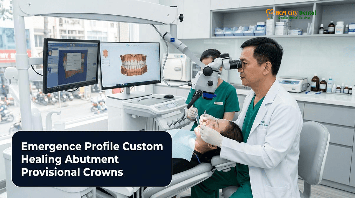
The evolution of modern implant dentistry has shifted the clinical paradigm from merely achieving successful osseointegration to mastering the intricate architecture of the peri-implant soft tissues. The transition zone—where the rigid, circular titanium platform of a dental implant meets the dynamic, scalloped, and highly vascularized gingival mucosa—dictates both the long-term biological stability and the biomimetic aesthetic success of the final restoration. At HCMC Dental Clinic in District 1, Ho Chi Minh City, the implementation of a comprehensive emergence profile custom healing abutment provisional crown implants protocol is recognized not as an optional luxury, but as the definitive standard of care for preventing peri-implant disease and achieving indistinguishable natural aesthetics.
Under the clinical direction of Dr. Nguyen Van Cuong, DDS (Clinical Director — MOH License 0003265/BL-CCHN), the advanced prosthodontic protocols utilized at HCMC Dental Clinic focus heavily on soft-tissue conditioning. By moving away from generic, mass-produced stock healing abutments and embracing CAD/CAM milled PMMA provisional crowns and custom PEEK/Ti-Base socket-sealing abutments, clinicians can actively sculpt the gingival architecture. This meticulous approach ensures that the final 1,100–1,200 MPa monolithic 3Y-TZP zirconia crown emerges from the gums with the exact anatomical contours of a natural tooth, effectively eliminating food impaction, preserving the interdental papillae, and establishing a robust mucosal seal against bacterial infiltration.
Clinical Summary:
The creation of a natural emergence profile around dental implants requires the active manipulation of peri-implant soft tissues using custom healing abutments and PMMA provisional crowns. Standard circular stock healing abutments fail to replicate the complex triangular or trapezoidal cross-sections of natural teeth, often resulting in “mushroom-shaped” final crowns that trap food and harbor bacteria. By applying Su and Gonzalez-Martin’s concepts of critical and subcritical contours, clinicians can use flowable composite on temporary cylinders or CAD/CAM milled PMMA provisionals to dynamically compress, stretch, and sculpt the gingival tissues. This progressive conditioning creates a biologically stable, anatomically correct mucosal tunnel that is subsequently captured via digital intraoral scanning and transferred to the dental laboratory for the fabrication of the final custom zirconia abutment and crown.
Key Takeaways:
- Anatomical Discrepancy: Natural teeth possess triangular (anterior) or trapezoidal (posterior) cervical cross-sections, whereas standard implant platforms and stock healing abutments are perfectly circular, necessitating custom soft-tissue sculpting.
- Critical vs. Subcritical Contours: The critical contour (1 mm subgingival to the gingival margin) dictates the zenith and height of the gingival margin, while the subcritical contour (apical to the critical zone) provides physical space for soft-tissue volume and thickness.
- Immediate Socket Sealing: Placing a custom healing abutment immediately after extraction and implant placement locks in the interdental papillae, contains bone graft materials (such as Autogenous Demineralized Dentin Matrix), and prevents tissue collapse.
- Dynamic Tissue Conditioning: Screw-retained CAD/CAM milled PMMA provisional crowns allow for progressive, chairside modifications using flowable composite to gradually stretch and shape the peri-implant mucosa over several weeks.
- Digital Transfer Protocol: Because peri-implant tissues collapse within seconds of removing a provisional crown, customized digital scan bodies or rapid 3Shape Trios 5D intraoral scanning must be utilized to accurately communicate the sculpted emergence profile to the dental laboratory.
Why Teeth Are Triangular or Trapezoidal at the Gumline While Standard Stock Healing Abutments Are Narrow Circles
To understand the necessity of a custom healing abutment emergence profile, one must first examine the fundamental anatomical differences between a natural tooth and a titanium dental implant. At the cementoenamel junction (CEJ)—the point where the anatomical crown meets the root—natural teeth exhibit highly complex, non-circular cross-sections. Maxillary central incisors are distinctly triangular at the cervical margin, providing the necessary physical support for the interdental papillae. Maxillary and mandibular molars exhibit broad, trapezoidal, or rhomboidal cross-sections, featuring distinct furcation concavities and wide interproximal contact areas.
In stark contrast, the restorative platform of a dental implant is a perfect, narrow circle. When a surgeon places a standard, factory-manufactured stock healing abutment (which is also perfectly circular and typically 4.0 mm to 5.5 mm in diameter) onto the implant, the surrounding gingival tissues heal and mature around this narrow cylinder. The resulting soft-tissue tunnel is a small, rigid, circular aperture that bears absolutely no resemblance to the natural cross-section of the tooth that was lost.
This anatomical mismatch creates a severe prosthodontic dilemma when it is time to fabricate the final crown. If the dental laboratory must transition from a narrow 4.5 mm circular tissue tunnel to a wide 10.5 mm molar occlusal table over a vertical distance of only 2 to 3 millimeters, the resulting crown will inevitably feature a drastic, convex flare. This phenomenon is commonly referred to in implant prosthodontics as a “mushroom crown” or “ridge-lap” design. The severe overhangs created by this un-sculpted emergence profile act as deep, un-cleansable undercuts. Patients frequently complain that these restorations trap meat fibers, seeds, and plaque, leading to chronic peri-implant mucositis and, eventually, peri-implantitis[1]. Therefore, to prevent food trapping around molar dental implants and to achieve Misch’s FP1 (white-only natural emergence) aesthetic classification, the clinician must actively convert the circular implant platform into a natural, tooth-shaped soft-tissue tunnel using custom healing abutments.
Su & Gonzalez-Martin’s ‘Critical Contour’ vs. ‘Subcritical Contour’ Concept in Peri-Implant Soft-Tissue Architecture
The systematic approach to sculpting peri-implant soft tissues was revolutionized by the landmark concepts introduced by Su, Gonzalez-Martin, Weisgold, and Lee in 2010. Their framework divides the subgingival emergence profile of an implant restoration into two distinct, highly influential zones: the critical contour and the subcritical contour. Understanding and manipulating these two zones is the cornerstone of the critical and subcritical contour implant crown protocol utilized by advanced prosthodontists.
The critical contour is defined as the area of the implant abutment and crown located immediately subgingival (approximately 1.0 mm apical to the free gingival margin). This zone acts as the direct physical support for the most coronal portion of the gingiva. Alterations to the critical contour have a direct, 1:1 impact on the height and zenith (the highest point) of the gingival margin. If the clinician adds flowable composite to make the critical contour more convex (bulky), the gingival margin will be pushed apically (upward in the maxilla), creating the illusion of a longer tooth. Conversely, if the critical contour is made concave (under-contoured), the gingival margin is allowed to migrate coronally, which is highly beneficial when attempting to correct minor soft-tissue recessions or mask the gray hue of a titanium abutment.
“The peri-implant mucosa is highly responsive to physical pressure. By meticulously adjusting the critical contour of a PMMA provisional crown, we can literally push the gingival zenith apically by fractions of a millimeter, achieving perfect symmetry with the adjacent natural teeth without the need for invasive periodontal plastic surgery.”
— Dr. Nguyen Van Cuong, DDS (Clinical Director, HCMC Dental Clinic)
The subcritical contour is located apical to the critical contour, extending down to the implant platform itself. The primary function of the subcritical contour is to dictate the volume and thickness of the surrounding connective tissue. In the aesthetic zone, a concave subcritical contour is almost always preferred. A concave profile creates a physical “void” or “running room” that allows the soft tissue to thicken and pool. Thicker peri-implant tissue is more robust, more resistant to mechanical trauma, and highly effective at masking the underlying titanium implant, preventing the unsightly gray shadowing often seen in thin-biotype patients. By mastering the interplay between a concave subcritical contour (for tissue volume) and a precisely calibrated critical contour (for margin height), clinicians can achieve unparalleled aesthetic outcomes[2].
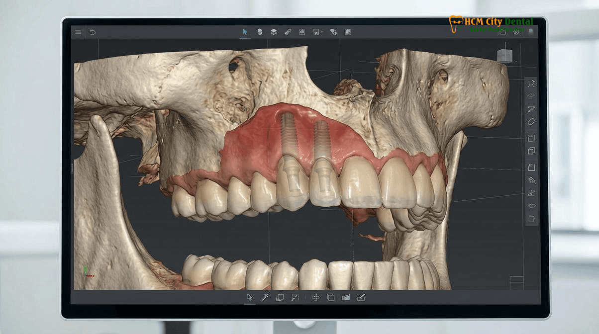
Immediate Socket-Sealing Custom Healing Abutments (Chairside Flowable Composite on PEEK/Ti-Base): Locking In the Extraction Papillae
The most opportune moment to preserve and shape the peri-implant soft tissue is immediately following tooth extraction. When a tooth is removed, the bundle bone that lines the socket begins to resorb rapidly, and the interdental papillae—deprived of the physical support previously provided by the tooth’s anatomical crown—will inevitably collapse. To counteract this biological degradation, the fabrication of an anatomical socket sealing healing abutment is performed at the time of immediate implant placement.
This technique, often referred to as the “dual-zone therapeutic concept,” involves placing the implant into the correct 3D spatial position (often utilizing Dynamic Optical Navigation or 3D-printed surgical guides) and immediately fabricating a custom healing abutment chairside. The clinician begins by attaching a temporary titanium cylinder (Ti-Base) or a PEEK (Polyether Ether Ketone) abutment to the implant. Using a sterile Teflon matrix or by directly injecting light-cured flowable composite resin around the cylinder, the clinician replicates the exact cervical cross-section of the extracted tooth.
This custom socket-sealing abutment serves multiple critical functions. First, it acts as a perfect “cork,” physically sealing the extraction socket. This is particularly vital when bone grafting materials are utilized. For instance, when utilizing a chairside Smart Dentin Grinder to process the extracted tooth into 300–1,200 µm Autogenous Demineralized Dentin Matrix (which releases native BMP-2 for rapid osteogenesis), the custom abutment prevents the graft particles from washing out into the oral cavity. Second, the anatomical shape of the abutment physically supports the interdental papillae and the mid-facial gingival margin, preventing the soft-tissue collapse that typically occurs during the first 8 to 12 weeks of healing. Finally, the highly polished, convex subgingival surface of the composite resin provides an ideal substrate for hemidesmosomal attachment, promoting a robust biological seal that protects the underlying bone from bacterial ingress[3].
For patients seeking 3D-Guided Dental Implants & Specialist Oral Surgery in Vietnam, this immediate socket-sealing protocol significantly reduces the total number of surgical interventions required, as the soft tissue is preserved from day one, eliminating the need for complex soft-tissue grafting procedures during the uncovery phase.
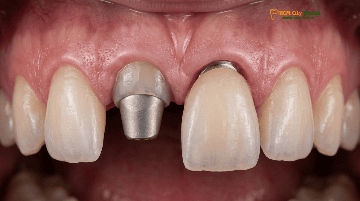
Clinical Case Note — Dr. Nguyen Van Cuong, DDS (Vietnam MOH License 0003265/BL-CCHN): When restoring an anterior or premolar single-tooth dental implant at HCMC Dental Clinic in District 1, Ho Chi Minh City, we never transition directly from a narrow cylindrical stock healing cap to a final ceramic crown. By utilizing a chairside-milled PMMA provisional crown or a customized PEEK anatomic healing abutment with a concave subcritical contour and convex critical contour, we mold 3.0mm of thick keratinized peri-implant mucosa that supports interdental papillae and prevents buccal food impaction over decades.
Progressive Cervical Tissue Conditioning Using Screw-Retained CAD/CAM Milled PMMA Provisional Crowns
While immediate custom healing abutments are excellent for preserving existing tissue architecture, there are many clinical scenarios—such as delayed implant placement or cases involving severe tissue deficiency—where the gingival contours must be actively regenerated and sculpted from scratch. In these instances, the cervical gingival sculpting pmma provisional implant protocol is employed. This process is known as dynamic tissue conditioning.
Following the integration of the implant (or during immediate loading protocols if insertion torque exceeds 45 Ncm, such as with bicortical Pterygoid implants or 6mm–8mm Straumann Roxolid splinted short implants), a screw-retained provisional crown is fabricated. At HCMC Dental Clinic, these provisionals are typically CAD/CAM milled from high-density PMMA (Polymethyl Methacrylate). PMMA is vastly superior to traditional chairside bis-acryl materials because it is highly dense, non-porous, and can be polished to a mirror-like finish, which is essential for minimizing plaque accumulation and promoting soft-tissue health.
The dynamic conditioning process is an iterative, progressive technique. The clinician intentionally designs the initial PMMA provisional to be slightly under-contoured in the subcritical zone to allow the soft tissue to heal and thicken. Once the tissue has matured (typically after 4 to 6 weeks), the patient returns for the sculpting phase. The provisional crown is unscrewed, and the clinician adds small increments of flowable composite to the critical contour to apply targeted pressure to the gingival margin. When the modified provisional is screwed back into the implant, the patient will experience a temporary blanching (ischemia) of the gums as the tissue is stretched. This blanching should resolve within 10 to 15 minutes as normal blood flow returns. By repeating this process every 2 to 3 weeks, the clinician can literally “orthodontically” move the gingival margin, coaxing the interdental papillae to grow and perfectly framing the future restoration[4].
Clinical Case Study: Anterior Aesthetic Zone Sculpting
A 34-year-old Australian expat presented to HCMC Dental Clinic with a failing maxillary right central incisor due to external root resorption. Following atraumatic extraction and immediate placement of a Straumann BLA-Active implant, a custom PEEK healing abutment was utilized to seal the socket and contain an L-PRF Sticky Bone graft. After 3 months of osseointegration, a screw-retained CAD/CAM milled PMMA provisional crown was delivered. Over a period of 6 weeks, Dr. Cuong progressively added flowable composite to the mesial and distal critical contours of the PMMA provisional, applying dynamic compression to regenerate the interdental papillae. The resulting soft-tissue architecture perfectly mirrored the contralateral central incisor, allowing for the delivery of a highly aesthetic, custom CAD/CAM hybrid zirconia abutment and an IPS E.max all-ceramic crown that was indistinguishable from the natural dentition.
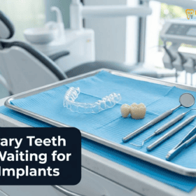
This meticulous approach is particularly crucial for anterior implant papilla sculpting in Vietnam, where international patients demand world-class aesthetic outcomes that rival or exceed the standards of their home countries. Furthermore, understanding the various temporary teeth options between Stage 1 and Stage 2 implants—such as Essix retainers, flippers, and fixed provisionals—is essential for managing patient expectations during the tissue conditioning phase.
Transferring the Sculpted Soft-Tissue Emergence Profile to the Dental Lab Without Tissue Collapse (Digital Intraoral Scan Protocol)
The ultimate success of the emergence profile custom healing abutment provisional crown implants protocol hinges entirely on the clinician’s ability to accurately communicate the newly sculpted soft-tissue architecture to the dental laboratory. Peri-implant mucosa is highly elastic and lacks the rigid periodontal ligament fibers found around natural teeth. Consequently, the moment a custom healing abutment or PMMA provisional crown is unscrewed, the soft-tissue tunnel begins to collapse rapidly—often losing its shape within 30 to 60 seconds.
If a standard digital scan body is placed into the collapsed tissue and scanned, the laboratory will design a final crown based on an inaccurate, constricted emergence profile. When the clinician attempts to seat this final crown, it will cause severe tissue blanching, extreme pain for the patient, and potential apical migration (recession) of the gingival margin due to excessive pressure.
To prevent this, advanced digital transfer protocols must be employed. The most precise method involves the fabrication of a customized digital scan body. The clinician unscrews the PMMA provisional crown and immediately attaches it to an implant analog outside the mouth. The apical half of the provisional is embedded in a rigid silicone matrix. Once the silicone sets, the provisional is removed, leaving a perfect negative replica of the sculpted emergence profile in the silicone. A standard PEEK scan body is then attached to the analog, and flowable composite is injected into the void space between the scan body and the silicone matrix. The result is a customized scan body that perfectly replicates the subgingival contours of the provisional crown.
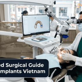
When this customized scan body is seated in the patient’s mouth, it physically supports the soft tissue, preventing collapse while the 3Shape Trios 5D intraoral scanner captures the exact 3D geometry. This digital data is then transmitted to the laboratory, where technicians design a Custom CAD/CAM Hybrid Zirconia vs. Titanium Implant Abutment that perfectly matches the conditioned tissue, ensuring a passive, frictionless seating of the final restoration[5].
Clinical Precaution: Recognizing Peri-Implant Mucositis
Even with a perfectly sculpted emergence profile, patients must maintain rigorous oral hygiene. If you experience bleeding upon brushing, localized swelling, or a bad taste around your dental implant, these are early signs of peri-implant mucositis. Left untreated, this can progress to peri-implantitis and bone loss. HCMC Dental Clinic utilizes Lang’s CIST Protocol (Cumulative Interceptive Supportive Therapy), employing erythritol air-polishing (Swiss EMS AirFlow GBT), PEEK ultrasonic tips, and diode laser decontamination to arrest inflammation. Contact Dr. Nguyen Van Cuong, DDS immediately via WhatsApp (+84 853 020 003) if you suspect peri-implant inflammation.
2026 Custom Healing Abutment, PMMA Provisional & Custom Zirconia Crown Fee Schedule at HCMC Dental Clinic (District 1)
For international patients, expats, and dental travelers seeking world-class implant prosthodontics, transparency in pricing is paramount. HCMC Dental Clinic offers an Official 2026 Dental Price List & -40% Online Booking Fee Schedule, providing access to premium European and Korean implant systems, custom soft-tissue sculpting protocols, and monolithic zirconia restorations at a fraction of the cost found in Australia, the United States, or Europe.
The clinic’s fee schedule is strictly regulated, ensuring that patients receive genuine, traceable biomaterials (such as Straumann BLT-SLA, Dentium, and IPS E.max) without hidden surcharges. The following table outlines the comprehensive costs associated with single implant therapy, including the fabrication of custom abutments and final zirconia crowns.
| Implant & Prosthodontic Procedure (2026) | Online Booking Price (USD) | Walk-In / Standard Price (USD) | Equivalent in VND |
|---|---|---|---|
| Single Korean Implant (Dentis / Neo Biotech / Dentium fixture + abutment) | $462 – $654 | $769 – $1,090 | 12,000,000 – 17,000,000 VND |
| Single European / Swiss Implant (Ritter / B&B / Straumann BLT-SLA / Straumann BLA-Active) | $962 – $1,346 | $1,603 – $2,244 | 25,000,000 – 35,000,000 VND |
| 100% Metal-Free Zirconia Ceramic Implant | $1,154 | $1,923 | 30,000,000 VND |
| Implant Zirconia HT Crown + Custom Abutment | $154 | $256 | 4,000,000 VND |
| Full Zirconia Crown (Cercon Standard / HT Multilayer) | $154 – $192 / tooth | $256 – $321 / tooth | 4,000,000 – 5,000,000 VND |
| IPS E.max All-Ceramic Crown / Inlay / Onlay / Overlay | $231 / tooth | $385 / tooth | 6,000,000 VND |
| 3D CBCT Scan (Diagnostic Imaging) | $18 | $30 | 480,000 VND |
By combining state-of-the-art digital workflows, rigorous EN 13060 Class B infection control standards, and the clinical expertise of Dr. Nguyen Van Cuong, DDS, HCMC Dental Clinic ensures that every implant restoration not only achieves absolute biomechanical stability but also seamlessly integrates into the patient’s natural gingival architecture.
Frequently Asked Questions
Why do molar implant crowns made on round stock healing caps often trap meat fibers and food underneath them?
Natural molars have a wide, trapezoidal, or rhomboidal cross-section at the gumline. Standard stock healing abutments are perfectly round and narrow. If the gum tissue heals around a narrow circle, the final crown must flare out drastically (a ‘mushroom’ shape) to meet the adjacent teeth. This severe overhang creates deep, un-cleansable undercuts where meat fibers and food debris become trapped, leading to peri-implant inflammation.
How does a custom socket-sealing healing abutment protect the bone graft placed inside a fresh extraction socket?
An anatomical socket-sealing healing abutment acts as a custom-fitted ‘cork’ for the extraction socket. By perfectly matching the shape of the extracted tooth’s root at the gingival level, it physically contains the bone graft material (such as Autogenous Demineralized Dentin Matrix), prevents the ingress of oral bacteria and food particles, and supports the delicate interdental papillae, preventing them from collapsing during the initial healing phase.
Does sculpting the gum tissue with a custom PMMA provisional crown hurt or require local anesthesia?
No, the progressive cervical tissue conditioning process is generally painless and rarely requires local anesthesia. The peri-implant mucosa has fewer pain receptors than the periodontal ligament of a natural tooth. When Dr. Nguyen Van Cuong, DDS adds flowable composite to the PMMA provisional crown to expand the critical contour, patients may feel a temporary sensation of pressure or mild ischemia (blanching of the gums), which subsides within 10 to 15 minutes as the tissue adapts.
How does the 3Shape Trios 5D scanner capture the exact subgingival gum tunnel within 15 seconds of unscrewing the provisional?
Peri-implant soft tissue is highly elastic and begins to collapse almost immediately after a provisional crown is removed. To capture the exact sculpted emergence profile, the clinician must work rapidly. The 3Shape Trios 5D scanner is used to digitally capture the soft tissue architecture within 10 to 15 seconds of unscrewing the provisional. Alternatively, a customized digital scan body—created by replicating the subgingival contours of the provisional crown with flowable composite—can be used to transfer the exact 3D geometry to the dental laboratory without rushing.
Are custom healing abutments and provisional crowns available for international implant patients at HCMC Dental Clinic?
Yes. HCMC Dental Clinic in District 1, Ho Chi Minh City, provides comprehensive custom soft-tissue sculpting protocols for international patients. Under the clinical direction of Dr. Nguyen Van Cuong, DDS, the clinic utilizes CAD/CAM milled PMMA provisionals, PEEK/Ti-Base custom healing abutments, and 1,100–1,200 MPa monolithic 3Y-TZP zirconia final crowns to ensure optimal aesthetic and functional outcomes for dental travelers and expats.
References
- Su H, Gonzalez-Martin O, Weisgold A, Lee E. Considerations of Implant Abutment and Crown Contour: Critical Contour and Subcritical Contour. International Journal of Periodontics & Restorative Dentistry. (2010).
- Chu SJ, Salama MA, Salama H, Garber DA, Saito H, Sarnachiaro GO, Tarnow DP. The dual-zone therapeutic concept of managing immediate implant placement and provisional restoration in anterior extraction sockets. Compendium of Continuing Education in Dentistry. (2012).
- Gómez-Meda R, Esquivel J, Blatz MB. The esthetic biological contour concept for implant restoration emergence profile design. Journal of Esthetic and Restorative Dentistry. (2021).
- Wittneben JG, Buser D, Belser UC, Brägger U. Peri-implant soft tissue conditioning with provisional restorations in the esthetic zone: the dynamic compression technique. International Journal of Periodontics & Restorative Dentistry. (2013).
- Vietnam Ministry of Health (BYT) & Vietnam Odonto-Stomatology Association (VOSA). National Clinical Guidelines on Specialist Dental Care, Biomaterial Traceability & EN 13060 Class B Infection Control. (2024).