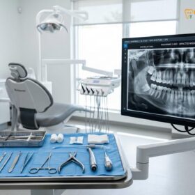
The global success rate of osseointegrated dental implants is exceptionally high; however, biological complications surrounding the peri-implant tissues remain a significant clinical challenge. For international patients, expatriates, and dental travelers seeking advanced implantology in Vietnam, understanding the critical distinction between reversible soft-tissue inflammation and irreversible crestal bone loss is paramount. The clinical management of peri-implant mucositis vs peri-implantitis laser decontamination gbr protocols requires a highly specialized, evidence-based approach. At HCMC Dental Clinic in District 1, Ho Chi Minh City, our clinical team, led by Dr. Nguyen Van Cuong, DDS (Clinical Director — MOH License 0003265/BL-CCHN), utilizes internationally recognized frameworks, such as Lang’s Cumulative Interceptive Supportive Therapy (CIST), combined with state-of-the-art laser decontamination and Guided Bone Regeneration (GBR) to rescue failing implants and restore peri-implant health.
Clinical Summary:
Peri-implant mucositis is a reversible inflammatory lesion confined to the soft tissues surrounding a dental implant, characterized by bleeding on probing without radiographic bone loss. If left untreated, it can progress to peri-implantitis, an irreversible condition marked by progressive crestal bone destruction and deep peri-implant pocketing. Successful intervention relies on early diagnosis and a staged therapeutic approach. Non-surgical treatments utilize erythritol air-polishing and laser decontamination, while advanced peri-implantitis requires surgical open-flap debridement, implantoplasty, and Guided Bone Regeneration (GBR) using L-PRF and autogenous or alloplastic bone grafts to arrest disease progression and regenerate lost supporting structures.
Key Takeaways:
- Diagnostic Distinction: Peri-implant mucositis involves soft-tissue inflammation (BOP positive) without bone loss, whereas peri-implantitis involves crater-like crestal bone destruction.
- Primary Etiologies: Subgingival residual excess cement, lack of keratinized mucosa, and non-passive prosthetic frameworks are leading triggers for peri-implant bone loss.
- CIST Protocol: Treatment is staged from mechanical debridement (Protocol A) to surgical regenerative rescue (Protocol D) based on pocket depth and radiographic findings.
- Advanced Decontamination: Safe detoxification of the titanium surface requires PEEK ultrasonic tips, erythritol air-polishing, and Er:YAG or diode laser therapy to eradicate biofilm without altering the implant macro-geometry.
- Surgical Rescue (GBR): Severe cases necessitate open-flap surgery, titanium brush cleaning, implantoplasty, and the application of L-PRF Sticky Bone or autogenous dentin grafts to regenerate lost bone.
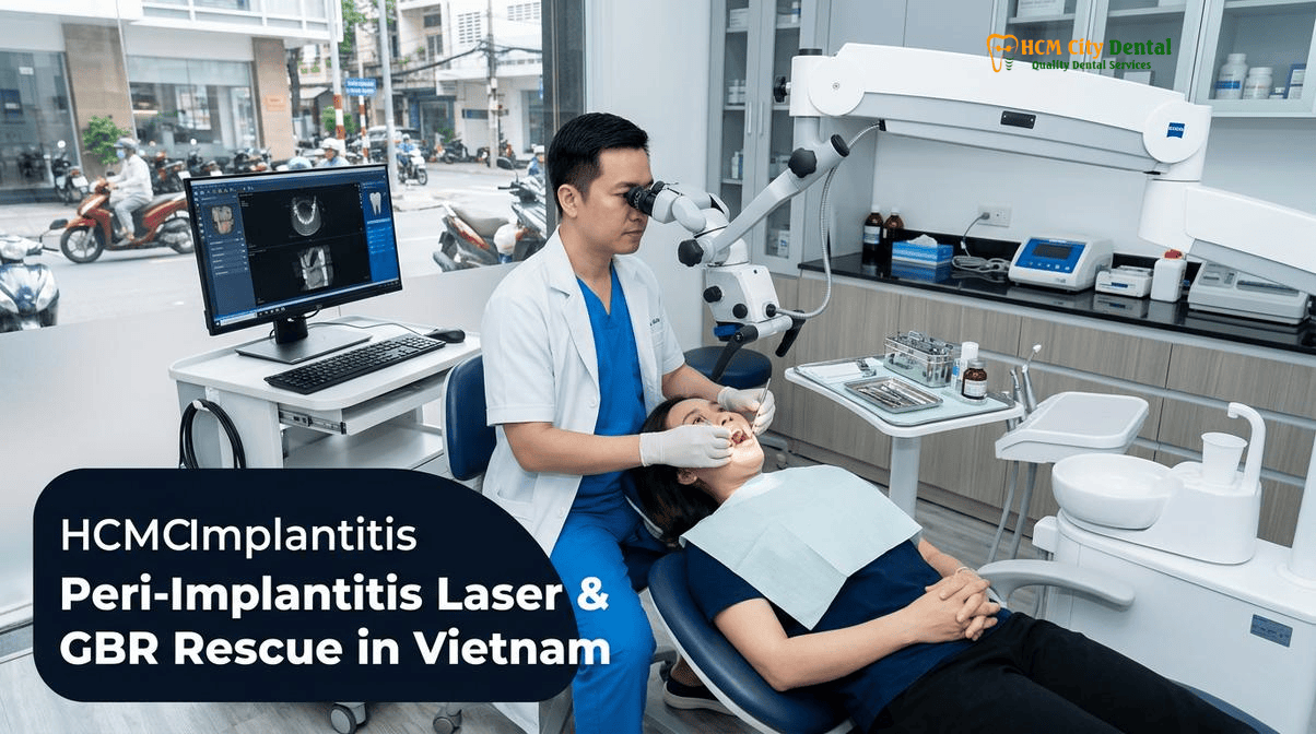
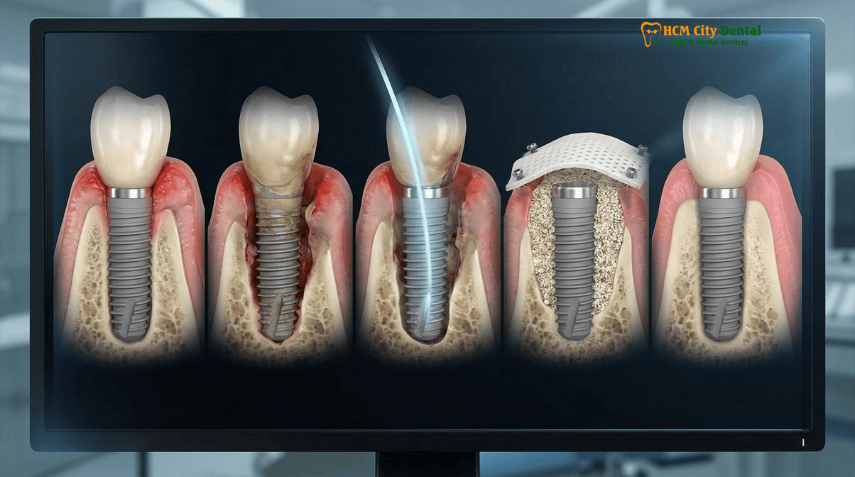
Reversible Peri-Implant Mucositis (Soft-Tissue Bleeding) vs. Destructive Peri-Implantitis (Crater-Like Crestal Bone Loss)
The biological seal around a dental implant differs fundamentally from the periodontium of a natural tooth. Natural teeth possess perpendicular inserting Sharpey’s fibers that create a robust physical barrier against bacterial invasion. In contrast, the peri-implant mucosa relies on parallel-oriented collagen fibers that offer less resistance to probing and bacterial penetration. This anatomical vulnerability makes implants highly susceptible to plaque-induced inflammatory diseases.
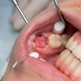
Peri-Implant Mucositis is defined as an inflammatory lesion residing exclusively in the soft tissues surrounding an endosseous implant. Clinically, it presents with erythema (redness), edema (swelling), and most importantly, Bleeding on Probing (BOP). Crucially, there is no evidence of progressive crestal bone loss beyond the initial physiological remodeling that occurs after implant placement. Because the underlying bone remains intact, peri-implant mucositis is entirely reversible with appropriate professional intervention and optimized home care. The primary goal at this stage is the mechanical disruption of the submucosal biofilm[1].
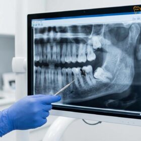
If peri-implant mucositis is ignored, the inflammatory infiltrate extends apically, leading to Peri-Implantitis. This is a destructive, irreversible condition characterized by inflammation of the peri-implant mucosa accompanied by progressive, crater-like loss of supporting crestal bone. Clinical signs include deep probing depths (typically ≥ 6mm), profuse bleeding, and frequently, suppuration (pus) upon palpation. Radiographically, peri-implantitis presents as a saucer-shaped or circumferential bone defect around the implant fixture. The progression of peri-implantitis is often non-linear and can occur in rapid, acute bursts, threatening the survival of the implant if surgical intervention is not promptly initiated.
“The transition from peri-implant mucositis to peri-implantitis marks a critical threshold in implant dentistry. Once the crestal bone begins to resorb, the exposed micro-rough titanium threads become a highly retentive sanctuary for anaerobic pathogens, making non-surgical eradication nearly impossible. Early interception is the cornerstone of implant longevity.”
— Dr. Nguyen Van Cuong, DDS
Primary Etiologies: Why Subgingival Residual Excess Cement, Lack of Keratinized Mucosa, and Non-Passive Prosthetics Trigger Bone Loss
Understanding the etiology of peri-implant diseases is essential for both prevention and treatment. While bacterial biofilm is the primary initiating factor, several iatrogenic and anatomical co-factors significantly amplify the risk of disease progression.
Subgingival Residual Excess Cement
One of the most common and preventable causes of peri-implantitis is residual excess cement. When a cement-retained implant crown is seated, hydraulic pressure can force the luting agent deep into the peri-implant sulcus. Because implants lack the perpendicular connective tissue attachment of natural teeth, the cement can easily migrate apically to the crestal bone. This rough, porous material acts as a permanent nidus for plaque accumulation. Studies have shown a direct correlation between subgingival cement and rapid, severe peri-implant bone loss. To mitigate this risk, modern implantology heavily favors screw-retained restorations or the use of custom abutments with supragingival cement margins[2].
Lack of Keratinized Mucosa and Soft Tissue Phenotype
The presence of an adequate band of attached, keratinized mucosa (KM) is vital for the long-term stability of dental implants. A lack of KM often results in mucosal mobility, patient discomfort during brushing, and a compromised biological seal, facilitating bacterial ingress. Furthermore, the emergence profile of the prosthetic crown plays a critical role. Utilizing the concepts of Su & Gonzalez-Martin regarding critical and subcritical emergence contours, custom PEEK or composite healing abutments are used to sculpt the soft tissue. Over-contoured restorations (often seen in Misch’s FP1 designs placed without adequate space) can create hygiene-impossible niches, whereas properly designed FP2 or FP3 prostheses allow for optimal access for interdental cleaning.
Biomechanical Overload and Non-Passive Prosthetics
While biofilm is the primary driver of inflammation, biomechanical overload can exacerbate bone loss, particularly in the presence of active infection. Non-passive prosthetic frameworks—where the bridge or crown does not seat perfectly without tension—introduce continuous micro-strain to the implant-bone interface. In full-arch rehabilitations, distinguishing between an RP4 (100% implant-borne CAD/CAM milled titanium bar overdenture) and an RP5 (tissue-supported, implant-retained overdenture) is crucial for load distribution. Heavy bruxism, if not managed with a hard Michigan splint or monolithic 3Y-TZP zirconia (1,100–1,200 MPa), can lead to micro-fractures in the crestal bone, accelerating the peri-implantitis process.
Lang’s Cumulative Interceptive Supportive Therapy (CIST Protocol A–D): Staging Treatment by Pocket Depth and Radiographic Bone Loss
To standardize the management of peri-implant diseases, the dental community widely utilizes the Cumulative Interceptive Supportive Therapy (CIST) protocol, originally developed by Lang and Mombelli. This evidence-based algorithm dictates a step-wise, cumulative approach based on clinical parameters such as probing depth (PD), bleeding on probing (BOP), suppuration, and radiographic bone loss.
Protocol A: Mechanical Debridement (PD ≤ 3mm, BOP+)
For early peri-implant mucositis, the focus is purely on mechanical plaque control. Standard stainless-steel scalers are strictly contraindicated as they scratch the titanium surface. Instead, clinicians utilize carbon fiber curettes, titanium scalers, or specialized PEEK ultrasonic tips to safely remove biofilm and calculus. Oral hygiene instructions are reinforced, emphasizing the use of interdental brushes and water flossers.
Protocol B: Antiseptic Therapy (PD 4–5mm, BOP+)
If pocket depths increase, mechanical debridement (Protocol A) is supplemented with antiseptic therapy. This involves subgingival irrigation with antimicrobial agents such as 0.12% to 0.2% chlorhexidine digluconate or localized application of chlorhexidine gels. The goal is to chemically reduce the bacterial load in areas that are difficult to access mechanically[3].
Protocol C: Antibiotic Therapy (PD ≥ 6mm, BOP+, Radiographic Bone Loss < 2mm)
When probing depths reach 6mm or greater, and minor crestal bone loss is evident radiographically, the condition has transitioned to early peri-implantitis. Protocols A and B are performed, followed by the administration of systemic or local antibiotics. Local delivery systems (e.g., minocycline microspheres) can provide high concentrations of the drug directly into the peri-implant pocket. Systemic antibiotics (often a combination of amoxicillin and metronidazole) may be prescribed to target tissue-invasive anaerobic pathogens.
Protocol D: Surgical Rescue (PD ≥ 6mm, BOP+, Suppuration, Radiographic Bone Loss ≥ 2mm)
In cases of advanced peri-implantitis with significant bone destruction, non-surgical therapy alone is insufficient. The micro-rough surface of the exposed implant threads cannot be adequately decontaminated in a closed environment. Protocol D mandates surgical intervention. This involves raising a mucoperiosteal flap for direct visual access, aggressive surface decontamination, and, depending on the defect morphology, either resective surgery (implantoplasty) or regenerative surgery (Guided Bone Regeneration – GBR).
Non-Surgical Decontamination: Subgingival Erythritol Air-Polishing, PEEK Ultrasonic Tips, and Photodynamic Laser Disinfection
Before any surgical intervention is considered, or for the definitive treatment of peri-implant mucositis, advanced non-surgical decontamination technologies are employed to maximize biofilm removal while preserving the integrity of the titanium implant surface.
Erythritol Air-Polishing (Guided Biofilm Therapy)
Traditional sodium bicarbonate air-polishing powders are highly abrasive and can damage the implant surface. At HCMC Dental Clinic, we utilize the Swiss EMS AirFlow system with Erythritol powder (14 microns). Erythritol is minimally abrasive, highly effective at disrupting biofilm, and safe for use subgingivally around implants. Guided Biofilm Therapy (GBT) ensures comprehensive decontamination of the peri-implant sulcus without altering the titanium oxide layer.
PEEK Ultrasonic Tips
Ultrasonic scaling is highly efficient, but standard metal tips will gouge titanium. Polyether ether ketone (PEEK) is a high-performance polymer that is softer than titanium but rigid enough to dislodge calculus. PEEK-coated ultrasonic tips allow for the safe and effective debridement of the implant abutment and exposed threads during non-surgical therapy.
Photodynamic Laser Disinfection (aPDT) and Er:YAG Lasers
Laser technology has revolutionized peri-implant decontamination. Antimicrobial Photodynamic Therapy (aPDT) involves applying a photosensitizer dye (such as phenothiazine chloride) into the peri-implant pocket, which binds to bacterial cell walls. A low-level diode laser is then used to activate the dye, producing reactive oxygen species that selectively destroy the bacteria without generating heat that could damage the bone. For more robust decontamination, Er:YAG lasers are utilized. The Er:YAG wavelength is highly absorbed by water, allowing it to effectively ablate calculus and biofilm from the micro-rough titanium surface while providing a bactericidal effect, all without overheating the implant fixture[4].
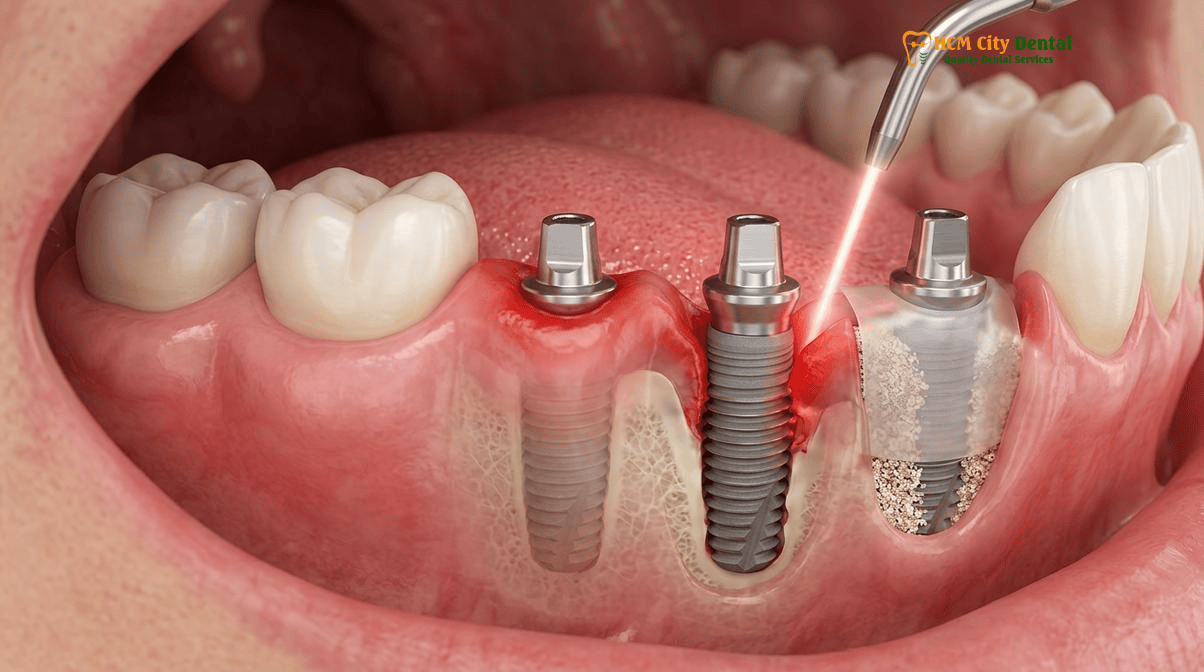
Surgical Rescue Protocol: Open-Flap Titanium Brush Detoxification, Implantoplasty, and L-PRF Sticky Bone Guided Bone Regeneration (GBR)
When non-surgical methods fail to resolve deep peri-implant pockets, or when radiographic evidence shows significant crater-like bone loss, surgical intervention is mandatory to save the implant. The surgical rescue protocol is highly technique-sensitive and requires a deep understanding of bone biology and biomaterials.
Open-Flap Debridement and Granulation Tissue Removal
The procedure begins with the elevation of a full-thickness mucoperiosteal flap to expose the bony defect and the contaminated implant surface. All inflamed granulation tissue must be meticulously removed using titanium or plastic curettes. This tissue is highly vascular and harbors dense concentrations of inflammatory cytokines and bacteria; its complete removal is critical for successful regeneration.
Titanium Brush Detoxification and Chemical Decontamination
Once the defect is cleared, the exposed implant threads must be decontaminated. Rotary titanium brushes, operating at low speeds, are used to mechanically scrub the micro-rough surface, removing residual biofilm and calculus. This is followed by chemical decontamination using agents such as 3% hydrogen peroxide, sterile saline, or citric acid to further detoxify the titanium oxide layer.
Implantoplasty: Modifying the Surface
In cases where the bone defect is horizontal or supracrestal, regenerating bone over the exposed threads is biologically improbable. In these scenarios, implantoplasty is performed. This involves using high-speed diamond and carbide burs to completely grind away the implant threads, polishing the surface until it is perfectly smooth. A smooth surface is far less susceptible to future plaque accumulation and allows the soft tissue to adapt closely, reducing pocket depth.
Guided Bone Regeneration (GBR) with L-PRF and Autogenous Dentin
For contained, crater-like (intra-bony) defects, regenerative therapy is the treatment of choice. After thorough decontamination, the defect is grafted to rebuild the lost bone. At HCMC Dental Clinic, we utilize advanced biomaterials, including Leukocyte-Platelet Rich Fibrin (L-PRF) derived from the patient’s own blood. L-PRF provides a dense fibrin matrix rich in growth factors that accelerates angiogenesis and soft tissue healing.
For the bone graft itself, we often employ a chairside Smart Dentin Grinder to process extracted teeth into a 300–1,200 µm Autogenous Demineralized Dentin Matrix. This material is rich in native Bone Morphogenetic Protein-2 (BMP-2) and offers exceptional osteoinductive properties. The graft is mixed with L-PRF exudate to create “Sticky Bone,” which is highly stable and easy to adapt into the defect. A resorbable collagen membrane is then placed over the graft to exclude fast-growing epithelial cells, allowing the slower-growing osteoblasts to repopulate the area and integrate with the implant surface[5].
Clinical Case Study: Advanced Peri-Implantitis Rescue
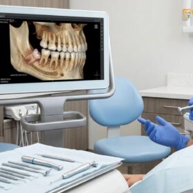
A 58-year-old expatriate patient presented to HCMC Dental Clinic with a history of recurrent swelling and a bad taste around a lower right molar implant placed five years prior in another country. Clinical examination revealed an 8mm probing depth, profuse bleeding, and suppuration. A 3D CBCT scan confirmed a severe 4mm circumferential crater-like bone defect (peri-implantitis). Dr. Nguyen Van Cuong initiated CIST Protocol D. Following open-flap debridement, the implant surface was detoxified using rotary titanium brushes and Er:YAG laser decontamination. The intra-bony defect was grafted using a combination of particulate allograft and L-PRF Sticky Bone, covered with a resorbable membrane. At the 6-month follow-up, CBCT imaging demonstrated complete radiographic bone fill of the defect, and probing depths were reduced to a healthy 3mm with no bleeding. For more information on patient safety during complex procedures, review our International Patient Medical Safety & Post-Op Recovery Guides.
Clinical Warning: When to Seek Immediate Intervention
Peri-implantitis can progress rapidly and silently. If you experience any of the following symptoms around a dental implant, contact a specialist immediately: spontaneous bleeding when brushing, a persistent bad taste or odor (indicating suppuration/pus), swelling or redness of the gums, or any sensation of implant mobility. Do not attempt to treat these symptoms with over-the-counter mouthwashes alone. Delaying professional diagnosis and treatment significantly reduces the probability of saving the implant through GBR rescue protocols.
2026 Peri-Implantitis Laser Rescue, GBR Regeneration & Implant Removal Fee Schedule at HCMC Dental Clinic (District 1)
For international patients seeking transparent, high-quality care, HCMC Dental Clinic offers a comprehensive and highly competitive fee structure. By utilizing our online booking system, patients can access specialist-led peri-implantitis treatments at significantly reduced rates compared to walk-in prices or standard fees in Australia, the US, or Europe. For a complete overview of all procedures, please consult our Official 2026 Dental Price List & -40% Online Booking Fee Schedule.
| Clinical Procedure / Biomaterial | 2026 Online Booking Fee (-40% USD) | Standard Walk-In Price (USD) | VND Equivalent (Online) |
|---|---|---|---|
| 3D CBCT Scan (Diagnostic Imaging) | $18 USD | $30 USD | 480,000 VND |
| Swiss EMS AirFlow Stain Removal (Erythritol GBT) | $58 USD | $96 USD | 1,500,000 VND |
| Single Korean Implant (Dentis / Neo Biotech / Dentium) | $462 – $654 USD | $769 – $1,090 USD | 12,000,000 – 17,000,000 VND |
| Single European / Swiss Implant (Straumann BLT-SLA) | $962 – $1,346 USD | $1,603 – $2,244 USD | 25,000,000 – 35,000,000 VND |
| Implant Zirconia HT Crown + Custom Abutment | $154 USD | $256 USD | 4,000,000 VND |
| Surgical Wisdom Tooth Extraction (Piezotome) | $58 – $115 USD / tooth | $96 – $192 USD | 1,500,000 – 3,000,000 VND |
Note: The exact cost of surgical peri-implantitis rescue (GBR, L-PRF, laser decontamination) is highly dependent on the size of the bone defect and the specific biomaterials required. A personalized quote will be provided following a comprehensive clinical examination and 3D CBCT analysis by Dr. Cuong. If an implant cannot be saved and requires removal, patients may also need to consider the Wisdom Tooth Extraction Cost Vietnam: Clinical Price Guide | HCMC Dental for comparative surgical extraction baseline costs, or read about managing acute oral infections in our Pericoronitis: Wisdom Tooth Gum Infection Guide | HCMC Dental.
“Transparency in clinical protocols and pricing is fundamental to building trust with our international patients. By providing clear, evidence-based treatment pathways for peri-implant diseases, we empower patients to make informed decisions about their oral health investments.”
— Dr. Nguyen Van Cuong, DDS
Frequently Asked Questions
Can a dental implant that is bleeding or losing bone around the top threads still be saved without removing it?
Yes, depending on the severity of the bone loss. If the condition is diagnosed early as peri-implant mucositis, it is entirely reversible through non-surgical mechanical debridement and laser decontamination. If it has progressed to peri-implantitis with moderate bone loss, the implant can often be saved using Lang’s CIST Protocol D, which involves surgical open-flap debridement, implantoplasty (smoothing the exposed threads), and Guided Bone Regeneration (GBR) using L-PRF and bone grafts. However, if bone loss exceeds two-thirds of the implant length or the implant is mobile, removal is usually required.
Why must hygienists never use standard stainless-steel scalers on titanium implant abutments?
Standard stainless-steel scalers are harder than the titanium used for dental implants and abutments. Using them will scratch and gouge the smooth titanium surface. These microscopic scratches create an ideal retentive surface for pathogenic biofilm (plaque) to accumulate, which significantly increases the risk of peri-implant mucositis and subsequent peri-implantitis. Instead, clinicians must use specialized PEEK (polyether ether ketone) ultrasonic tips, carbon fiber curettes, or titanium curettes that safely clean the implant without altering its surface topography.
How does excess dental cement trapped under a cement-retained implant crown cause peri-implantitis years after placement?
When a cement-retained implant crown is seated, excess cement can be extruded deep into the subgingival peri-implant sulcus. Because implants lack the perpendicular Sharpey’s fibers found in natural teeth, the cement can easily be pushed down to the crestal bone. This residual cement acts as a highly retentive, rough nidus for anaerobic bacteria. Over months or even years, this localized biofilm triggers a severe inflammatory immune response, leading to rapid and destructive crestal bone loss (peri-implantitis). This is why screw-retained prosthetics or custom abutments with supragingival margins are clinically preferred.
When is it better to atraumaticly remove a failing implant with a reverse-torque kit rather than attempting bone grafting?
Atraumatic implant removal using a reverse-torque explantation kit is indicated when the implant exhibits clinical mobility (which means osseointegration has completely failed), when radiographic bone loss extends beyond the apical third of the fixture, or when the patient has suffered recurrent acute peri-implant abscesses despite previous surgical interventions. Attempting GBR on a mobile implant is futile. Removing the implant atraumatically preserves the remaining buccal and lingual bone walls, allowing the site to be grafted and a new implant to be placed after a 4-to-6-month healing period.
How much does non-surgical or surgical peri-implantitis rescue treatment cost at HCMC Dental Clinic in District 1?
At HCMC Dental Clinic in District 1, non-surgical decontamination using Swiss EMS AirFlow (Erythritol GBT) costs $58 USD online. For advanced surgical rescue, procedures such as open-flap debridement with laser decontamination and Guided Bone Regeneration (GBR) vary based on the extent of the defect and the biomaterials required (e.g., L-PRF, autogenous dentin graft, or particulate allograft). A comprehensive clinical assessment and 3D CBCT scan ($18 USD online) are required to provide an exact surgical quote. Patients can view the Official 2026 Dental Price List & -40% Online Booking Fee Schedule for detailed baseline costs.
References
- Lang, N. P., & Berglundh, T. (2011). Periimplant diseases: where are we now? Consensus of the Seventh European Workshop on Periodontology. Journal of Clinical Periodontology, 38(s11), 178-181.
- Schwarz, F., Derks, J., Monje, A., & Wang, H. L. (2018). Peri-implantitis. Journal of Periodontology, 89, S267-S290.
- Heitz-Mayfield, L. J., & Mombelli, A. (2014). The therapy of peri-implantitis: a systematic review. International Journal of Oral & Maxillofacial Implants, 29(Suppl), 325-345.
- Romanos, G. E., & Nentwig, G. H. (2008). Regenerative therapy of deep peri-implant-related bone defects after CO2 laser implant surface decontamination. International Journal of Periodontics & Restorative Dentistry, 28(3), 245-255.
- Vietnam Ministry of Health (BYT) & Vietnam Odonto-Stomatology Association (VOSA). National Clinical Guidelines on Specialist Dental Care, Biomaterial Traceability & EN 13060 Class B Infection Control. (2024).