Root resorption is one of the most insidious and clinically challenging pathologies in modern endodontics and restorative dentistry. It is a progressive, destructive process where the body’s own immune cells (odontoclasts) begin to dissolve the mineralized structures of the tooth—specifically the dentin and cementum. For international patients, expats, and dental travelers seeking advanced care in Vietnam, understanding the critical differences between internal and external resorption is the first step toward saving a compromised tooth. At HCMC Dental Clinic, located in District 1, Ho Chi Minh City, we utilize hospital-grade microscopic endodontics and high-resolution 3D CBCT imaging to accurately diagnose and execute internal and external cervical root resorption MTA repair. Under the clinical direction of Dr. Nguyen Van Cuong (Dr. Cuong), DDS, our biomimetic protocols focus on halting the resorptive process, regenerating lost tissue with bioactive ceramics, and providing honest, evidence-based guidance on when a tooth can be saved versus when it requires an immediate 3D-guided dental implant.
Clinical Summary:
Root resorption is broadly categorized into internal root resorption (originating within the pulp canal) and external cervical resorption (originating from the periodontal ligament on the outside of the root). Accurate diagnosis relies entirely on 0.075mm 3D CBCT imaging, as standard 2D X-rays cannot reliably differentiate the two. Treatment for internal resorption involves microscopic root canal therapy using ultrasonic irrigation and bioceramic obturation. External cervical root resorption treatment depends heavily on the Heithersay classification (Class 1 through 4). Early-stage lesions (Class 1 and 2) can be predictably repaired surgically using 90% Trichloroacetic Acid (TCA) and bioactive cements like ProRoot MTA or Septodont Biodentine. However, advanced lesions (Class 3 and 4) severely compromise the structural integrity of the tooth, making extraction and immediate 3D-guided implant placement the most predictable, long-term clinical solution.
Key Takeaways:
- Diagnostic Necessity: 2D periapical X-rays are insufficient for diagnosing resorption. A 0.075mm high-resolution 3D CBCT scan is mandatory to determine the exact location, portal of entry, and circumferential spread of the defect.
- Internal Resorption: Often asymptomatic until it presents as a pink discoloration on the crown, internal resorption is halted 100% by removing the inflamed pulp tissue via microscopic endodontics and sealing the canal with bioceramics.
- External Cervical Resorption (ECR): Triggered by trauma, internal bleaching, or orthodontic forces, ECR requires surgical intervention. The Heithersay classification dictates the prognosis.
- Bioceramic Repair: Heithersay Class 1 and 2 defects are highly savable using 90% TCA to coagulate the invasive tissue, followed by MTA and Biodentine root resorption repair.
- Implant Transition: Heithersay Class 3 and 4 defects have a poor restorative prognosis. Immediate extraction and placement of a 3D-guided dental implant prevent catastrophic root fracture and preserve vital alveolar bone.
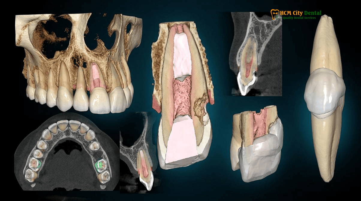
Internal Root Resorption (‘Pink Tooth of Mummery’) vs. External Cervical Invasive Resorption (ECR): 3D CBCT Differential Diagnosis
The clinical management of root resorption begins with a definitive differential diagnosis. Because the treatment protocols for internal and external resorption are fundamentally different, misdiagnosis inevitably leads to treatment failure, persistent infection, and eventual tooth loss. The primary challenge is that both types of resorption can appear radiographically similar on standard 2D X-rays, presenting as a radiolucent (dark) enlargement within the root structure. However, their etiology, pathophysiology, and anatomical origins are entirely distinct.
The Pathophysiology of Internal Root Resorption
Internal root resorption is a relatively rare condition that originates within the root canal system. It is triggered by chronic pulpal inflammation, usually resulting from dental trauma, deep caries, or extreme heat generated during restorative procedures. The inflamed pulp tissue undergoes a metaplastic change, transforming normal pulp cells into multinucleated dentinoclastic cells. These cells begin to resorb the internal dentinal walls of the root canal, creating a balloon-like expansion of the canal space. Clinically, internal root resorption, often presenting as the ‘pink tooth of Mummery’, becomes visible when the highly vascular, inflamed granulation tissue within the pulp chamber resorbs the coronal dentin to such an extent that the pink tissue shows through the translucent enamel. If left untreated, the internal resorption will eventually perforate the external root surface, complicating the prognosis significantly [1].
The Pathophysiology of External Cervical Resorption (ECR)
External Cervical Resorption (ECR), also known as invasive cervical resorption, is a much more aggressive and complex pathology. It originates on the external surface of the root, typically just below the epithelial attachment at the cervical margin (the gumline). ECR is initiated when the protective layer of cementum on the root surface is damaged, allowing fibrovascular tissue from the periodontal ligament (PDL) to invade the dentin. Common predisposing factors include a history of dental trauma, non-vital internal bleaching, orthognathic surgery, and excessive orthodontic forces. In fact, orthodontic root resorption from braces or Invisalign is a well-documented risk factor when teeth are subjected to heavy, continuous forces that overwhelm the adaptive capacity of the periodontium. Unlike internal resorption, the pulp in a tooth with ECR often remains vital and healthy for a long period, protected by a thin layer of predentin that resists clastic attack [2].
The Mandatory Role of 0.075mm 3D CBCT Imaging
Differentiating between internal and external resorption using traditional 2D periapical radiographs is fraught with error due to the superimposition of anatomical structures. A lesion that appears to be inside the canal on a 2D X-ray may actually be an external defect superimposed over the pulp space. At HCMC Dental Clinic, we mandate the use of 0.075mm high-resolution 3D Cone Beam Computed Tomography (CBCT) for all suspected resorption cases. A 3D CBCT scan allows our specialists to view the tooth in axial, coronal, and sagittal planes. This reveals the exact portal of entry, the circumferential spread of the lesion, the thickness of the remaining dentin walls, and whether the pulp space has been breached. For international patients seeking a second opinion, you can explore our 2026 Clinical Dental Research & Statistical Reports Hub to understand how advanced imaging drastically improves endodontic success rates.
Heithersay Classification (Classes 1, 2, 3, and 4): The Honest Clinical Boundary Between Saving the Tooth and Extraction
When diagnosing External Cervical Resorption, the Heithersay classification (Class 1, 2, 3, and 4 resorption) is the universally accepted clinical standard used to determine the extent of the lesion and the prognosis for saving the tooth. Developed by Dr. Geoffrey Heithersay, this classification system provides an honest, evidence-based framework that guides our specialists at HCMC Dental Clinic in recommending either biomimetic repair or extraction.
Heithersay Class 1: Shallow Cervical Penetration
A Class 1 lesion is a small, shallow resorptive defect localized near the cervical area of the root, with minimal penetration into the dentin. These lesions are highly treatable. Because the defect is superficial, it can usually be accessed surgically, cleaned, and restored with a high degree of predictability. The structural integrity of the tooth remains largely uncompromised.
Heithersay Class 2: Deeper Penetration, Coronal Localization
A Class 2 lesion is a well-defined resorptive defect that has penetrated deeper into the dentin, often approaching the coronal pulp chamber, but it shows little to no extension down into the radicular (root) dentin. While more complex than Class 1, Class 2 lesions still carry a favorable prognosis. Treatment typically involves surgical exposure, chemical coagulation of the resorptive tissue, and sealing the defect. If the pulp is exposed during the procedure, concurrent microscopic root canal therapy is required.
Heithersay Class 3: Deep Radicular Invasion
A Class 3 lesion represents a critical turning point in the prognosis. In this stage, the resorptive defect has invaded deeply into the coronal third of the root dentin. The fibrovascular tissue often spreads circumferentially around the root canal, severely undermining the structural strength of the tooth. While heroic attempts can be made to save a Class 3 tooth using internal approaches and bioceramic materials, the long-term prognosis is guarded. The tooth is at a high risk of catastrophic vertical or horizontal root fracture under normal chewing forces.
Heithersay Class 4: Extensive Radicular Destruction
A Class 4 lesion is characterized by a massive, invasive resorptive process that extends beyond the coronal third of the root, often reaching the middle or apical thirds. The root structure is hollowed out and replaced by fibrovascular tissue. A Class 4 tooth is considered structurally hopeless. Attempting to repair a Class 4 lesion is clinically irresponsible, as it inevitably leads to failure, chronic infection, and the loss of surrounding alveolar bone. In these cases, extraction and immediate implant placement is the only viable medical recommendation.
“The Heithersay classification is not just an academic grading system; it is the definitive clinical boundary that protects patients from undergoing expensive, painful, and ultimately doomed procedures on structurally hopeless teeth. Our duty is to save teeth whenever predictably possible, but equally, to know exactly when to transition to a 3D-guided implant to preserve the patient’s bone volume.”
— Dr. Nguyen Van Cuong, DDS (Clinical Director — MOH License 0003265/BL-CCHN)
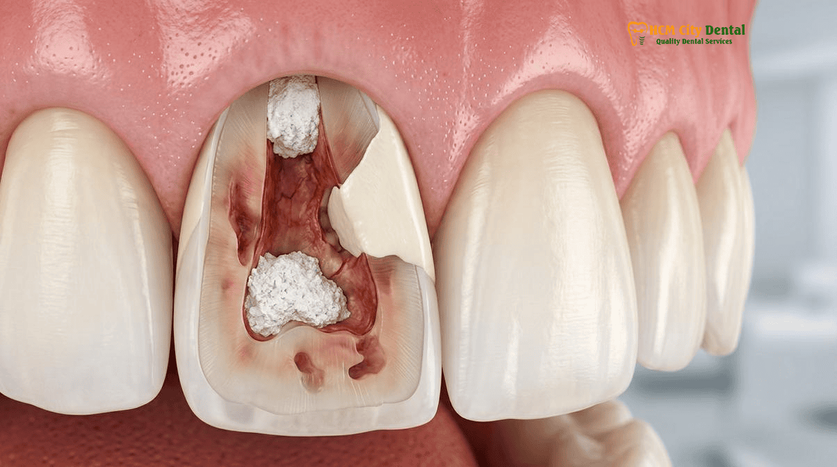
How Internal Root Resorption Is Halted 100% by Microscopic Ultrasonic Irrigation, Calcium Hydroxide, and Warm Thermoplastic/Bioceramic Fill
Unlike external cervical resorption, internal root resorption is entirely dependent on the presence of vital, inflamed pulp tissue to sustain the clastic activity. Therefore, the definitive cure for internal resorption is the complete removal of the blood supply and the inflamed tissue driving the process. Once the pulp is extirpated, the odontoclasts die, and the resorptive process is halted 100%. However, the anatomical challenges created by the resorptive defect require highly specialized endodontic techniques.
The Role of the Dental Operating Microscope and Rubber Dam Isolation
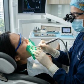
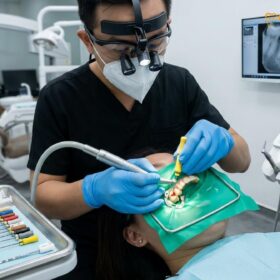
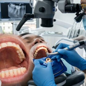
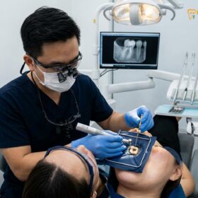
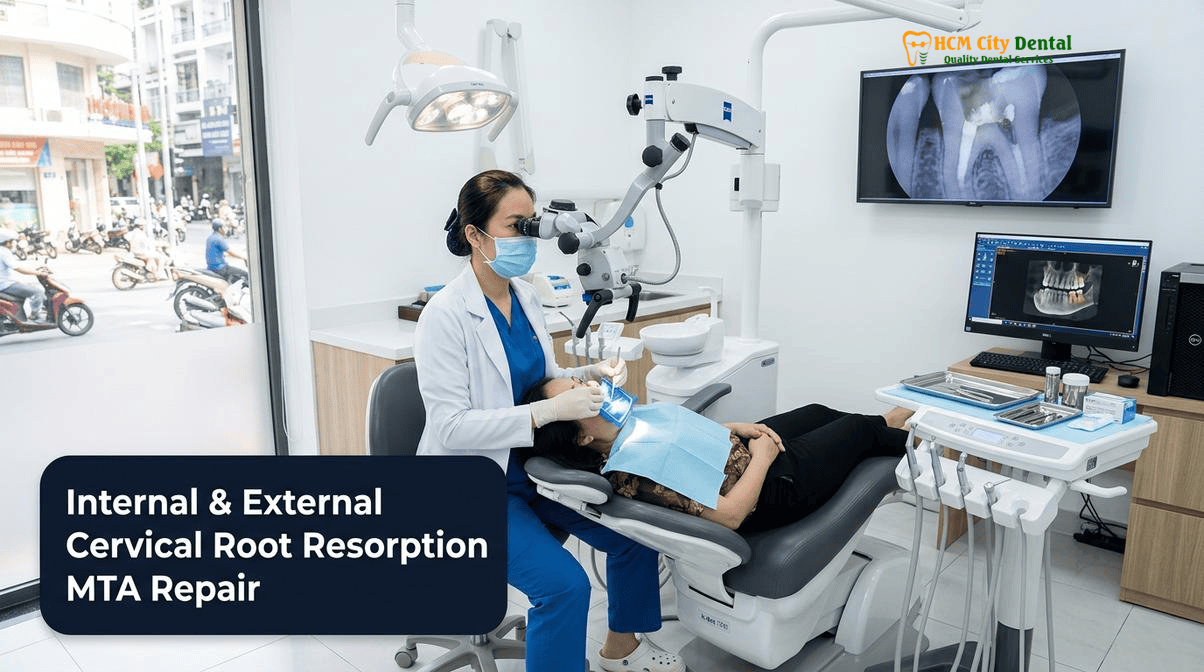
At HCMC Dental Clinic, internal resorption is treated exclusively under a 25x Carl Zeiss Extaro 300 dental operating microscope. The magnification and coaxial illumination allow the endodontist to visualize the internal resorptive lacunae (the ballooned-out areas of the canal) that are invisible to the naked eye. Furthermore, the procedure is performed under strict, 100% latex-free heavy-gauge Nitrile Rubber Dam isolation. This prevents salivary contamination and protects the patient from the strong antimicrobial irrigants used during the procedure. You can read more about our strict isolation protocols in our guide on Rubber Dam Isolation in Biomimetic Dentistry & Root Canal Therapy: Safety & Clinical Protocol in Vietnam.
Advanced Chemical Debridement: Ultrasonic Activation
The primary challenge in treating internal resorption is that standard endodontic files cannot physically touch or clean the irregular, ballooned-out walls of the resorptive defect. If any inflamed tissue is left behind, the resorption may continue, or the necrotic remnants will cause an apical abscess. To overcome this, we utilize Satelec/Acteon Titanium Ultrasonic Micro-Tips (ET20/ET25) to activate our chemical irrigants (typically 5.25% Sodium Hypochlorite and 17% EDTA). The ultrasonic energy creates acoustic microstreaming and cavitation, driving the irrigants deep into the resorptive lacunae to chemically dissolve the granulation tissue and disinfect the space [3].
Calcium Hydroxide Therapy and Bioceramic Obturation
Once the canal is thoroughly debrided, we often place an intracanal medicament of pure Calcium Hydroxide for 1 to 2 weeks. Calcium Hydroxide has a highly alkaline pH (around 12.5), which effectively necrotizes any remaining clastic cells, neutralizes bacterial endotoxins, and helps control bleeding within the defect. In the final visit, the canal and the resorptive defect must be densely obturated (filled) to prevent reinfection. Because the defect is irregular, standard cold gutta-percha techniques are inadequate. We utilize warm vertical compaction of gutta-percha combined with advanced bioceramic sealers, or in cases of massive internal defects, we fill the entire space with flowable bioactive ceramics. For a deeper understanding of these materials, review our Bioceramic Root Canal Sealer: Clinical Guide | HCMC Dental.
Clinical Case Study: Saving a ‘Pink Tooth’ for an Australian Expat
Patient Profile: A 34-year-old Australian expat residing in Thao Dien, District 2, presented to HCMC Dental Clinic after noticing a distinct pink spot on his upper right central incisor. He reported a history of a sports-related impact to the mouth five years prior.
Diagnosis: A 0.075mm 3D CBCT scan revealed a massive internal resorptive defect in the middle third of the root. The highly vascular granulation tissue had resorbed the dentin from the inside out, causing the ‘pink tooth of Mummery’ effect. Fortunately, the defect had not yet perforated the external root surface.
Treatment Protocol: Dr. Nguyen Van Cuong isolated the tooth with a Nitrile Rubber Dam and accessed the canal under 25x Zeiss magnification. The bleeding granulation tissue was removed using copious Sodium Hypochlorite activated by Satelec ultrasonic micro-tips. Calcium Hydroxide was placed for two weeks to ensure complete hemostasis and disinfection. In the second visit, the massive internal defect was backfilled using a warm thermoplastic injection technique and a bioceramic sealer, completely sealing the lacunae.
Outcome: The pink discoloration faded immediately as the vascular tissue was removed. At the 12-month follow-up, the tooth was completely asymptomatic, structurally sound, and the surrounding periapical bone showed perfect health.
Surgical & Non-Surgical Repair of Heithersay Class 1–2 External Cervical Resorption: 90% Trichloroacetic Acid (TCA) + Biodentine/Geristore
External cervical root resorption treatment requires a completely different approach than internal resorption. Because the clastic cells originate from the periodontal ligament on the outside of the tooth, root canal therapy alone will not stop the process. The invasive fibrovascular tissue must be physically and chemically eradicated from the external root surface, and the resulting defect must be sealed with a biocompatible material that the gums and bone will tolerate.
The Surgical Approach and the Role of 90% Trichloroacetic Acid (TCA)
For Heithersay Class 1 and Class 2 lesions, the standard of care involves a minor periodontal surgical flap to expose the resorptive defect at the gumline. Once exposed, the highly vascular, bleeding resorptive tissue is visible. Mechanical curettage alone is insufficient, as microscopic clastic cells will remain in the dentinal tubules and cause the resorption to recur. To ensure complete eradication, we apply 90% Trichloroacetic Acid (TCA) directly to the resorptive tissue. TCA is a powerful chemical agent that causes immediate coagulative necrosis of the fibrovascular tissue, turning it white and avascular. This not only destroys the clastic cells but also provides excellent hemostasis (bleeding control), creating a dry, clean field for the restorative material [4].
MTA and Biodentine Root Resorption Repair
Once the defect is chemically debrided and cleaned, it must be sealed. Traditional dental composites are highly sensitive to moisture and are not well-tolerated by the deep periodontal tissues. Instead, we utilize advanced calcium silicate-based cements, specifically ProRoot MTA (Mineral Trioxide Aggregate) or Septodont Biodentine. These materials are hydrophilic (they set in the presence of moisture) and are highly bioactive. When placed against the periodontium, MTA and Biodentine release calcium and hydroxide ions, creating an alkaline environment that promotes the regeneration of cementum and periodontal ligament cells directly over the restorative material. This MTA and Biodentine root resorption repair protocol ensures a biologically sound seal that prevents future bacterial microleakage and halts the resorptive process permanently.
Restoring the Cervical Margin
While MTA and Biodentine are excellent for subgingival (below the gum) repair, they do not possess the aesthetic properties or wear resistance required for the visible portions of the tooth. Therefore, a “sandwich technique” is often employed. The deep, subgingival portion of the defect is filled with Biodentine, and once set, the superficial, visible portion of the cavity is restored with a resin-modified glass ionomer (like Geristore) or a highly polished biomimetic composite resin. This dual-layered approach ensures both biological compatibility and optimal aesthetics.
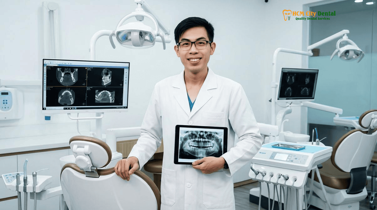
Why Heithersay Class 3–4 Resorption Should Be Replaced by an Immediate 3D-Guided Dental Implant Before Catastrophic Crown Fracture
One of the most difficult conversations in clinical dentistry is explaining to a patient when to extract a tooth with root resorption. While modern biomimetic dentistry can achieve incredible results, the laws of biomechanics cannot be ignored. When a tooth presents with Heithersay Class 3 or Class 4 external cervical resorption, the structural damage is so extensive that the tooth is essentially a hollow shell.
The Biomechanical Reality of Severe Resorption
In Class 3 and Class 4 lesions, the resorptive process has destroyed the critical cervical dentin—the area of the tooth that bears the highest stress during chewing and biting. Even if the resorptive tissue could be completely removed and the massive defect filled with MTA or composite, the tooth lacks the necessary tensile strength to survive. Furthermore, these teeth lack a “ferrule”—a solid band of healthy dentin above the gumline required to support a crown. Without a minimum 2mm ferrule, any restorative attempt will inevitably fail. The tooth will suffer a catastrophic horizontal fracture at the gumline, or a vertical root fracture that splits the tooth in half.
The Danger of Waiting: Bone Loss and Infection
Many patients, understandably wanting to avoid extraction, ask if they can “just wait and see.” Waiting is clinically dangerous. As the resorption progresses, it triggers chronic localized inflammation in the surrounding bone. If the tooth fractures subgingivally, it becomes a nidus for acute bacterial infection. More importantly, the chronic inflammation and eventual fracture lead to the rapid destruction of the buccal bone plate (the thin bone on the outside of the jaw). Losing this bone makes future implant placement significantly more complex, often requiring expensive and invasive bone grafting procedures.
The Immediate 3D-Guided Implant Solution
At HCMC Dental Clinic, our protocol for Heithersay Class 3 and 4 resorption is atraumatic extraction followed by immediate 3D-guided dental implant placement. By extracting the tooth carefully using Piezosurgery (ultrasonic bone surgery), we preserve the delicate socket walls. A titanium or zirconia implant is then placed immediately into the socket, guided by a surgical stent fabricated from the patient’s 3D CBCT scan. This immediate placement supports the surrounding bone and gingival architecture, preventing the collapse of the soft tissues. For patients interested in the success rates of our advanced protocols, we invite you to review our Single Visit Root Canal Success Rate Study 2026, which highlights our commitment to evidence-based outcomes.
“Extracting a tooth is never our first choice, but in cases of Class 4 resorption, it is the only ethical choice. By removing the compromised tooth and placing an immediate implant, we stop the bone destruction instantly and provide the patient with a permanent, structurally sound, and highly aesthetic replacement that will last a lifetime.”
— Dr. Nguyen Van Cuong, DDS (Clinical Director — MOH License 0003265/BL-CCHN)
Clinical Precaution: Do Not Ignore a ‘Pink Spot’ on Your Tooth
If you notice a pinkish discoloration near the gumline of any tooth, or if you experience unexplained sensitivity or bleeding from a specific area of the gums, do not ignore it. Root resorption is a silent, painless, and rapidly progressive disease. The difference between a simple, predictable repair and the total loss of the tooth is often just a matter of months. Contact Dr. Nguyen Van Cuong immediately via WhatsApp at +84 853 020 003 to arrange an urgent 3D CBCT scan and clinical evaluation.
2026 Diagnostic 3D CBCT, Bioceramic Repair & Immediate Implant Pricing at HCMC Dental Clinic (District 1)
At HCMC Dental Clinic, we believe in absolute financial transparency. International patients, expats, and dental travelers can access world-class microscopic endodontics, bioceramic resorption repair, and premium dental implants at a fraction of the cost of clinics in Australia, the US, or the UK. By utilizing our Official 2026 Dental Price List & -40% Online Booking Fee Schedule, patients receive guaranteed, upfront pricing with zero hidden fees.
| Clinical Procedure (Resorption Repair & Implants) | 2026 Online Booking Fee (USD) | 2026 Online Booking Fee (VND) | Walk-In / Standard Price (USD) |
|---|---|---|---|
| High-Resolution 3D CBCT Scan (0.075mm) | $18 USD | 480,000 VND | $30 USD |
| Microscopic Endodontic Therapy (Anterior/Premolar) | $77 – $115 USD / tooth | 2,000,000 – 3,000,000 VND | $128 – $192 USD |
| Microscopic Endodontic Therapy (Complex Molar) | $115 – $154 USD / tooth | 3,000,000 – 4,000,000 VND | $192 – $256 USD |
| Bioactive ProRoot MTA / Septodont Biodentine Resorption Repair | $58 – $96 USD | 1,500,000 – 2,500,000 VND | $96 – $160 USD |
| Biomimetic Quartz/Glass Fiber Post & Core Buildup | $38 – $58 USD / tooth | 1,000,000 – 1,500,000 VND | $63 – $96 USD |
| Single Korean Implant (Dentis / Neo Biotech / Dentium + Abutment) | $462 – $654 USD | 12,000,000 – 17,000,000 VND | $769 – $1,090 USD |
| Single European / Swiss Implant (Straumann BLT-SLA / BLA-Active) | $962 – $1,346 USD | 25,000,000 – 35,000,000 VND | $1,603 – $2,244 USD |
| Implant Zirconia HT Crown + Custom Abutment | $154 USD | 4,000,000 VND | $256 USD |
Frequently Asked Questions
Why can’t a standard 2D periapical X-ray accurately distinguish internal resorption from external cervical resorption?
Standard 2D periapical X-rays compress three-dimensional dental anatomy into a single flat image, causing superimposition. This makes it nearly impossible to determine if a resorptive defect originates from inside the pulp canal (internal) or from the external periodontal ligament space (external). A high-resolution 0.075mm 3D CBCT scan is mandatory to visualize the exact portal of entry, the circumferential spread, and the true Heithersay classification of the lesion.
What causes External Cervical Resorption (ECR) in healthy patients—is it linked to past braces, trauma, or internal bleaching?
External Cervical Resorption (ECR) is often triggered by damage to the protective cementum layer on the root surface. Common predisposing factors include a history of dental trauma, non-vital internal bleaching (which allows bleaching agents to seep through dentinal tubules), and excessive orthodontic forces. In fact, orthodontic root resorption from braces or Invisalign is a well-documented risk factor when teeth are moved too rapidly, stimulating clastic cells to attack the root structure.
Can a tooth with Heithersay Class 1 or Class 2 resorption be predictably saved with Biodentine or MTA?
Yes. When diagnosed early via 3D CBCT, Heithersay Class 1 and Class 2 external cervical resorption can be predictably treated. The protocol involves surgical access, application of 90% Trichloroacetic Acid (TCA) to coagulate the invasive fibrovascular tissue, and sealing the defect with bioactive materials like ProRoot MTA or Septodont Biodentine. These calcium silicate-based cements promote cementogenesis and periodontal healing.
Why is waiting until a Class 3–4 resorbed tooth snaps off at the gumline bad for future dental implant bone volume?
When a tooth with severe Heithersay Class 3 or Class 4 resorption is left untreated, the resorptive process destroys the root structure and triggers chronic localized inflammation. If the tooth eventually fractures at the gumline, the surrounding buccal bone plate often collapses. Extracting the tooth early and placing an immediate 3D-guided dental implant preserves the critical alveolar bone volume and gingival architecture, ensuring a highly aesthetic and stable long-term result [5].
How fast can Dr. Nguyen Van Cuong review my 3D CBCT scan on WhatsApp (+84 853 020 003) to tell me if my resorbed tooth is savable?
International patients and dental travelers can send their existing 3D CBCT scans or digital X-rays directly to Dr. Nguyen Van Cuong via WhatsApp at +84 853 020 003. Dr. Cuong typically provides a comprehensive clinical triage, diagnostic review, and transparent treatment plan within 5 to 10 minutes, helping you understand exactly when to extract a tooth with root resorption versus when it can be saved with microscopic endodontics.
References
- Patel, S., et al. (2018). External cervical resorption: part 1 – histopathology, distribution and presentation. International Endodontic Journal, 51(11), 1205-1223.
- Heithersay, G. S. (1999). Invasive cervical resorption: an analysis of potential predisposing factors. Quintessence International, 30(2), 83-95.
- Torabinejad, M., & Parirokh, M. (2010). Mineral trioxide aggregate: a comprehensive literature review—part II: leakage and biocompatibility investigations. Journal of Endodontics, 36(2), 190-202.
- Mavridou, A. M., et al. (2016). Understanding external cervical resorption in vital teeth. Journal of Endodontics, 42(12), 1737-1751.
- Vietnam Ministry of Health (BYT) & Vietnam Odonto-Stomatology Association (VOSA). National Clinical Guidelines on Specialist Dental Care, Biomaterial Traceability & EN 13060 Class B Infection Control. (2024).