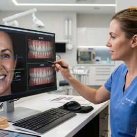
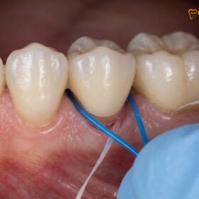
A broken crown tooth is a significant dental event that requires prompt clinical evaluation to prevent underlying tooth decay, nerve damage, or localized infection. While minor superficial chips may occasionally be repaired with advanced composite resin protocols, severe structural fractures typically necessitate a comprehensive diagnostic assessment and a complete crown replacement to restore optimal occlusal function and long-term biomechanical stability.
Clinical Summary:
A fractured or broken crown tooth compromises the essential protective barrier over the underlying dentin and pulpal tissues. Clinical management depends heavily on the extent of the fracture, the specific restorative material involved, and the biological health of the abutment tooth. Immediate patient actions should include retrieving any dislodged fragments, maintaining meticulous oral hygiene, and avoiding masticatory stress on the affected side. Definitive clinical treatment ranges from polishing minor porcelain irregularities to fabricating a completely new restoration, often utilizing high-strength modern materials. Prompt intervention at a professional facility ensures optimal functional recovery while mitigating the severe risk of secondary endodontic complications and structural tooth loss.
Key Takeaways:
- A broken crown exposes the vulnerable underlying tooth structure to rapid bacterial microleakage and acute thermal sensitivity.
- Minor porcelain fractures can occasionally be managed using specialized dental adhesives and composite bonding techniques.
- Severe structural failures or compromised subgingival margins mandate a complete crown replacement to ensure long-term stability.
- Patients must never attempt to reattach a broken crown with household adhesives, as this causes irreversible tissue toxicity.
- Immediate clinical evaluation is critical to assess potential traumatic damage to the abutment tooth or the root canal system.
Understanding the Clinical Causes of a Broken Crown Tooth
Dental crowns fracture due to a combination of excessive occlusal forces, material fatigue over time, underlying secondary caries, or acute maxillofacial trauma. Identifying the precise root cause is essential for preventing future restorative failures.
Dental crowns are engineered to withstand the immense forces of human mastication, but they are not entirely impervious to damage. The oral cavity presents a harsh biomechanical environment characterized by constant moisture, extreme temperature fluctuations, and significant mechanical stress. When a patient presents with a broken crown tooth, the clinician must first determine the etiology of the failure to ensure that the subsequent restoration does not suffer the same fate. Understanding these underlying causes is a fundamental aspect of modern prosthodontics.
One of the most prevalent causes of crown fracture is bruxism, or chronic teeth grinding. Patients who clench or grind their teeth, particularly during sleep, subject their dental restorations to lateral excursive forces that far exceed normal chewing pressures. These non-axial forces can cause microscopic stress concentrations within the ceramic matrix, eventually leading to catastrophic crack propagation[1]. Over time, the constant friction wears down the occlusal anatomy of the crown, thinning the material and making it highly susceptible to sudden fracture when biting down on a relatively soft object.
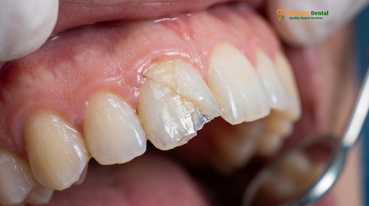
Material fatigue is another critical factor. Like any physical material subjected to cyclic loading, dental ceramics and metal alloys experience fatigue degradation over years of service. The constant thermal cycling—alternating between hot coffee and cold water—causes microscopic expansion and contraction of the crown materials. In porcelain-fused-to-metal crowns, this thermal stress can weaken the chemical and mechanical bond between the opaque porcelain layer and the underlying metal coping, leading to a specific type of failure known as delamination, where a large shard of porcelain shears off entirely.
Furthermore, the health of the underlying abutment tooth plays a vital role in the longevity of the crown. If secondary caries (tooth decay) develops at the crown margin—often due to inadequate oral hygiene or a poorly fitting restoration—the underlying dentin becomes soft and cavitated. As the foundational support weakens, the crown is left unsupported. When occlusal force is applied, the rigid crown material flexes into the void left by the decay, resulting in a fracture. This scenario highlights the critical importance of precise marginal adaptation during the initial custom dental restorations process.
“A broken crown is rarely just a material failure; it is often a symptom of an underlying biomechanical imbalance. Achieving precise occlusal harmony and selecting the appropriate restorative material based on the patient’s specific parafunctional habits are paramount to ensuring the long-term survival of any dental prosthesis.”
— Dr. Nguyen Van Cuong, Lead Prosthodontist
Acute trauma is also a frequent culprit. A sudden blow to the face during contact sports, a fall, or an automobile accident can generate impact forces that instantly shatter even the most robust dental ceramics. Similarly, biting down unexpectedly on a hard object—such as an olive pit, a popcorn kernel, or a piece of ice—can cause a localized point-loading fracture. In these instances, the damage is often immediate and highly visible, requiring urgent clinical intervention to protect the exposed tooth structure.
Immediate Steps: What to Do When Your Dental Crown Breaks
If your dental crown breaks, carefully remove any loose fragments to prevent aspiration, rinse your mouth with warm saline, and contact your dentist immediately for an emergency clinical evaluation.
Experiencing a broken crown tooth can be alarming, but taking the correct immediate steps can significantly mitigate pain and prevent further damage to the underlying tooth structure. The moment you realize your crown has fractured, the first priority is safety. Carefully run your tongue over the area to assess the damage, but avoid touching the sharp edges with your fingers to prevent lacerations. If a large piece of the crown has completely dislodged, carefully remove it from your mouth to eliminate the risk of accidental swallowing or aspiration into the airway.
Once the oral cavity is clear of loose debris, gently rinse your mouth with a warm saline solution. This helps to flush away any microscopic ceramic shards and provides a mild antibacterial effect to protect the newly exposed dentin. The underlying tooth structure is highly porous, containing thousands of microscopic dentinal tubules that communicate directly with the dental pulp (the nerve center of the tooth). Exposure of these tubules can lead to acute thermal and tactile hypersensitivity[2]. Therefore, it is crucial to avoid consuming extremely hot, cold, or highly acidic foods and beverages until you can be evaluated by a professional.
If you are experiencing significant discomfort, over-the-counter analgesics such as ibuprofen or acetaminophen can be utilized to manage the pain, provided there are no medical contraindications. In cases where the broken edge of the crown is exceptionally sharp and is traumatizing the buccal mucosa (cheek) or tongue, orthodontic wax can be applied over the jagged edge as a temporary protective barrier. Some pharmacies also sell temporary dental cement; while this can be used to cover exposed dentin in a pinch, it is strictly a stopgap measure and does not replace the need for professional care.
Contacting a dental professional promptly is the most critical step. Even if the broken crown tooth is not causing immediate pain, the loss of the protective coronal seal allows oral bacteria to rapidly colonize the underlying dentin. This microleakage can quickly lead to secondary caries or, if the tooth has previously undergone endodontic therapy, contamination of the root canal system. A timely visit to a facility like HCMC Dental Clinic in Ho Chi Minh City ensures that the tooth is properly stabilized and a definitive treatment plan is established before irreversible complications arise.
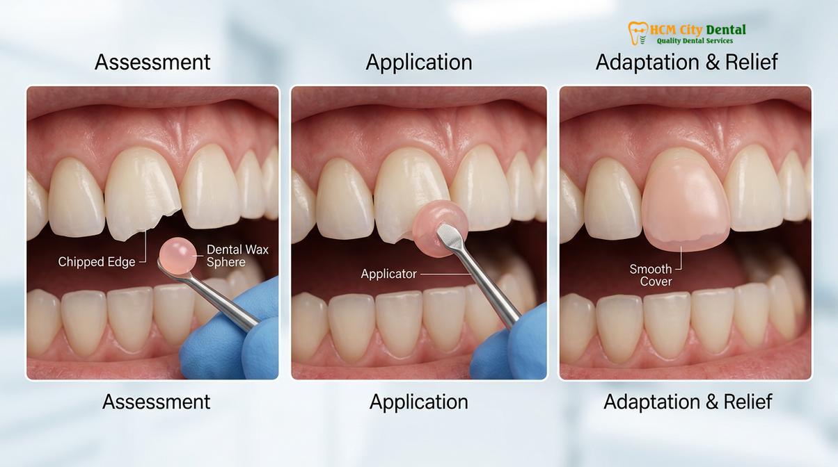
Diagnostic Evaluation and Treatment Workflows
Dentists utilize clinical probing, transillumination, and periapical radiography to assess the extent of the crown fracture and evaluate the structural integrity of the underlying abutment tooth before determining the appropriate treatment.
When a patient presents with a broken crown tooth, the clinical evaluation must be thorough and systematic. The dentist’s primary objective is to determine not only the extent of the damage to the prosthesis but also the biological status of the underlying tooth. The examination begins with a detailed visual inspection, often utilizing high-powered dental loupes and transillumination to identify the exact fracture lines and assess the remaining crown structure.
A critical component of the diagnostic workflow is radiographic imaging. High-resolution periapical X-rays, and occasionally Cone Beam Computed Tomography (CBCT), are employed to evaluate the root structure and the surrounding alveolar bone. The clinician must rule out catastrophic root fractures, which can occur simultaneously with crown fractures during traumatic impact. Furthermore, radiographs help assess the health of the periapical tissues and verify the integrity of any existing endodontic treatments. If the tooth requires a crown placement following endodontic therapy, ensuring the root canal system remains hermetically sealed is paramount.
The clinician will also meticulously evaluate the crown margins using a periodontal probe and a dental explorer. If the fracture extends subgingivally (below the gum line), it complicates the restorative process, as achieving a dry, isolated field for bonding or impression-taking becomes highly challenging. The concept of the “ferrule effect”—the presence of a continuous band of healthy tooth structure above the gum line—is evaluated to determine if the tooth has sufficient biomechanical strength to support a new crown.
| Clinical Parameter | Indications for Repair (Bonding) | Indications for Complete Replacement |
|---|---|---|
| Fracture Size | Minor superficial chipping; non-stress bearing areas. | Extensive structural loss; catastrophic failure. |
| Margin Integrity | Crown margins remain completely sealed and intact. | Fracture involves or compromises the marginal seal. |
| Occlusal Contact | Fracture does not alter the patient’s bite or centric stops. | Loss of occlusal vertical dimension or guiding contacts. |
| Underlying Tooth | Abutment tooth is healthy with no secondary caries. | Presence of recurrent decay or pulpal involvement. |
| Material Type | Feldspathic porcelain (amenable to hydrofluoric acid etching). | Monolithic zirconia or exposed metal frameworks. |
If the diagnostic evaluation reveals that the broken crown tooth can be repaired, the clinician will employ advanced adhesive dentistry protocols. This involves isolating the tooth, micro-etching the fractured ceramic surface with hydrofluoric acid, applying a silane coupling agent, and utilizing a high-quality dental veneer adhesive to bond a composite resin restoration to the defect. However, it is widely acknowledged in the dental literature that intraoral repairs of ceramics have lower long-term survival rates compared to definitive replacements, serving often as medium-term solutions.
Material Considerations: Porcelain, Zirconia, and PFM
The choice of restorative material significantly influences fracture resistance, with monolithic zirconia offering superior tensile strength compared to traditional porcelain-fused-to-metal or all-ceramic crowns.
When a broken crown tooth necessitates a complete replacement, selecting the appropriate restorative material is a critical clinical decision. The material must be tailored to the patient’s specific functional requirements, aesthetic expectations, and occlusal dynamics. The evolution of dental biomaterials has provided clinicians with a diverse array of options, each possessing distinct biomechanical properties and indications for use.
Traditional porcelain-fused-to-metal (PFM) crowns have been a staple in restorative dentistry for decades. They combine the high tensile strength of a cast metal substructure with the aesthetic appeal of an overlying feldspathic porcelain veneer. However, the primary mode of failure for PFM crowns is the cohesive fracture or delamination of the veneering porcelain[5]. Because the porcelain is baked onto the metal framework in layers, the bond is susceptible to fatigue from thermal cycling and occlusal stress. While highly durable, PFM crowns are increasingly being replaced by newer, monolithic materials that eliminate the risk of porcelain shearing.
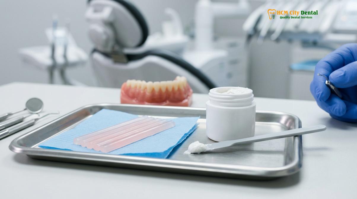
All-ceramic crowns, such as those made from lithium disilicate (e.g., IPS e.max), offer unparalleled aesthetics, mimicking the natural translucency and light-scattering properties of human enamel. These materials are frequently utilized for anterior (front) teeth and Porcelain Veneers, where cosmetic demands are paramount. While modern lithium disilicate is significantly stronger than older ceramics, it still possesses a lower flexural strength than metal or zirconia, making it more susceptible to fracture if placed in high-stress posterior regions, particularly in patients with active bruxism.
For maximum durability, monolithic zirconia crowns have become the gold standard in posterior restorations. Yttria-stabilized tetragonal zirconia polycrystal (Y-TZP) exhibits exceptionally high fracture toughness and flexural strength, often exceeding 1000 MPa[3]. Because monolithic zirconia crowns are milled from a single, solid block of material, there is no veneering porcelain to chip or delaminate. This makes them highly resistant to the catastrophic failures seen in PFM or layered ceramic crowns.
“The transition towards monolithic zirconia in posterior restorations has drastically reduced the incidence of catastrophic crown fractures. By eliminating the weak interface between metal and porcelain, we provide our patients with a restoration that can reliably withstand the most demanding occlusal forces.”
— Clinical Consensus on Advanced Prosthodontics
Dr. Nguyen Van Cuong frequently emphasizes that material selection must be a highly individualized process. A comprehensive occlusal analysis is required to determine the magnitude and direction of the forces the new crown will endure. By carefully matching the physical properties of the restorative material to the patient’s unique biomechanical profile, the risk of experiencing another broken crown tooth is minimized, ensuring a long-lasting and functional result.
Clinical Case Study: Restoring a Fractured Molar Crown
A detailed clinical case demonstrates the systematic approach to diagnosing a catastrophic crown failure and successfully restoring the tooth using advanced digital dentistry and high-strength ceramics.
To illustrate the clinical workflow for managing a broken crown tooth, consider a recent case managed at a professional facility. The presentation and subsequent treatment highlight the importance of precise diagnostics and modern material application in achieving a successful outcome.
Patient Presentation: A 45-year-old male patient presented to HCMC Dental Clinic in Ho Chi Minh City with a chief complaint of a broken crown tooth on the lower right first molar (tooth 46). The patient reported biting down on a hard piece of candy, resulting in an immediate, audible crack and subsequent thermal sensitivity.
Clinical Findings: Intraoral examination revealed a catastrophic cohesive fracture of the veneering porcelain on an existing PFM crown, exposing the underlying metal coping and a portion of the tooth margin. Periapical radiography confirmed that the root structure and surrounding alveolar bone were intact, with no signs of periapical pathology. The abutment tooth exhibited adequate ferrule, making it a viable candidate for a new restoration.
Treatment Protocol:
1. Removal and Preparation: Under local anesthesia, the remaining PFM crown was carefully sectioned and removed using specialized diamond burs to avoid traumatizing the underlying dentin.
2. Core Evaluation: The underlying composite core buildup was evaluated, refined, and smoothed to ensure ideal geometric retention and resistance form.
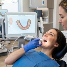
3. Digital Impression: An intraoral 3D scanner was utilized to capture highly accurate digital impressions of the prepared tooth, adjacent dentition, and the patient’s occlusal relationship.
4. Fabrication: A full-contour monolithic zirconia crown was digitally designed and milled, selected for its superior flexural strength to withstand the patient’s heavy posterior bite forces.
5. Delivery: The final zirconia crown was evaluated for marginal fit, proximal contacts, and occlusal harmony before being permanently cemented using a dual-cure resin cement.
Outcome: The patient experienced immediate relief from sensitivity and full restoration of masticatory function, with the monolithic zirconia providing a highly durable, fracture-resistant solution.
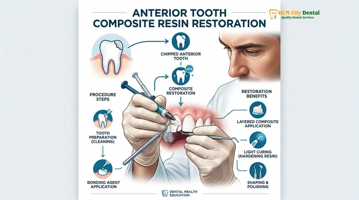
This case underscores the necessity of a methodical approach. Simply replacing the broken crown tooth without evaluating the underlying core or considering the occlusal forces that caused the initial failure would likely result in a recurrent fracture. By utilizing digital scanning and high-strength monolithic materials, clinicians can provide restorations that offer both precise marginal adaptation and exceptional long-term durability.
When to See a Doctor for a Broken Crown
While some minor chips may not cause immediate pain, a broken crown tooth should always be evaluated by a dental professional to rule out hidden complications. Patients must be vigilant in monitoring their symptoms and understanding when immediate clinical intervention is required to save the underlying tooth.
You should seek urgent dental care if the broken crown tooth is accompanied by severe, throbbing pain that is not relieved by over-the-counter analgesics. This type of pain often indicates that the fracture has propagated deep into the tooth structure, potentially exposing the dental pulp to bacterial contamination. Similarly, if you experience significant swelling in the gums surrounding the broken crown, or if you notice a purulent discharge (pus), this is a clear sign of an active localized infection or abscess that requires immediate antibiotic therapy and clinical management[4].
Another critical red flag is extreme sensitivity to hot or cold stimuli that lingers long after the stimulus is removed. While mild, transient sensitivity is common when dentin is exposed, lingering pain suggests irreversible pulpitis, meaning the nerve of the tooth is actively dying and may require endodontic intervention before a new crown can be placed. Additionally, if the broken crown leaves a sharp, jagged edge that is continuously lacerating your tongue or cheek, prompt smoothing or removal of the fragment is necessary to prevent painful mucosal ulcerations.
Even in the absence of pain, a broken crown tooth compromises the marginal seal. Delaying treatment allows saliva and bacteria to seep between the remaining crown and the tooth, leading to rapid, silent decay. By the time this decay causes pain, the tooth may be so structurally compromised that it cannot be saved. Therefore, scheduling an evaluation at a reputable facility like HCMC Dental Clinic in Ho Chi Minh City within 48 hours of the incident is highly recommended to ensure the best possible prognosis for your oral health.
Frequently Asked Questions
Is a broken crown tooth considered a dental emergency?
Yes, a broken crown tooth is often considered a dental emergency, especially if accompanied by pain, bleeding, or exposed dentin. Prompt clinical evaluation is necessary to prevent bacterial contamination of the underlying tooth structure, mitigate thermal sensitivity, and avoid potential damage to the root canal system or surrounding gingival tissues.
Can a dentist repair a chipped porcelain crown without replacing it?
Minor chips in a porcelain crown can sometimes be repaired using specialized composite resin bonding, provided the structural integrity of the crown remains intact. However, if the fracture compromises the crown’s margins, affects the occlusal contact points, or exposes the underlying metal framework, a complete replacement is typically required.
What should I do if I swallow a piece of my broken dental crown?
If you accidentally swallow a small fragment of a broken dental crown, remain calm, as small, smooth pieces usually pass through the digestive tract without complication. However, if you experience coughing, difficulty breathing, or abdominal pain, seek immediate medical attention, as the fragment may have been aspirated into the airway.
How long can I wait to fix a broken dental crown?
You should ideally have a broken dental crown evaluated by a professional within 24 to 48 hours to minimize the risk of complications. Delaying treatment can lead to rapid bacterial microleakage, severe dentinal hypersensitivity, shifting of adjacent teeth, and irreversible damage to the underlying abutment tooth.
Does repairing or replacing a broken crown hurt?
Repairing or replacing a broken crown is generally a comfortable procedure, as dentists utilize local anesthesia to numb the affected tooth and surrounding tissues. Patients may experience mild, transient sensitivity following the procedure, which typically resolves within a few days and can be managed with over-the-counter analgesics.
References
- Journal of Prosthetic Dentistry. Biomechanical failure modes and fracture resistance of all-ceramic restorations. (2021).
- International Endodontic Journal. Microleakage and pulpal response following catastrophic failure of coronal restorations. (2020).
- Dental Materials. Comparative flexural strength and fatigue degradation of monolithic zirconia versus lithium disilicate. (2022).
- Journal of the American Dental Association. Clinical guidelines for the management of fractured dental prostheses and emergency temporization. (2019).
- Clinical Oral Investigations. Long-term survival rates and complication profiles of porcelain-fused-to-metal versus all-ceramic crowns. (2023).