Finding the best dentist for wisdom teeth removal requires evaluating a practitioner’s surgical experience, diagnostic technology, and complication management protocols. Patients should prioritize clinics utilizing 3D CBCT imaging and minimally invasive techniques to ensure safe extractions and predictable postoperative recovery, even for asymptomatic cases that may pose future risks[4].
Clinical Summary:
Wisdom tooth extraction is a complex surgical procedure that demands specialized expertise, particularly for impacted or anatomically challenging third molars. Selecting a highly qualified dental professional ensures accurate preoperative assessment, appropriate anesthesia management, and the use of advanced modalities like Piezotome surgery. This comprehensive guide explores the clinical criteria for choosing an extraction specialist, surgical workflows, and essential recovery protocols to minimize risks such as nerve injury or alveolar osteitis. By prioritizing evidence-based practices and advanced diagnostics, patients can achieve optimal surgical outcomes and a smooth healing trajectory.
Key Takeaways:
- Impacted wisdom teeth require precise 3D CBCT diagnostics to map proximity to the inferior alveolar nerve and maxillary sinus.
- Oral and maxillofacial surgeons or highly trained general dentists offer the safest outcomes for complex, multi-rooted extractions.
- Minimally invasive techniques, including ultrasonic bone surgery (Piezotome), significantly reduce postoperative swelling and pain.
- Proper postoperative care, including clot preservation, is critical to preventing complications like dry socket and secondary infections.
- Timely clinical intervention is necessary when symptoms of pericoronitis, cyst formation, or adjacent tooth damage arise.
Why Specialized Expertise Matters for Wisdom Teeth Removal
The anatomical complexity of third molars, including variable root morphology and proximity to vital structures, necessitates advanced surgical proficiency to ensure safe and atraumatic removal.
The extraction of third molars, commonly known as wisdom teeth, is rarely a straightforward procedure. Unlike anterior teeth or premolars, wisdom teeth develop late in life and frequently lack adequate space within the dental arch to erupt into a functional, hygienic position. This lack of space leads to various degrees of impaction, where the tooth remains partially or completely encased within the jawbone or submerged beneath the gingival tissue. The biomechanical and anatomical challenges presented by these impactions underscore the necessity of seeking the best dentist for wisdom teeth removal, one who possesses a deep understanding of maxillofacial anatomy and surgical principles.
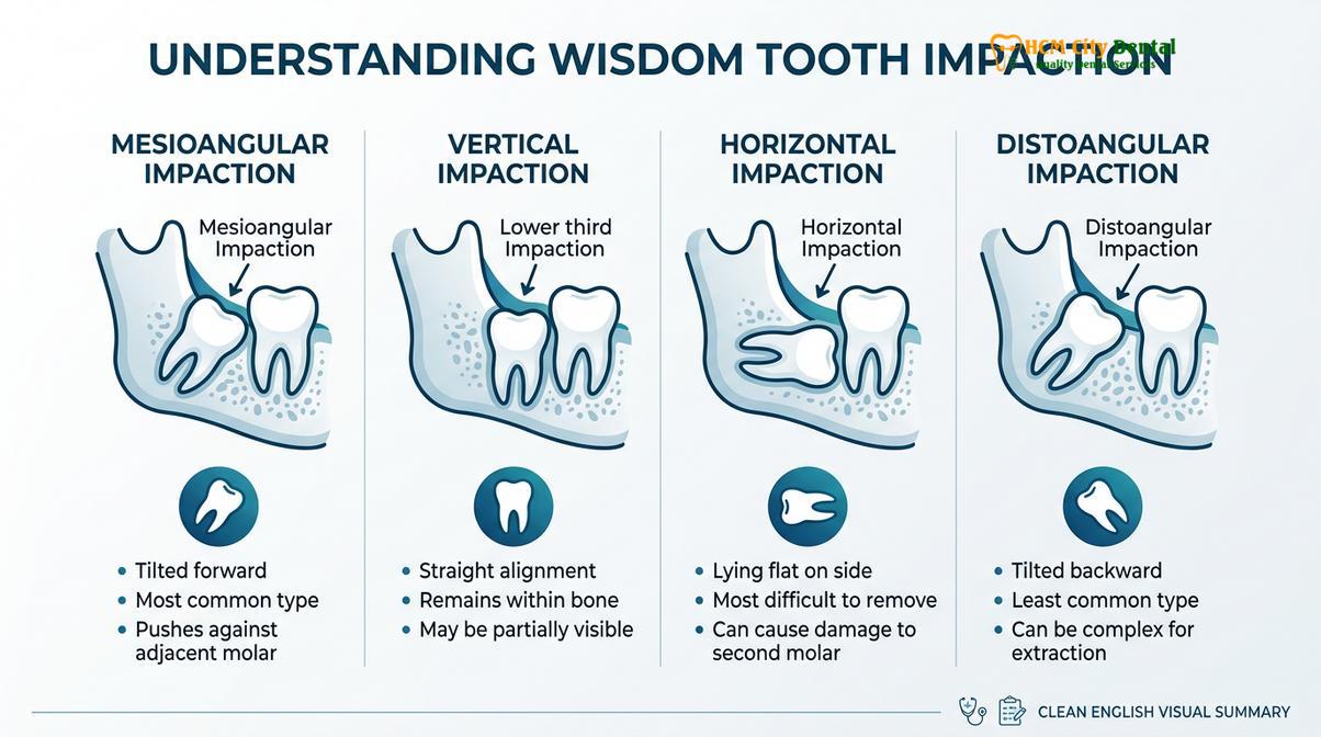
Clinically, impactions are classified based on their angulation relative to the adjacent second molar. Mesioangular impactions, where the tooth is tilted forward, are the most common and often create a plaque-retentive trap that leads to localized periodontal defects. Horizontal impactions present a more severe challenge, as the crown of the wisdom tooth directly abuts the roots of the second molar, potentially causing root resorption. Vertical and distoangular impactions carry their own unique surgical difficulties, often requiring significant bone removal to establish an adequate path of draw. A practitioner highly skilled in Tooth Extraction tại HCMC Dental Clinic protocols will accurately classify the impaction and formulate a surgical strategy that minimizes trauma to the surrounding periodontium.
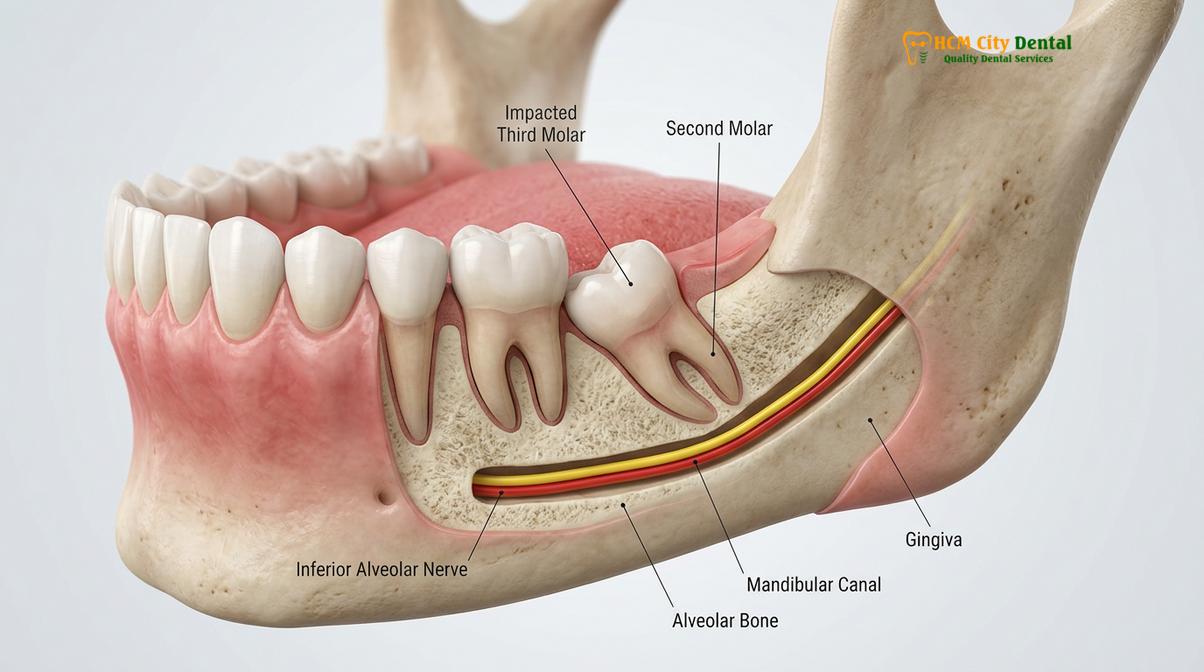
Beyond angulation, the root morphology of third molars is notoriously unpredictable. Roots may be fused, widely divergent, or exhibit severe dilaceration (sharp curvature). In the mandible, these roots frequently develop in close proximity to, or even wrap around, the inferior alveolar nerve (IAN)—a major sensory nerve supplying the lower lip, chin, and teeth. Iatrogenic injury to this nerve during extraction can result in temporary or permanent paresthesia (numbness) or dysesthesia (altered sensation). In the maxilla, the roots of upper wisdom teeth often approximate or protrude into the maxillary sinus cavity, creating a risk for oroantral communication upon removal. Navigating these critical anatomical landmarks requires not just theoretical knowledge, but extensive tactile experience and spatial awareness[1].
Evaluating the Best Dentist for Wisdom Teeth Removal: Key Clinical Criteria
Selecting the optimal provider involves assessing their surgical volume, utilization of 3D imaging technology, and adherence to strict sterilization and sedation protocols.
When determining who is the most qualified professional to perform a third molar extraction, patients must look beyond basic credentials and evaluate the specific clinical environment and technological capabilities of the practice. The distinction between a simple extraction and a complex surgical extraction is profound, and the chosen clinician must be equipped to handle intraoperative complications seamlessly. A general dentist may be perfectly suited for fully erupted, straightforward wisdom teeth, but deeply impacted cases generally require the expertise of an oral and maxillofacial surgeon or a general dentist with extensive, focused training in dentoalveolar surgery.
One of the most critical criteria in modern oral surgery is the routine use of Cone Beam Computed Tomography (CBCT). Traditional 2D panoramic radiographs provide a flat overview of the jaws but suffer from magnification errors and superimposition of structures. They cannot definitively show the buccal-lingual (side-to-side) relationship between a tooth root and the inferior alveolar nerve. A practitioner utilizing 3D CBCT imaging can virtually map the exact trajectory of the nerve, measure the thickness of the overlying bone, and plan the surgical approach with millimeter precision. This diagnostic foresight is a hallmark of a top-tier surgical practice and is non-negotiable for high-risk impactions.
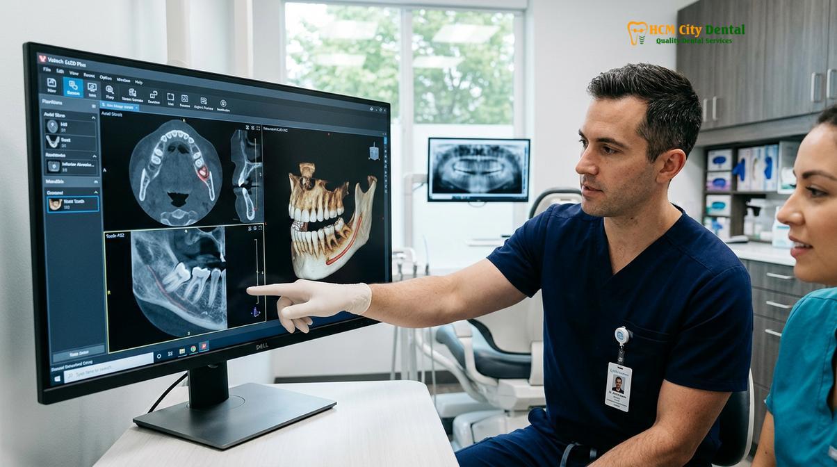
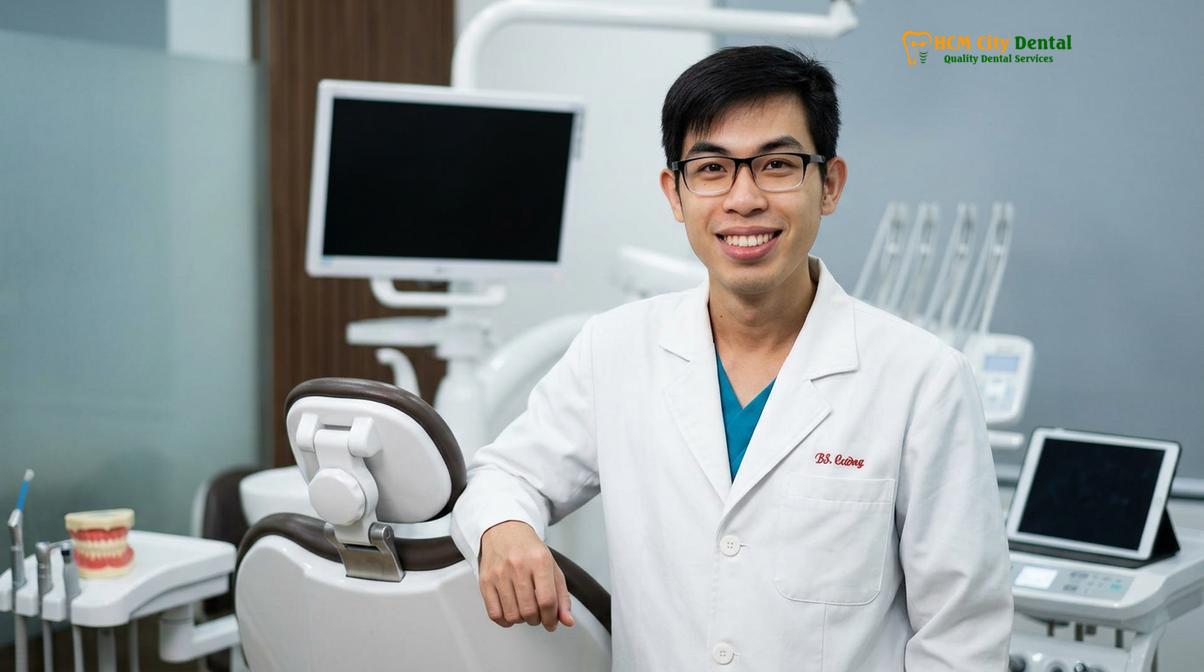
Dr. Nguyen Van Cuong, a leading oral surgery expert at HCMC Dental Clinic, emphasizes that the success of a surgical extraction is largely determined before the scalpel ever touches the tissue. His extensive clinical experience in managing complex impactions ensures that patients receive the highest standard of care, minimizing risks and optimizing recovery. By integrating advanced diagnostics with meticulous surgical planning, Dr. Cuong and his team provide a predictable and comfortable experience for every patient.
“The integration of 3D CBCT imaging has fundamentally transformed dentoalveolar surgery. It shifts our approach from reactive problem-solving during the procedure to proactive, precision-guided surgical planning, drastically reducing the risk of neurological complications.”
Another crucial factor is the availability of comprehensive pain management and sedation options. Wisdom tooth extraction can be an anxiety-inducing experience. The best clinics offer a spectrum of anesthesia, ranging from profound local anesthesia with advanced buffering techniques to nitrous oxide analgesia, oral conscious sedation, and intravenous (IV) sedation. Furthermore, reviewing Dentist Reviews and Feedback: Clinical Guide | HCMC Dental can provide valuable insights into a practitioner’s bedside manner, their ability to manage patient anxiety, and the overall efficiency of their clinical team.
Advanced Surgical Workflows and Technologies
Modern extraction protocols integrate ultrasonic instruments and biologic modifiers to enhance surgical precision, minimize tissue trauma, and accelerate hard and soft tissue healing.
The methodology of tooth extraction has evolved significantly from the traditional reliance on high-speed rotary burs and heavy elevation forces. Today, the leading practitioners employ minimally invasive workflows designed to preserve as much native bone as possible and respect the delicate architecture of the soft tissues. This paradigm shift is driven by the understanding that surgical trauma is the primary catalyst for postoperative pain, swelling, and delayed healing.
A cornerstone of advanced surgical extraction is the use of Piezosurgery, or ultrasonic bone cutting. Unlike conventional drills that cut both bone and soft tissue indiscriminately, a Piezotome utilizes micro-vibrations at ultrasonic frequencies to selectively cut osseous tissue while leaving adjacent soft tissues—such as nerves, blood vessels, and the sinus membrane—completely unharmed. This selective cutting capability is invaluable when removing bone overlying an impacted wisdom tooth near the inferior alveolar nerve. Furthermore, the cavitation effect of the ultrasonic coolant spray creates a relatively bloodless surgical field, enhancing visibility and precision[2].
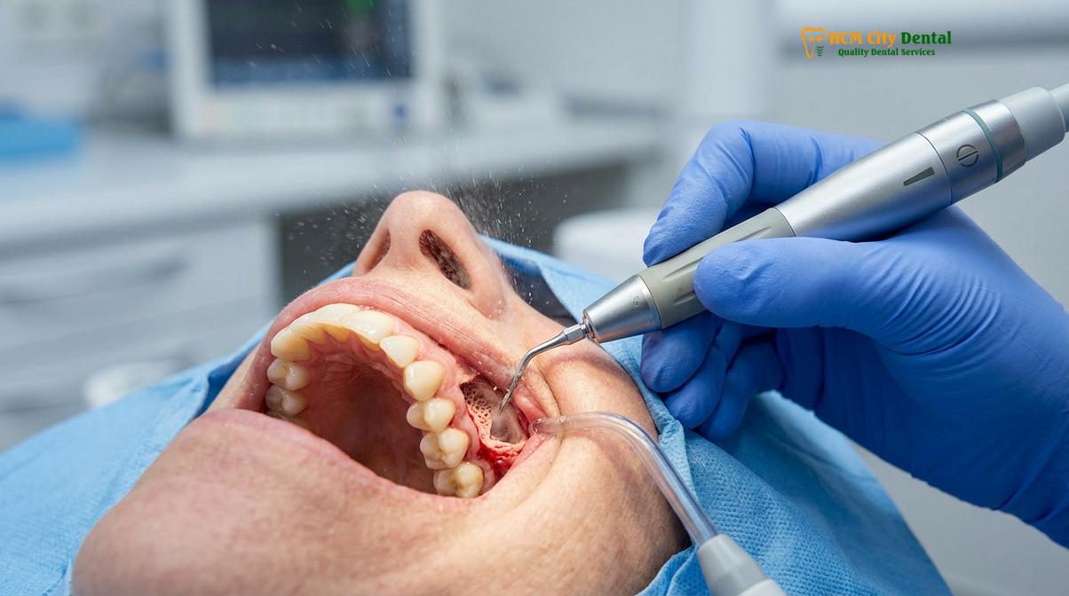
In addition to advanced instrumentation, the application of biologic modifiers has revolutionized postoperative healing. Platelet-Rich Fibrin (PRF) is an autologous matrix derived from a small sample of the patient’s own blood, drawn immediately prior to surgery. The blood is centrifuged to isolate a concentrated membrane of platelets, leukocytes, and growth factors. When this PRF membrane is placed into the extraction socket, it acts as a bioactive scaffold, accelerating angiogenesis (new blood vessel formation), promoting rapid soft tissue closure, and significantly reducing the risk of alveolar osteitis (dry socket)[3].
| Clinical Parameter | Traditional Rotary Extraction | Advanced Minimally Invasive (Piezotome + PRF) |
|---|---|---|
| Bone Cutting Mechanism | High-speed rotary bur (macro-trauma) | Ultrasonic micro-vibrations (selective cutting) |
| Soft Tissue Risk | Higher risk of collateral damage to nerves/vessels | Soft-tissue sparing; minimal risk of iatrogenic injury |
| Intraoperative Bleeding | Moderate to high | Minimal (cavitation effect improves visibility) |
| Postoperative Swelling | Typically moderate to severe | Significantly reduced due to decreased surgical trauma |
| Healing Acceleration | Standard physiological healing timeline | Accelerated via autologous growth factors (PRF) |
Prior to any surgical intervention, maintaining optimal oral hygiene is essential to reduce the bacterial load in the oral cavity, thereby minimizing the risk of postoperative infection. Patients are often advised to undergo a professional prophylaxis. Understanding How Much Does a Teeth Cleaning Cost Without Insurance | HCMC Dental can help patients budget for this important preparatory step. A clean surgical environment is a fundamental prerequisite for successful dentoalveolar surgery.
Clinical Case Study: Complex Impacted Mandibular Third Molar
A detailed case review demonstrates how comprehensive diagnostic imaging and specialized surgical techniques successfully resolve high-risk mandibular impactions while preserving neurological function.
To illustrate the practical application of these advanced protocols, we examine a recent clinical case managed at HCMC Dental Clinic in Ho Chi Minh City. The patient, a 24-year-old male, presented with recurrent episodes of dull, throbbing pain in the lower right quadrant, accompanied by mild trismus (restricted mouth opening). A preliminary panoramic radiograph revealed a deeply impacted, horizontally positioned mandibular right third molar. The crown of the impacted tooth was intimately engaged with the distal root of the adjacent second molar, posing a severe risk of root resorption.
Case Study: Horizontal Impaction with IAN Proximity
Patient Profile: 24-year-old male visiting HCMC Dental Clinic (Ho Chi Minh City), presenting with recurrent pericoronitis and localized pain.
Diagnostic Findings: 3D CBCT imaging revealed a horizontal impaction. The apical third of the roots exhibited severe dilaceration and were positioned directly adjacent to the superior cortical border of the inferior alveolar nerve canal.
Surgical Intervention: The extraction was performed under IV sedation. A full-thickness mucoperiosteal flap was elevated. Piezosurgery was utilized to carefully remove the overlying buccal bone without endangering the nerve. The tooth was sectioned into three distinct fragments using a surgical handpiece, allowing for atraumatic elevation of each piece along its specific path of draw. PRF was placed in the osteotomy site prior to primary closure with resorbable sutures.
Clinical Outcome: The patient experienced minimal postoperative swelling and required only over-the-counter analgesics after 48 hours. Normal sensory function of the lower lip and chin was fully preserved, confirming the success of the nerve-sparing techniques.
This case highlights the critical importance of spatial awareness and controlled force application. Had traditional elevation techniques been attempted without sectioning the tooth, the leverage applied could have easily fractured the mandible or crushed the inferior alveolar nerve. The meticulous approach taken by the surgical team ensured a predictable and safe outcome. It is important to note that while functional teeth lost to decay or trauma often require replacement, such as a 4 Tooth Dental Bridge: Clinical Guide, Costs & Procedure | HCMC Dental, extracted wisdom teeth are not replaced, as they are non-essential for masticatory function and their absence eliminates the risk of future pathology.
When to See a Doctor for Wisdom Tooth Complications
Recognizing the early warning signs of wisdom tooth pathology is crucial for preventing severe infections and irreversible damage to adjacent dental structures.
While some wisdom teeth remain asymptomatic and pathology-free throughout a patient’s life, many develop complications that necessitate prompt clinical evaluation. Waiting until pain becomes unbearable is a flawed strategy, as significant structural damage to adjacent teeth or bone may have already occurred by the time symptoms manifest. Regular radiographic monitoring is essential for early detection.
One of the most frequent acute complications is pericoronitis, an infection of the operculum—the flap of gum tissue that partially covers an erupting wisdom tooth. This tissue creates a deep, inaccessible pocket where food debris and anaerobic bacteria accumulate. The resulting infection causes severe inflammation, localized pain, foul breath, and purulent exudate (pus). If left untreated, the infection can spread to the fascial spaces of the head and neck, becoming a life-threatening medical emergency. Any signs of pericoronitis require immediate evaluation by a qualified dentist wisdom teeth removal provider.
Important Clinical Considerations: Red Flag Symptoms
Patients should seek immediate emergency dental or medical care if they experience any of the following symptoms associated with their wisdom teeth:
- Severe Trismus: Inability to open the mouth more than a few millimeters, indicating spreading infection into the masticatory muscles.
- Facial Cellulitis: Rapidly expanding swelling of the cheek, jawline, or neck, which may compromise the airway.
- Systemic Symptoms: High fever, chills, or lethargy accompanying oral pain.
- Prolonged Bleeding: Uncontrolled bleeding from an extraction site that does not stop with firm pressure.
Following an extraction, patients must also be vigilant for signs of alveolar osteitis, commonly known as dry socket. This painful condition occurs when the protective blood clot at the extraction site fails to form or is prematurely dislodged, exposing the underlying bone and nerves to the oral environment. According to guidelines from the Vietnam Odonto-Stomatology Association (VOSA), strict adherence to postoperative instructions—such as avoiding smoking, spitting, and using straws—is paramount in preventing this complication[5]. If severe, radiating pain develops three to five days post-surgery, patients should return to their clinician for palliative treatment, which typically involves placing a medicated dressing within the socket.
“Preserving the blood clot within the extraction socket is the single most critical factor in preventing alveolar osteitis. Patient compliance with postoperative instructions directly dictates the trajectory of their healing process.”
In cases where adjacent teeth have been damaged by an impacted wisdom tooth, restorative interventions may be necessary once the extraction site has healed. For instance, if a second molar is lost due to severe root resorption caused by a horizontal impaction, patients might need to explore replacement options and understand How Much Does a Dental Bridge Cost? Clinical Guide | HCMC Dental. Furthermore, patients undergoing orthodontic treatment should have their wisdom teeth evaluated, as erupting third molars can sometimes exert pressure and disrupt alignment. Understanding the Invisalign vs Braces Cost: Clinical Guide & Pricing | HCMC Dental or the Damon System Braces Cost: Clinical Guide & Pricing | HCMC Dental is important for comprehensive treatment planning.
References
- Journal of Oral and Maxillofacial Surgery. Anatomical considerations and risk factors in third molar extractions. (2021).
- International Journal of Oral and Maxillofacial Surgery. Efficacy of piezosurgery versus conventional rotary instruments in impacted mandibular third molar extraction. (2020).
- British Dental Journal. The role of Platelet-Rich Fibrin (PRF) in accelerating post-extraction wound healing. (2019).
- Journal of the American Dental Association. Prophylactic removal of asymptomatic impacted wisdom teeth: a clinical review. (2022).
- Vietnam Odonto-Stomatology Association (VOSA). National clinical guidelines on the management and prevention of alveolar osteitis following third molar surgery. (2023).
Frequently Asked Questions
How do I know if I need an oral surgeon or a general dentist for my wisdom teeth?
The choice depends on the complexity of the impaction, root anatomy, and proximity to vital nerves. A clinical examination and 3D CBCT scan will determine if a specialized oral surgeon is required for safe removal.
Is it necessary to remove wisdom teeth if they are not currently causing pain?
Not always, but asymptomatic wisdom teeth can still develop silent pathologies such as cysts, root resorption of adjacent teeth, or chronic periodontal pockets. Prophylactic removal is often recommended based on radiographic evidence of future risk.
What is the typical recovery timeline after a surgical wisdom tooth extraction?
Initial soft tissue healing takes approximately one to two weeks, while complete bone regeneration within the extraction socket can take several months. Most patients resume normal daily activities within three to five days.
How can I effectively prevent dry socket after my extraction procedure?
To prevent dry socket, avoid smoking, using straws, vigorous rinsing, and spitting for at least 72 hours post-surgery. These actions create negative pressure that can dislodge the critical blood clot forming in the socket.
Does the use of 3D CBCT imaging increase the safety of the extraction?
Yes, 3D CBCT imaging significantly enhances surgical safety by providing a precise, multi-planar view of the tooth’s roots in relation to the inferior alveolar nerve and the maxillary sinus, minimizing the risk of iatrogenic injury.
For personalized advice and to consult with the best dentist for wisdom teeth removal, schedule a comprehensive evaluation at HCMC Dental Clinic, Ho Chi Minh City today. Our specialized team utilizes advanced 3D imaging and minimally invasive techniques to ensure your safety and comfort.
![Tooth Replacement Cost: [Find Your Best Option] – Dr. Cuong](https://hcmcdental.com/wp-content/uploads/2025/07/tooth-replacement-cost-1-dr-cuong-hcmc-dental-clinic-280x280.jpg)