Experiencing tooth crown pain chewing sensitivity is a clinical scenario that frequently causes anxiety for patients who have recently undergone restorative dental procedures. While a dental crown is designed to protect and strengthen a compromised tooth, the complex biological and mechanical processes involved in its placement can sometimes lead to post-operative discomfort. This discomfort can range from mild, transient thermal sensitivity to sharp, localized pain specifically triggered by biting pressure. Understanding the underlying etiology—whether it stems from a hyperocclusion (a high bite), transient nerve inflammation, or exposed dentinal tubules at the crown margin—is essential for determining the appropriate clinical intervention. While mild sensitivity to hot and cold is often a normal part of the healing process immediately following cementation, sharp pain upon mastication requires a thorough clinical evaluation to adjust the occlusion or assess the health of the underlying pulpal tissue.
Clinical Summary:
Post-operative discomfort following the placement of a full-coverage dental restoration is a common clinical presentation with a multifactorial etiology. The primary causes include occlusal discrepancies (hyperocclusion), pulpal hyperemia resulting from the mechanical trauma of tooth preparation, and microleakage at the cementation margin. Advanced diagnostic protocols, including digital occlusal analysis, percussion testing, and thermal vitality assessments, are critical to differentiate between reversible conditions that require conservative management—such as simple bite adjustments—and irreversible pulpitis that necessitates endodontic intervention. Utilizing precision CAD/CAM technology, immediate dentin sealing techniques, and highly biocompatible materials like monolithic Zirconia and Lithium Disilicate significantly reduces the incidence of long-term sensitivity and promotes optimal integration with the surrounding periodontal tissues.
Key Takeaways:
- Mild sensitivity to hot and cold stimuli is a normal physiological response that typically resolves within a few weeks post-cementation as the pulp heals.
- Sharp, localized pain specifically triggered by biting pressure strongly indicates a hyperocclusion, requiring a minor mechanical bite adjustment by the dentist.
- Spontaneous, throbbing pain that wakes a patient at night or lingers long after a thermal stimulus is removed is a hallmark sign of irreversible nerve inflammation.
- Modern digital workflows, including advanced intraoral scanners, enhance margin precision and drastically reduce the risk of post-operative microleakage.
- Choosing highly biocompatible, low-thermal-conductivity materials minimizes temperature transfer and protects the underlying pulpal tissue from environmental shock.
Common Causes of Pain Under a Dental Crown
Discomfort beneath a new restoration typically stems from occlusal discrepancies, cement washout, or transient pulpal hyperemia following tooth preparation. Identifying the exact trigger is critical for accurate diagnosis and targeted intervention.
The placement of a dental crown is a highly precise prosthodontic procedure designed to restore the structural integrity, function, and aesthetics of a severely decayed, fractured, or endodontically treated tooth. However, the journey from a damaged tooth to a fully restored, asymptomatic bite involves complex biological and mechanical interactions. When a patient reports discomfort, understanding the underlying pathophysiology is the first step toward effective resolution. The causes of pain under a dental restoration can be broadly categorized into mechanical, biological, and chemical factors, each requiring a distinct diagnostic approach.
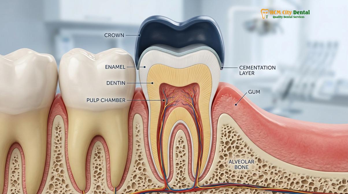
Mechanically, the process of tooth preparation involves the strategic removal of enamel and a portion of the underlying dentin to create sufficient restorative space for the crown material. This reduction is absolutely necessary to ensure the final restoration is not bulky, maintains proper emergence profile, and aligns harmoniously with the adjacent dentition. However, dentin is a highly porous living tissue composed of thousands of microscopic tubules that communicate directly with the central pulp chamber. When these tubules are exposed and severed during preparation, the underlying nerve tissue undergoes a localized inflammatory response known as pulpal hyperemia. This transient inflammation is a normal physiological reaction to the mechanical trauma and vibration of the high-speed dental handpiece, even when copious water coolant is utilized to mitigate heat generation.
Biologically, the health of the gingival tissues and the concept of the biologic width play a crucial role in post-operative comfort. The biologic width is the natural dimension of the soft tissue attached to the portion of the tooth above the alveolar bone crest. If a tooth preparation margin is placed too deeply beneath the gum line—encroaching upon this sacred biological space—the body will mount a chronic inflammatory response. This results in persistent gingival tenderness, localized swelling, bleeding upon probing, and discomfort that closely mimics deep tooth pain. In such cases, a functional crown lengthening procedure or a margin revision may be required to re-establish a healthy periodontal architecture and alleviate the chronic irritation.
Chemically, the cementation process itself can induce temporary sensitivity. Modern dental cements, particularly resin-modified glass ionomers and dual-cure adhesive resin cements, require specific protocols for tooth conditioning, including acid etching and the application of bonding agents. The acidic nature of some primers can temporarily irritate the dentinal tubules. Furthermore, if the tooth is overly desiccated (dried out) prior to cementation, the sudden movement of dentinal fluid upon the application of the cement can cause a sharp, transient pain known as hydrodynamic shock. Strict adherence to immediate dentin sealing techniques—where a bonding agent is applied immediately after tooth preparation—significantly mitigates this chemical irritation, seals the tubules, and protects the delicate pulp complex from bacterial ingress[1].
| Type of Discomfort | Typical Clinical Cause | Expected Duration | Recommended Action |
|---|---|---|---|
| Mild to moderate thermal sensitivity (Cold/Heat) | Transient pulpal hyperemia, dentinal tubule exposure | A few weeks, gradually decreasing | Use desensitizing toothpaste; monitor symptoms closely. |
| Sharp pain specifically upon biting or chewing | Hyperocclusion (High bite), periodontal ligament sprain | Persists until mechanically adjusted | Schedule a clinical bite adjustment immediately. |
| Spontaneous, throbbing pain or lingering heat sensitivity | Irreversible pulpitis, nerve infection | Worsens over time; does not resolve naturally | Immediate endodontic evaluation (Root Canal). |
| Localized gum tenderness and bleeding around the margin | Biologic width violation, retained excess cement | Chronic until the irritant is removed | Professional scaling or margin revision. |
Sensitivity to Cold and Heat Post-Placement
Thermal sensitivity after crown placement usually results from transient nerve irritation or exposed dentinal tubules near the margin. This reaction typically subsides as the pulp heals and secondary dentin forms.
It is exceedingly common for patients to experience sensitivity to hot and cold temperatures immediately following the final cementation of a dental crown. To understand why this occurs, one must look at the hydrodynamic theory of tooth sensitivity. According to this widely accepted clinical model, fluid resides within the microscopic dentinal tubules that radiate from the pulp to the dentinoenamel junction. When a thermal stimulus—such as a sip of ice water or hot coffee—is applied to the tooth, it causes this fluid to rapidly expand or contract. This sudden fluid movement stimulates the mechanoreceptors on the odontoblast cells located at the periphery of the pulp, which the brain instantly interprets as a sharp, zinging pain.
The choice of restorative material significantly influences the degree of thermal conductivity and, consequently, the level of post-operative sensitivity a patient might experience. Historically, Porcelain-Fused-to-Metal (PFM) crowns and full-cast gold crowns were the standard of care. However, the underlying metal substructure in these restorations is an excellent conductor of temperature. When a patient consumes a cold beverage, the temperature change is rapidly transmitted through the metal coping directly to the underlying dentin. This often results in a more pronounced thermal shock compared to modern all-ceramic alternatives, requiring a longer adaptation period for the pulp.
“The transition from traditional metal-based restorations to highly biocompatible ceramics like monolithic zirconia has dramatically reduced the incidence of post-operative thermal sensitivity. By acting as a thermal insulator, these advanced materials allow the traumatized pulpal tissue the necessary time to lay down reparative secondary dentin without continuous environmental irritation.”
Today, advanced material science has revolutionized prosthodontics. Materials such as Zirconia HT (High Translucency) and Lithium Disilicate (E.max) are not only superior in aesthetics but also act as excellent thermal insulators. Zirconia, a crystalline dioxide of zirconium, possesses a very low thermal conductivity rate. When a monolithic Zirconia crown is placed, it effectively shields the underlying tooth structure from extreme temperature fluctuations in the oral cavity. As the tooth heals over the subsequent weeks, the pulp naturally defends itself by depositing a layer of reparative secondary dentin along the inner walls of the pulp chamber, further insulating the nerve and gradually eliminating the thermal sensitivity[2].
Pain When Chewing: High Bite vs. Nerve Issues
Sharp pain specifically triggered by biting pressure is most frequently caused by a hyperocclusion (high bite) that traumatizes the periodontal ligament. If adjusting the bite does not resolve the discomfort, underlying pulpal pathology must be investigated.
When a patient reports that their tooth hurts exclusively upon mastication (chewing), the diagnostic focus immediately shifts to the biomechanics of occlusion. The human masticatory system is incredibly sensitive and finely tuned; the proprioceptive nerve endings within the periodontal ligament (PDL)—the fibrous connective tissue that anchors the tooth root to the surrounding alveolar bone—can detect interferences as minute as 10 to 20 microns. If a newly placed crown is even fractionally taller than the adjacent teeth, it will bear the brunt of the biting force before the rest of the teeth come together in maximum intercuspation.
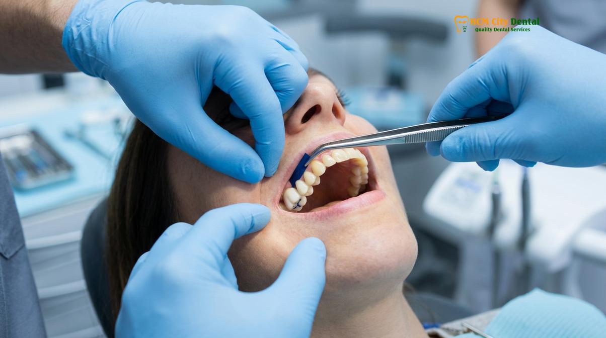
This condition, clinically termed hyperocclusion or a “high bite,” results in localized occlusal trauma. Every time the patient swallows, speaks, or chews, the excessive force compresses the PDL, leading to acute inflammation and localized edema within the ligament space. Because the fluid within the inflamed PDL cannot easily escape, subsequent biting pressure causes severe, sharp pain. In these scenarios, a dental crown bite adjustment is the definitive and immediate solution. The dentist will use ultra-thin articulating paper to mark the premature contacts and gently polish the ceramic surface until the bite is perfectly harmonized. Once adjusted, the PDL inflammation typically subsides within 48 to 72 hours[3].
Clinical Case Study: Resolving Occlusal Trauma
A 45-year-old dental tourism patient visited HCMC Dental Clinic in Ho Chi Minh City complaining of severe pain when chewing on a newly placed 3-unit Zirconia bridge fabricated at a different overseas clinic. The patient was unable to masticate on the right side and feared the teeth needed to be extracted. Clinical examination utilizing digital occlusal analysis revealed a severe premature contact on the mesial-lingual cusp of the posterior abutment crown. A precise bite adjustment was performed, and the ceramic was re-polished to a high gloss. The patient reported a significant reduction in pain within 24 hours and complete resolution shortly after, successfully avoiding the need for unnecessary endodontic treatment.
However, if the bite has been verified as perfectly balanced and the patient still experiences sharp pain upon pressure, the clinician must consider alternative diagnoses. One distinct possibility is Cracked Tooth Syndrome. If the underlying tooth structure had a pre-existing micro-fracture that was not visible during the initial preparation, the flexural forces of chewing can cause the crack to propagate, stimulating the pulpal nerves. Differentiating between PDL inflammation from a high bite and periapical periodontitis from a necrotic nerve or a structural crack requires comprehensive radiographic imaging, percussion testing, and vitality assessments.
Nerve Inflammation (Pulpitis) and Root Canals
When the nerve inside a crowned tooth becomes irreversibly inflamed or infected, conservative adjustments are no longer sufficient. Endodontic therapy (a root canal) is required to remove the diseased tissue and save the foundation of the tooth.
The dental pulp is a delicate neurovascular bundle housed within the rigid walls of the dentin. Because it is encased in hard tissue, it has a very limited capacity to expand when inflamed. When a tooth undergoes the trauma of deep decay removal or extensive crown preparation, the pulp responds with inflammation, known clinically as pulpitis. The critical diagnostic challenge for the dentist is determining whether this inflammation is reversible or irreversible.
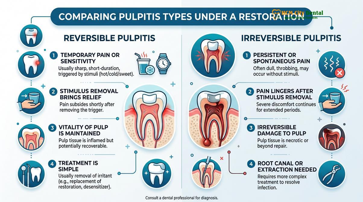
Reversible pulpitis is characterized by mild to moderate sensitivity to cold or sweet stimuli that dissipates quickly once the stimulus is removed. This indicates that the pulp is irritated but still vital and capable of healing. In contrast, irreversible pulpitis occurs when the inflammatory process has progressed to a point where the pulpal tissue begins to necrose (die). Symptoms of irreversible pulpitis are far more severe: spontaneous throbbing pain that occurs without any trigger, pain that wakes the patient up from a deep sleep, or lingering, excruciating pain following exposure to heat. When irreversible pulpitis is diagnosed, the tooth will not heal on its own, and endodontic intervention is mandatory to prevent the formation of a periapical abscess.
Clinical Warning:
Ignoring the signs of irreversible pulpitis can lead to severe complications. If the infection spreads beyond the apex of the tooth root into the surrounding alveolar bone, it can cause significant facial swelling, systemic infection, and ultimately result in the loss of the tooth. Prompt endodontic evaluation is crucial if you experience spontaneous, throbbing pain.
A common concern among patients is whether a newly placed crown must be removed and discarded if a root canal becomes necessary. Fortunately, in most cases, an endodontist or a highly skilled general dentist can perform the root canal treatment directly through the existing crown. This procedure involves using specialized diamond burs to create a small, precise access hole through the occlusal (biting) surface of the crown to reach the pulp chamber. The infected nerve tissue is meticulously cleaned, shaped, and disinfected using rotary files and antimicrobial irrigants. Once the root canal system is completely sealed with gutta-percha, the access hole in the crown is restored using a high-strength composite resin filling. This conservative approach preserves the structural integrity and aesthetics of the crown while effectively resolving the underlying infection.
Advanced Prosthodontic Solutions and Material Selection
Utilizing state-of-the-art digital scanning and premium certified materials ensures precise marginal adaptation and optimal biocompatibility. This meticulous approach significantly reduces the risk of post-operative sensitivity and structural failure.
The long-term success of a dental restoration and the minimization of post-operative discomfort are heavily dependent on the precision of the clinical workflow and the quality of the materials utilized. Traditional silicone impressions, while historically effective, are inherently prone to dimensional distortion, tearing, and patient discomfort (often triggering the gag reflex). Inaccuracies in the impression phase inevitably lead to poorly fitting crowns with open margins. An open margin acts as a microscopic gateway for oral bacteria and acidic fluids to seep underneath the crown, leading to microleakage, recurrent decay, and chronic sensitivity that can ultimately destroy the underlying tooth.
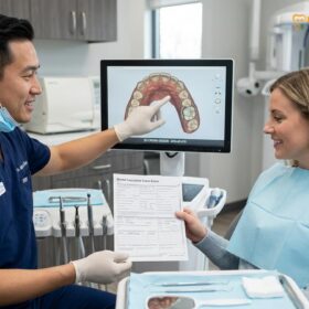
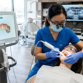
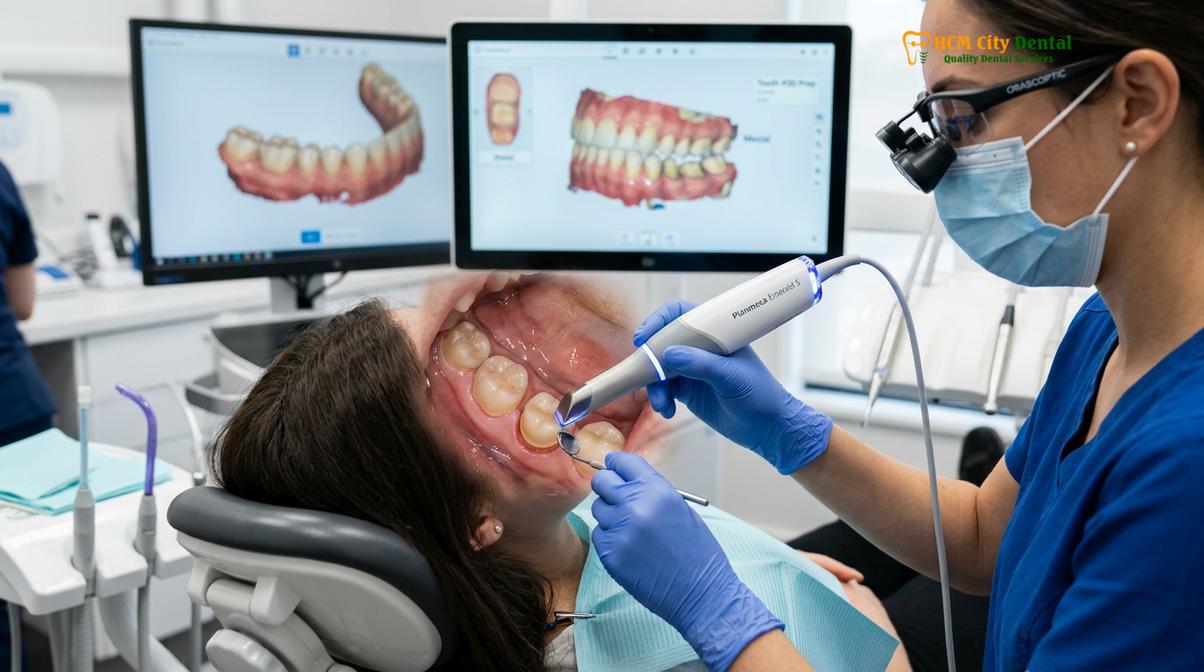
To eliminate these variables, modern prosthodontic practices employ advanced digital dentistry. The integration of Intraoral Scanners allows clinicians to capture highly accurate, 3D digital impressions of the prepared tooth, adjacent dentition, and the opposing arch in real-time. This technology provides maximum comfort for the patient and allows the dentist to magnify the preparation margin on a high-definition monitor, ensuring absolute precision before the data is transmitted to the dental laboratory. The digital file is then used to design the crown using CAD (Computer-Aided Design) software, and the final restoration is milled from a solid block of ceramic using CAM (Computer-Aided Manufacturing) technology.
“Precision in prosthodontics is not merely about aesthetics; it is fundamentally about biological integration. A perfectly sealed margin and a harmonized occlusal scheme are the most critical factors in preventing bacterial microleakage, minimizing periodontal inflammation, and ensuring the long-term vitality of the underlying tooth.”
Furthermore, the selection of the restorative material plays a pivotal role in the biological response of the tooth. Lithium Disilicate (commonly known as E.max) offers exceptional aesthetics and high flexural strength, making it ideal for anterior restorations where translucency is paramount. For posterior teeth that endure heavy masticatory forces, monolithic Zirconia provides unparalleled durability and fracture resistance. Both of these modern ceramics are highly biocompatible, meaning they are well-tolerated by the gingival tissues and do not provoke the allergic reactions or galvanic responses sometimes associated with base metal alloys[4].
When to See a Dentist for Crown Pain
While mild, transient sensitivity is a normal part of the healing process, certain symptoms serve as red flags that require prompt professional assessment to prevent further complications and ensure the longevity of the restoration.
Patients should monitor their symptoms closely following the placement of a new restoration. It is entirely normal to experience some mild tenderness in the gums where the retraction cord was placed, or slight sensitivity to cold liquids for a few weeks. However, if the pain is spontaneous—meaning it occurs without any stimulus like eating or drinking—or if it wakes the patient up from sleep, this is a strong indicator of irreversible nerve damage. According to clinical guidelines from the Vietnam Odonto-Stomatology Association (VOSA), lingering pain that lasts for more than a few minutes after a hot or cold stimulus is removed also suggests that the pulpal tissue is failing to recover and may require endodontic therapy[5].
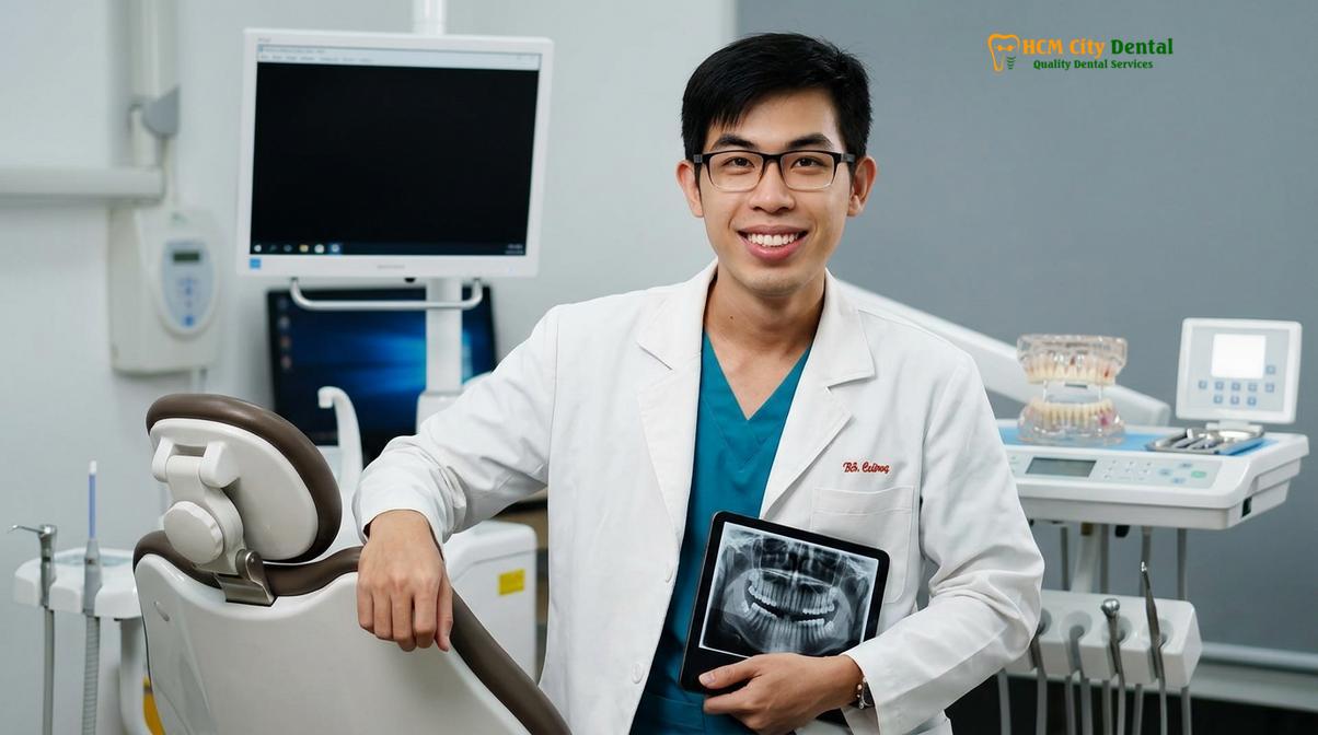
At HCMC Dental Clinic in Ho Chi Minh City, Dr. Nguyen Van Cuong emphasizes the importance of precise occlusal harmony and meticulous tissue management to prevent post-operative discomfort. Dr. Cuong utilizes advanced digital scanning and comprehensive bite adjustment protocols to ensure every restoration integrates seamlessly with the patient’s natural masticatory system. By prioritizing biological principles and utilizing premium materials, the clinical team strives to minimize the risk of chewing sensitivity and protect the long-term health of the tooth.
Additionally, patients should seek immediate clinical evaluation if they notice visible swelling in the gums around the crowned tooth, a pimple-like bump on the gums (known as a fistula or sinus tract), or a foul taste and odor in the mouth. These are definitive signs of an active, purulent infection. Delaying treatment in these scenarios can lead to the rapid spread of infection to the surrounding alveolar bone, potentially compromising adjacent teeth. Prompt clinical evaluation allows the dental team to accurately diagnose the source of the pain through radiographic imaging and implement the appropriate restorative or endodontic solution to restore oral health.
References
- Journal of Prosthodontic Research. Flexural strength and clinical outcomes of monolithic zirconia crowns.
- Journal of Endodontics. Pulpal response to full coverage restorations and cementation protocols.
- International Journal of Prosthodontics. Occlusal trauma and periodontal ligament inflammation in fixed prosthodontics.
- Journal of Esthetic and Restorative Dentistry. Thermal conductivity of lithium disilicate versus metal-ceramic restorations.
- Vietnam Odonto-Stomatology Association (VOSA). National clinical guidelines for the management of post-operative sensitivity in fixed prosthodontics.
If you are experiencing persistent discomfort, lingering thermal sensitivity, or sharp pain when biting down on a new restoration, do not wait for the symptoms to worsen. Contact HCMC Dental Clinic in Ho Chi Minh City for a comprehensive evaluation and precise bite adjustment. Learn more about our advanced Dental Crowns & Bridges tại HCMC Dental Clinic to restore your smile comfortably and predictably.