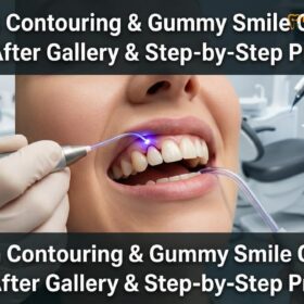
When comparing laser vs scalpel gingivectomy, diode lasers offer advanced clinical outcomes through instant tissue coagulation, sutureless cuts, and a rapid recovery phase. Conversely, traditional scalpel incisions require sutures, induce more bleeding, and demand a longer healing period, though they remain highly effective and necessary for extensive osseous recontouring procedures.
Clinical Summary:
The surgical management of gingival hyperplasia and altered passive eruption has evolved significantly with the introduction of soft tissue diode lasers. While the traditional surgical blade incision remains a foundational technique in periodontology—particularly when flap reflection and bone recontouring are required—diode lasers (such as the 810nm wavelength) have revolutionized soft tissue excision. By utilizing targeted photothermal energy, lasers provide instant cauterization, sterilize the surgical field, and generally eliminate the need for sutures. This technological shift can reduce the standard recovery associated with scalpels to a fraction of the time, minimizing postoperative edema and patient discomfort while ensuring precise calibration of gingival zenith points.
Key Takeaways:
- Diode lasers (810nm) instantly coagulate blood vessels, resulting in a virtually bloodless and sutureless procedure.
- Scalpel gingivectomy requires physical incisions, manual hemostasis, and typically a longer inflammatory healing phase.
- Laser technology sterilizes the surgical margin, which can significantly reduce the risk of postoperative bacterial infection.
- Respecting the biological width (minimum 2mm supracrestal tissue attachment) is mandatory in both techniques to help prevent bone resorption.
- Severe gummy smiles may require multidisciplinary approaches, including orthognathic surgery or lip repositioning, beyond simple gingivectomy.
Surgical Scalpels: The Traditional Incision and Bleeding Mechanics
Traditional scalpel gingivectomy utilizes physical steel blades to excise excess gingival tissue, requiring manual hemostasis, periodontal dressings, and sutures to manage intraoperative bleeding and protect the raw wound.
For decades, the conventional approach to reshaping the gingival architecture has relied on the surgical blade incision. Utilizing specialized instruments such as Kirkland and Orban periodontal knives, or standard No. 15 and No. 12 scalpel blades, the periodontist physically severs the fibrotic gingival tissue. This mechanical cutting process, while highly effective for bulk tissue removal, inherently triggers a robust physiological response. The severing of capillaries and larger blood vessels results in immediate hemorrhage, which obscures the surgical field and necessitates continuous suction and manual hemostasis techniques[1].
From a histological perspective, a cold steel cut leaves a raw, open wound margin. The body’s natural hemostatic cascade must initiate, forming a fibrin clot to seal the exposed connective tissue. Because the wound is left to heal by secondary intention, the surgeon must often place a protective periodontal dressing over the surgical site to shield the delicate forming clot from mechanical trauma during mastication and oral hygiene routines. Furthermore, if the incisions are extensive, sutures may be required to approximate the tissue and stabilize the gingival margin.
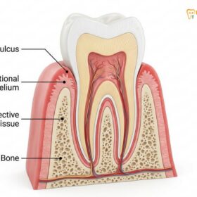
A critical consideration during scalpel gingivectomy is the precise measurement of the biological width, now clinically referred to as the supracrestal tissue attachment. This dimension comprises the junctional epithelium and the supracrestal connective tissue attachment. If a scalpel incision inadvertently encroaches upon this zone without concurrent bone removal, the body will respond with chronic inflammation and unpredictable crestal bone resorption in an attempt to re-establish the necessary biological space[3]. Therefore, meticulous bone sounding is a mandatory prerequisite before any blade touches the tissue.
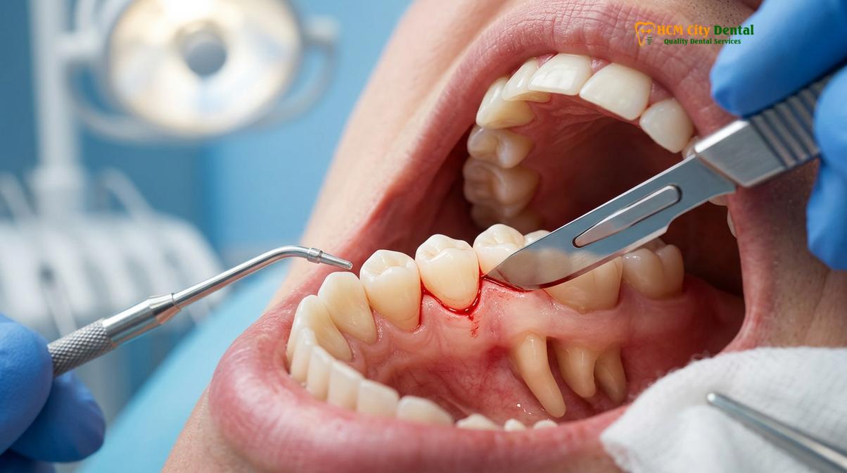
Despite its drawbacks regarding bleeding and patient comfort, the scalpel remains indispensable in specific clinical scenarios. When a patient presents with altered passive eruption where the alveolar bone crest is situated directly at the cementoenamel junction (CEJ), soft tissue excision alone is insufficient. In these cases, the surgeon must raise a full-thickness mucoperiosteal flap to perform an osteotomy (removal of supporting bone) and osteoplasty (reshaping of non-supporting bone). Scalpels provide the tactile feedback and precise flap reflection capabilities required for these complex osseous interventions.
Soft Tissue Lasers: Coagulation, Sterilization, and Sutureless Cuts
Soft tissue diode lasers utilize targeted light energy to vaporize gingival tissue while simultaneously sealing blood vessels and nerve endings, resulting in a sterile, bloodless, and highly precise surgical field.
The integration of the soft tissue diode laser into periodontal surgery represents a paradigm shift in patient comfort and clinical efficiency. Operating at specific wavelengths, these systems are highly absorbed by specific chromophores in the oral cavity—namely, melanin and hemoglobin. This selective absorption allows the laser energy to precisely target and vaporize the hyperplastic gingival tissue without causing collateral thermal damage to the underlying alveolar bone or tooth structure, provided the correct wattage and pulsing parameters are utilized.
The most profound advantage of the diode laser is its ability to achieve instant coagulation. As the laser tip glides across the gingival margin, the photothermal effect denatures proteins and instantly seals severed capillaries and lymphatic vessels. This cauterization creates a virtually bloodless surgical field, granting the clinician unparalleled visibility. The absence of bleeding not only accelerates the procedure but also generally eliminates the need for sutures or bulky periodontal dressings. Patients benefit immensely from this sutureless gum cutting technique, as it removes the anxiety and discomfort associated with stitch removal[2].
Furthermore, the laser beam acts as a powerful sterilizing agent. The high-energy light destroys bacterial cell walls upon contact, effectively decontaminating the surgical margin. This localized sterilization significantly reduces the risk of postoperative bacteremia and localized infections. According to the Vietnam Odonto-Stomatology Association (VOSA), the clinical application of diode lasers in periodontal therapy provides a safer, more controlled environment for soft tissue management, particularly for patients with compromised immune systems[6].
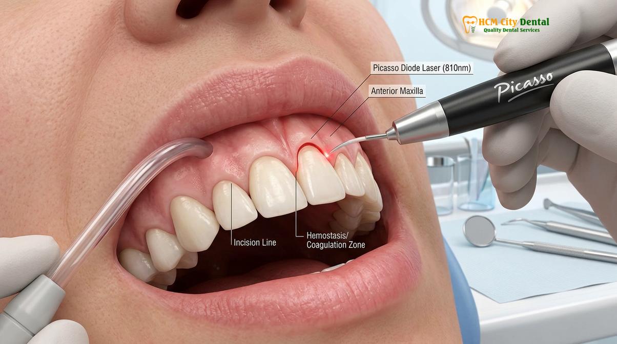
The precision afforded by the laser allows for micro-adjustments to the gingival architecture that are difficult to achieve with a scalpel. Clinicians can sculpt the tissue in fractions of a millimeter, carefully contouring the papillae and establishing ideal gingival zeniths. By utilizing a continuous or pulsed wave mode, the laser provides a sweeping motion, gently erasing excess tissue and revealing the natural anatomical crown of the tooth.
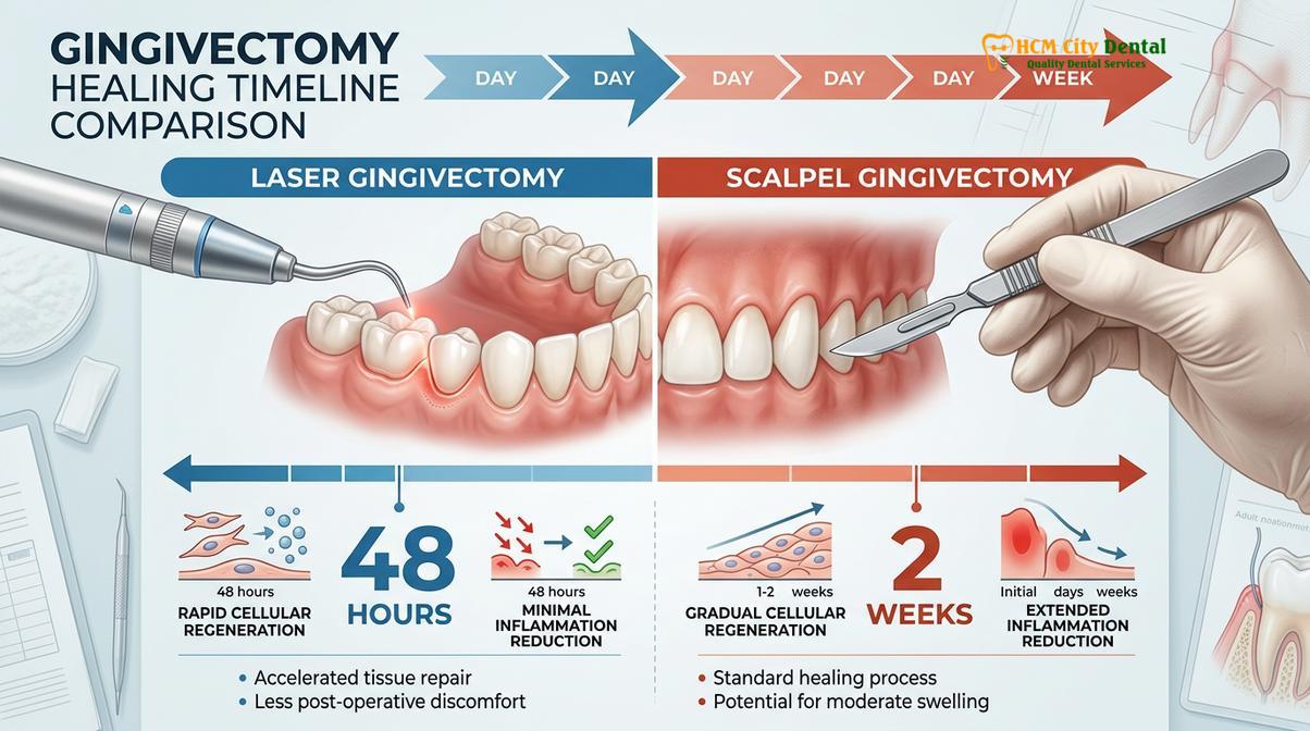
Healing Time Comparison: 48 Hours (Laser) vs. 2 Weeks (Scalpel)
Laser gingivectomy accelerates cellular regeneration by sealing the wound instantly, allowing patients to resume normal habits rapidly, whereas scalpel surgery necessitates a prolonged inflammatory healing phase.
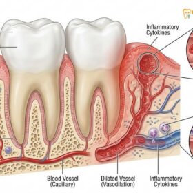
The postoperative recovery experience differs drastically between the two modalities, primarily due to the nature of the wound created. When a scalpel is used, the physical severing of tissue initiates a classic inflammatory cascade. The release of prostaglandins, histamines, and cytokines leads to vasodilation, increased capillary permeability, and subsequent edema. The wound must heal by secondary intention, meaning epithelial cells must migrate across a raw, open surface originating from the wound edges. Consequently, complete re-epithelialization and the maturation of the connective tissue following a scalpel gingivectomy typically require an extended period.
During this recovery period, patients often experience moderate discomfort, swelling, and dietary restrictions. The presence of sutures or a periodontal pack can trap plaque, making oral hygiene challenging and increasing the risk of localized gingivitis. Patients are usually advised to consume a soft diet and avoid mechanical brushing in the surgical area until the initial healing phase is complete.
In stark contrast, the healing trajectory following a laser gingivectomy is remarkably accelerated. Because the laser instantly seals the blood vessels and nerve endings, the initial inflammatory response is significantly blunted. The thermal energy creates a thin, sterile layer of denatured protein over the wound surface. This coagulum protects the underlying connective tissue and provides a stable scaffold for rapid epithelial cell migration.
| Clinical Parameter | Scalpel Gingivectomy | Laser Gingivectomy (Diode) |
|---|---|---|
| Intraoperative Bleeding | Moderate to High; requires manual hemostasis | Virtually None; instant coagulation |
| Sutures / Dressings | Often required (periodontal packs, sutures) | Generally not required (Sutureless) |
| Initial Healing Phase | Prolonged (often up to 2 weeks) | Rapid (often within 48 hours) |
| Postoperative Pain | Moderate; often requires analgesics | Minimal to None; rarely requires medication |
| Infection Risk | Moderate; open wound susceptible to bacteria | Very Low; laser sterilizes the surgical margin |
Patients undergoing laser treatment report minimal to no postoperative pain, often requiring no analgesic medication beyond over-the-counter options for the first day. The absence of sutures and dressings allows patients to resume gentle, normal oral hygiene practices almost immediately, which further promotes a healthy healing environment.
Reducing Post-Operative Swelling and Scars with Laser Tech
The photothermal effect of dental lasers minimizes the release of inflammatory mediators and seals lymphatic vessels, significantly reducing postoperative edema and helping prevent the formation of fibrotic scar tissue.
Aesthetic outcomes in periodontal plastic surgery are judged not only by the final position of the gingival margin but also by the quality and texture of the healed tissue. Traditional scalpel surgery, due to the intense inflammatory response it provokes, carries a higher risk of fibrotic healing. If the wound is subjected to trauma or excessive tension during the healing phase, the body may overproduce collagen, leading to thick, rolled gingival margins or visible scar tissue that detracts from the natural aesthetics of the smile.
Laser technology mitigates these risks through its unique interaction with biological tissues. By sealing the lymphatic vessels concurrently with the blood vessels, the diode laser helps prevent the accumulation of interstitial fluid, which is the primary cause of postoperative swelling. The reduction in swelling is not merely a matter of patient comfort; it is critical for maintaining the precise contours established during the surgery.
“The true artistry of aesthetic periodontics lies in the invisible healing. By utilizing diode laser wavelengths, we bypass the aggressive inflammatory cascade, allowing the gingival architecture to regenerate seamlessly without the fibrotic scarring often associated with cold steel incisions.”
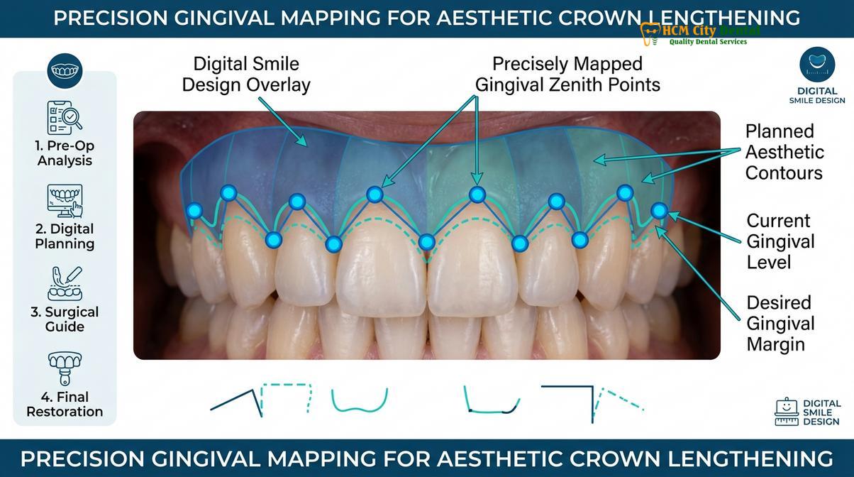
Furthermore, the precision of the laser is instrumental in executing a Digital Smile Design (DSD). Before the procedure, the clinician maps out the ideal gingival zenith points—the most apical point of the gingival scallop. Using the laser, the surgeon can trace these exact digital parameters onto the soft tissue with micro-millimeter accuracy. The resulting tissue architecture is smooth, naturally tapered, and free of the jagged edges that can sometimes occur with a scalpel blade[4].
Advanced Surgical Interventions and Multidisciplinary Care
While lasers excel in soft tissue management, severe skeletal discrepancies or hyperactive lip muscles require advanced surgical interventions, such as orthognathic surgery or lip repositioning, to achieve optimal aesthetic results.
It is crucial to understand that a gingivectomy, whether performed via laser or scalpel, is exclusively a soft tissue procedure. It is highly effective for treating gingival hyperplasia, altered passive eruption, and minor aesthetic asymmetries. However, when a gummy smile is the result of severe skeletal deformities—such as Vertical Maxillary Excess (VME)—soft tissue excision alone will not yield satisfactory results. In these complex cases, the underlying bone structure must be addressed through orthognathic surgery.
Another non-gingival factor contributing to a gummy smile is a hyperactive levator muscle of the upper lip. If the lip retracts excessively upon smiling, exposing a wide band of gingiva despite normal tooth proportions, alternative treatments are indicated. Surgical Lip Repositioning involves removing a strip of mucosa from the maxillary vestibule and suturing the lip in a lower position, restricting its upward mobility. Clinical studies indicate that this procedure offers predictable longevity and high patient satisfaction when correctly diagnosed and executed[5].
Clinical Case Review: Comprehensive Smile Design
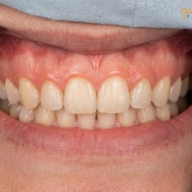
A patient visited HCMC Dental Clinic in Ho Chi Minh City presenting with a severe gummy smile and short clinical crowns. Clinical evaluation revealed a combination of altered passive eruption and a hyperactive upper lip. The treatment plan utilized a multidisciplinary approach: an initial full-arch laser gingivectomy to establish ideal zenith points and expose the anatomical crowns, followed by a surgical lip repositioning procedure to restrict excessive lip elevation. The integration of laser technology ensured a rapid soft tissue recovery, allowing the patient to achieve a harmonious, balanced smile with minimal downtime.
When to Consult a Periodontist
Patients experiencing excessive gingival display, uneven gum lines, or prolonged inflammation should seek a professional periodontal evaluation to determine the most appropriate surgical or laser intervention.
Determining the right approach for gum contouring requires a comprehensive clinical assessment. If you notice that your teeth appear unusually short, or if a significant amount of gum tissue is visible when you smile, a consultation with a dental specialist is highly recommended. A periodontist can accurately diagnose whether the issue stems from excess soft tissue, underlying bone structure, or lip mobility.
“Accurate diagnosis is the cornerstone of successful periodontal plastic surgery. Whether utilizing a scalpel for complex osseous recontouring or a diode laser for precise soft tissue sculpting, the chosen modality must align perfectly with the patient’s unique anatomical presentation.”
Dr. Nguyen Van Cuong, a leading specialist at HCMC Dental Clinic, emphasizes that thorough periodontal evaluations are essential before any tissue modification. His clinical approach integrates advanced laser protocols to ensure optimal aesthetic outcomes while prioritizing patient comfort and safety. By carefully assessing the biological width and gingival biotype, Dr. Cuong ensures that every procedure is tailored to provide long-lasting, natural-looking results.
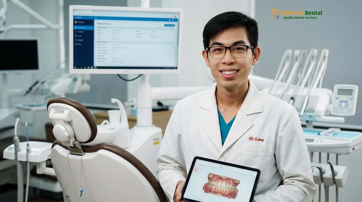
If you are considering aesthetic gum improvements, contact HCMC Dental Clinic in Ho Chi Minh City. Explore our comprehensive Gum Contouring & Gummy Smile services to learn how modern laser technology and expert clinical care can safely transform your smile.
References
- Journal of Clinical Periodontology. Histological evaluation of laser versus scalpel incisions.
- International Journal of Periodontics & Restorative Dentistry. Diode laser applications in aesthetic gingival recontouring.
- Journal of Aesthetic and Restorative Dentistry. Biological width and supracrestal tissue attachment around restorations.
- Journal of Oral and Maxillofacial Surgery. Le Fort I osteotomy stability in vertical maxillary excess.
- Clinical Oral Investigations. Surgical lip repositioning longevity and patient outcomes.
- Vietnam Odonto-Stomatology Association (VOSA). Guidelines on the clinical application of diode lasers in periodontal therapy.