A smile design gum line refers to the precise architectural contouring of gingival tissues to frame the teeth harmoniously. By calibrating the gingival zenith and respecting biological width, clinicians can correct gummy smiles, resolve asymmetry, and establish optimal aesthetic proportions for a balanced, natural appearance.
Clinical Summary:
The aesthetic success of any smile makeover relies heavily on the soft tissue framework. A meticulously planned smile design gum line addresses discrepancies in gingival height, altered passive eruption, and hypermobile lips. Utilizing advanced digital smile design (DSD) software, clinicians map the ideal gingival contours before executing the treatment with precision diode lasers. By adhering to strict biological principles—specifically the supracrestal tissue attachment—dentists ensure that cosmetic enhancements do not compromise periodontal health. Treatments range from minimally invasive laser gingivectomy to complex orthognathic surgery for skeletal discrepancies, offering tailored solutions for every degree of gingival display.
Key Takeaways:
- Laser Precision: AMD Picasso Diode lasers (810nm) provide bloodless, sutureless contouring with rapid 48-hour recovery.
- Biological Width: A minimum of 2mm of supracrestal tissue attachment must be maintained to prevent chronic inflammation and bone loss.
- Digital Mapping: Digital Smile Design (DSD) allows for precise preoperative visualization and surgical guide fabrication.
- Multidisciplinary Care: Severe skeletal gummy smiles (Vertical Maxillary Excess) are managed via Le Fort I osteotomy in collaboration with maxillofacial specialists.
- Cost-Effective Solutions: Laser gingivectomy starts at 1,500,000 VND per tooth, with comprehensive full-arch options available.
The Elements of a Balanced Smile: Lips, Teeth, and Gums
A harmonious smile requires a delicate equilibrium between the upper lip mobility, the exposed clinical crowns, and the framing gingival architecture.
When evaluating the aesthetics of a smile, clinicians look far beyond the color and alignment of the teeth. The “pink aesthetic score” is just as critical as the “white aesthetic score.” A truly balanced smile is the result of a dynamic interplay between three primary components: the lips, the teeth, and the gums. If any one of these elements is disproportionate, the entire visual harmony is disrupted. The fundamental smile architecture rules dictate that the upper lip should rest gracefully at or just above the gingival margins of the central incisors during a full, uninhibited smile.[1]
The upper lip serves as the curtain that reveals the dental stage. Its length, volume, and mobility are critical diagnostic factors. In some patients, a hyperactive levator muscle—specifically the levator labii superioris alaeque nasi—causes the lip to elevate excessively during smiling, exposing a significant band of gingival tissue. This condition, commonly referred to as a hypermobile lip, can turn an otherwise normal dental and skeletal relationship into a pronounced gummy smile. Diagnosing the exact etiology of the gingival display is the first and most crucial step in formulating a treatment plan.
The teeth themselves must exhibit specific macro-esthetic and micro-esthetic proportions. The central incisors are the focal point of the smile, and their width-to-length ratio should ideally fall between 75% and 80%. When the gums cover too much of the enamel—a condition known as altered passive eruption—the teeth appear short, square, and childlike. Conversely, gingival recession can make the teeth appear excessively long and aged. The gums act as the frame for the teeth, and their contour, color, and texture must exude health. Stippled, firm, coral-pink gingiva that fills the interdental spaces completely (forming sharp papillae) is the hallmark of periodontal health and aesthetic perfection.
Defining the Gingival Zenith: The Highest Point of the Gum Margin
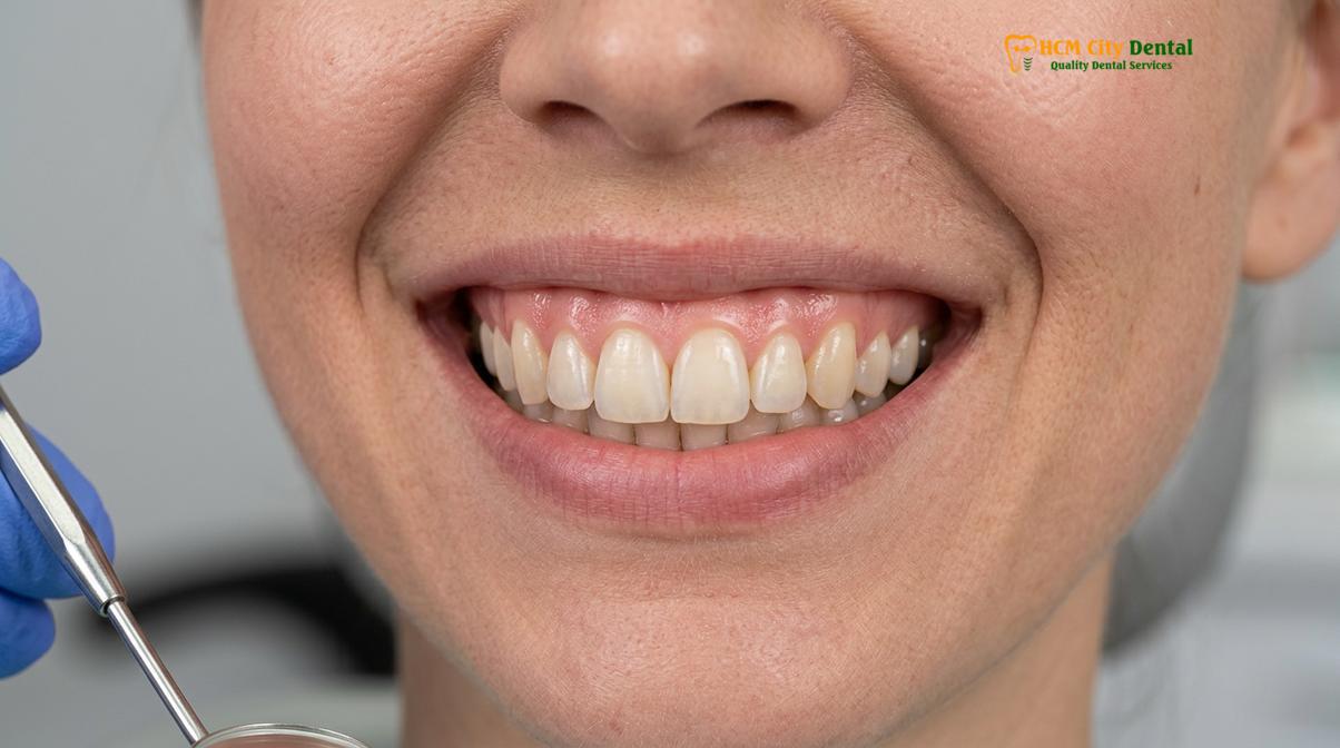
The gingival zenith is the most apical point of the gum tissue scallop, typically positioned slightly distal to the tooth’s vertical midline.
In the realm of periodontal plastic surgery and cosmetic dentistry, the concept of the gingival zenith is paramount. The zenith is the highest point of the free gingival margin along the facial aspect of a tooth. It is not merely a random curve; its exact placement is dictated by strict anatomical and aesthetic guidelines. For the maxillary central incisors, the ideal zenith point is located approximately 1 millimeter distal (towards the back of the mouth) to the tooth’s long axis. This subtle distal displacement gives the tooth a dynamic, natural appearance rather than a static, artificial look.[2]
The lateral incisors, being smaller and more delicate, typically have their zenith points centered directly on the long axis of the tooth. The canines, which are robust and anchor the corners of the smile, follow the pattern of the central incisors with a distally displaced zenith. When these points are connected, they form the Gingival Zenith Line (GZL). In an ideal aesthetic scenario, the zenith of the lateral incisor should sit approximately 0.5 to 1 millimeter below the line connecting the zeniths of the central incisor and the canine. This creates the highly desired scalloped gingival margins that characterize a youthful, attractive smile.
“Precise calibration of the gingival zenith is the secret to transforming a good smile into an exceptional one. Even a millimeter of asymmetry in the zenith position can subconsciously signal to the human eye that something is structurally unbalanced.”
— Dr. Nguyen Van Cuong, Lead Clinician
When performing a smile design gum line procedure, the clinician must meticulously map these zenith points before making any incisions. Failure to respect these micro-esthetic details can result in a smile that looks artificial or “chiclet-like.” Advanced advanced gum contouring procedures focus heavily on redefining these points, especially in cases where the natural eruption process has left the gingival margins flat, asymmetrical, or excessively coronal.
Symmetry and Balance: Restoring Harmony Across the Central Incisors
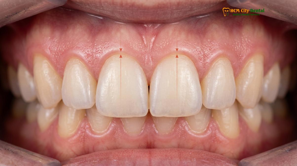
Achieving symmetry across the central incisors is the cornerstone of aesthetic dentistry, as these teeth dictate the visual midline and overall facial balance.
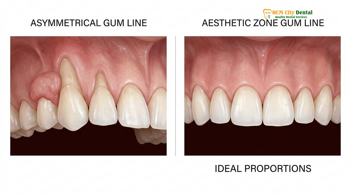
The human brain is hardwired to perceive symmetry as beautiful. In dental aesthetics, the central incisors are the most critical elements; they are the first structures noticed during social interaction. If the aesthetic zone gumline is asymmetrical—meaning the gum level on the right central incisor is higher or lower than on the left—the entire smile will appear canted or crooked, even if the incisal edges are perfectly aligned. Correcting this asymmetry is often the primary objective of cosmetic gingival contouring.
Symmetry is not just about horizontal alignment; it also involves proportional harmony. The Golden Proportion and Chu’s Aesthetic Gauges are frequently used to evaluate the relationship between the width and length of the teeth. When a patient presents with short clinical crowns due to excess gingival tissue, the clinician must determine the location of the cementoenamel junction (CEJ). If the CEJ is buried beneath the gum line, a simple gingivectomy can expose the hidden enamel, instantly restoring the correct proportions and achieving bilateral symmetry.[3]
| Aesthetic Parameter | Ideal Presentation | Asymmetrical/Abnormal Presentation | Clinical Solution |
|---|---|---|---|
| Gingival Margin Height | Symmetrical across central incisors; laterals 0.5-1mm lower. | Uneven heights causing a canted or crooked appearance. | Laser gingivectomy to level the margins. |
| Tooth Proportions | 75% – 80% width-to-length ratio. | Square teeth (>85% ratio) due to excess gum coverage. | Aesthetic crown lengthening to expose enamel. |
| Gingival Zenith Position | Distally displaced on centrals and canines; centered on laterals. | Centered on all teeth, creating a flat, artificial look. | Micro-contouring of the zenith points. |
| Interdental Papillae | Fills the embrasure space completely, sharp and coral pink. | Blunted, inflamed, or missing (black triangles). | Periodontal therapy, hyaluronic acid, or restorative closure. |
Restoring harmony requires a comprehensive understanding of the underlying bone architecture. The soft tissue generally follows the contour of the underlying alveolar bone. Therefore, achieving lasting symmetry in the soft tissue may sometimes require minor recontouring of the bone (osteoplasty) to ensure the gums do not rebound to their original asymmetrical position. This meticulous attention to detail ensures that the final result is both aesthetically pleasing and biologically stable.
How Digital Smile Design (DSD) Maps Your Gum Contouring Plan
Digital Smile Design utilizes dynamic videography and intraoral scanning to digitally superimpose ideal gingival contours onto the patient’s facial anatomy before treatment begins.
The era of guesswork in cosmetic dentistry is over. Today, the integration of digital smile design HCMC protocols allows clinicians to plan, visualize, and execute complex soft tissue modifications with unprecedented accuracy. DSD is a conceptual workflow that begins with comprehensive data acquisition. This includes high-resolution digital photography (capturing the patient at rest, during a natural smile, and during an exaggerated smile), dynamic video analysis to evaluate lip mobility during speech, and intraoral 3D scanning to capture the exact topography of the teeth and gums.
Once the data is imported into specialized DSD software, the clinician can begin the architectural planning. The software allows the dentist to draw reference lines—such as the facial midline, the interpupillary line, and the occlusal plane—to ensure that the new smile will be perfectly aligned with the patient’s facial features. The clinician can then digitally manipulate the gingival margins, calibrating the zenith points and adjusting the width-to-length ratios of the teeth until the ideal aesthetic zone gumline is achieved.[3]
One of the most powerful aspects of DSD is its role in patient communication. Before any irreversible procedures are performed, the patient can view a digital simulation of their proposed outcome. In many cases, a physical “mock-up” can be printed and temporarily placed over the patient’s teeth, allowing them to “test drive” their new smile in real life. Once the design is approved, the digital data is used to fabricate precise surgical guides. These guides are placed in the mouth during the procedure, directing the laser or scalpel to the exact millimeter, ensuring that the clinical execution perfectly matches the digital plan.
Clinical Workflows and Advanced Technologies in Gum Reshaping
Modern gingivectomy relies on diode lasers and precise bone sounding to alter the gum line without violating the critical supracrestal tissue attachment.
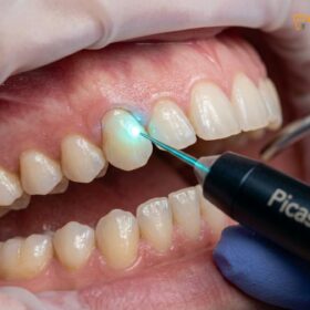
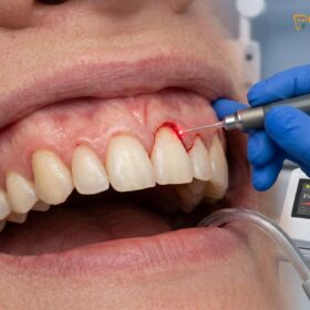
The clinical execution of a smile design gum line procedure has been revolutionized by the advent of soft tissue lasers. At the forefront of this technology are the AMD Picasso Lasers. Operating at an 810nm wavelength, these diode lasers are highly absorbed by melanin and hemoglobin, making them exceptionally efficient for soft tissue surgery. When the laser energy interacts with the gingival tissue, it simultaneously vaporizes the excess cells and coagulates the blood vessels. This results in a virtually bloodless surgical field, providing the clinician with maximum visibility and precision.[2]
The benefits of the diode laser extend far beyond the operating chair. Because the laser seals nerve endings and lymphatic vessels as it cuts, patients experience significantly reduced postoperative pain and swelling compared to traditional scalpel techniques. The procedure is sutureless, and the laser beam sterilizes the surgical site, drastically minimizing the risk of infection. Most patients undergoing laser gingivectomy experience a rapid 48-hour recovery, allowing them to return to their normal routines almost immediately.
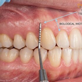
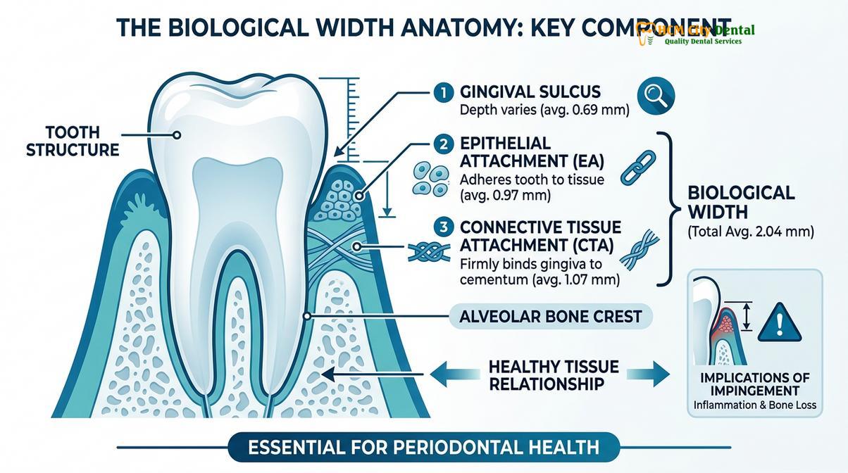
However, the use of advanced technology must be guided by strict biological principles. The most critical concept in gum contouring is the preservation of the biological width, now more accurately termed the supracrestal tissue attachment. This attachment consists of the junctional epithelium (approximately 0.97mm) and the supracrestal connective tissue (approximately 1.07mm), totaling roughly 2 millimeters of soft tissue that naturally exists between the base of the gingival sulcus and the crest of the alveolar bone.[1]
Clinical Warning: Biological Width Violation
If gum tissue is removed without respecting the 2mm supracrestal tissue attachment, the body will perceive this as a biological violation. The result is chronic gingival inflammation, unpredictable bone resorption, and eventual recession as the body attempts to re-establish the necessary space. Proper bone sounding is mandatory before any tissue excision.
To prevent this violation, the clinician performs “bone sounding” under local anesthesia to measure the exact distance from the gum margin to the bone crest. If there is more than 3 millimeters of tissue, a simple laser gingivectomy can be performed safely. However, if the bone level is too close to the desired new gum line, removing soft tissue alone will violate the biological width. In these cases, functional or esthetic crown lengthening is required. This involves reflecting a small flap and performing osteoplasty (recontouring the bone) to recreate the necessary 2mm space further up the root, ensuring long-term stability and health of the new gingival margin.
Comprehensive Treatment Options and Pricing Structure
From minimally invasive laser contouring to complex surgical lip repositioning, treatment modalities are tailored to the specific etiology of the gingival display.
Addressing a gummy smile or an uneven gum line requires a customized approach based on the underlying anatomical cause. Modern dental clinics offer a spectrum of treatments, ranging from simple soft tissue modifications to muscular and mucosal interventions. Understanding the etiology—whether it is altered passive eruption, a hypermobile lip, or dentoalveolar extrusion—dictates the appropriate clinical pathway.
For patients with excess gingival tissue but normal bone levels, a Laser Gingivectomy is the treatment of choice. This procedure simply sculpts the soft tissue to reveal the natural crown of the tooth. According to the latest clinic fee schedule, this highly precise procedure is priced at 1,500,000 VND (~$60 USD) per tooth, with a walk-in rate of 2,500,000 VND. For patients requiring extensive aesthetic zone modification, a Full-Arch Laser Gum Contouring (covering up to 10 teeth) is available for 12,000,000 VND (~$480 USD), offering a comprehensive transformation of the smile frame.
When bone recontouring is necessary to respect the supracrestal tissue attachment, Functional/Esthetic Crown Lengthening is performed. This more involved procedure, which includes osteoplasty, is priced at 3,000,000 VND (~$120 USD) per tooth. In cases where the teeth and gums are perfectly proportioned but the upper lip rises too high during smiling (hyperactive levator muscle), alternative treatments are required. Botox Gummy Smile Correction offers a temporary, non-surgical solution by relaxing the lip muscles, priced at 4,500,000 VND (~$180 USD) for both sides.
For a permanent solution to a hypermobile lip, Surgical Lip Repositioning (or Lip Lowering) is highly effective. This procedure involves removing a strip of mucosa from the inside of the upper lip and suturing the lip in a lower position, physically restricting its upward movement.[5] This advanced surgical intervention is priced at 15,000,000 VND (~$600 USD). Patients utilizing dental tourism services often take advantage of the -40% WhatsApp booking discount, making these world-class treatments highly accessible.
Clinical Case Review: Aesthetic Zone Transformation
A 28-year-old patient visited HCMC Dental Clinic in Ho Chi Minh City presenting with a severe gummy smile and short, square clinical crowns. Diagnostic bone sounding revealed altered passive eruption with thick gingival biotype. The treatment plan utilized DSD to map the ideal zenith points, followed by esthetic crown lengthening using the AMD Picasso Diode laser and minor osteoplasty. The procedure was completed in a single visit. At the 48-hour follow-up, the patient exhibited minimal swelling, and the final result yielded a perfectly proportioned, symmetrical smile with ideal width-to-length ratios.
Severe Skeletal Cases and Maxillofacial Collaboration
Vertical Maxillary Excess requires orthognathic intervention via Le Fort I osteotomy, performed in collaboration with specialized maxillofacial surgical partners.
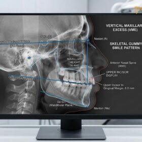
While soft tissue lasers and lip repositioning techniques can resolve the majority of gummy smile cases, they are insufficient for patients presenting with severe skeletal discrepancies. When the entire upper jaw (maxilla) has grown too far downward—a condition clinically diagnosed as Vertical Maxillary Excess (VME)—the excessive gingival display is a symptom of a deeper structural issue. In these cases, the patient’s face may appear elongated, and they may struggle with lip incompetence, meaning they cannot comfortably close their lips at rest.
Treating VME requires a multidisciplinary approach that extends beyond the scope of traditional cosmetic dentistry. To address these complex skeletal cases, elite dental practices collaborate with Surgical Jaw Partners. For instance, severe cases are often co-managed with maxillofacial specialists at the National Hospital of Odonto-Stomatology in Ho Chi Minh City (Bệnh viện Răng Hàm Mặt Trung Ương TP.HCM), which stands as the highest clinical authority for maxillofacial surgery in the region.[4]
“When we diagnose Vertical Maxillary Excess, we must look beyond the gums and address the skeletal foundation. Collaborating with top-tier maxillofacial surgeons ensures that our patients receive comprehensive care that corrects both the aesthetic concern and the underlying functional discrepancy.”
— Dr. Nguyen Van Cuong, Lead Clinician
The definitive treatment for VME is orthognathic surgery, specifically a Le Fort I osteotomy with superior impaction. During this major surgical procedure, the maxillofacial surgeon separates the entire upper jaw from the skull, removes a predetermined wedge of bone, and repositions the maxilla higher up. The jaw is then stabilized with titanium plates and screws. This physically shortens the midface, eliminates the skeletal gummy smile, and restores proper lip competence and facial harmony. Following the healing phase of the orthognathic surgery, the patient returns to the cosmetic dentist for final micro-esthetic refinements, such as laser contouring or veneer placement, to perfect the smile design gum line.
When to See a Doctor for Gum Line Irregularities
Determining the right time to seek professional intervention for a gummy smile or uneven gum line depends on both aesthetic desires and functional health. If you feel self-conscious about the amount of gum showing when you smile, or if your teeth appear disproportionately short or asymmetrical, a consultation with a cosmetic dentist is highly recommended. Early evaluation allows the clinician to diagnose the exact etiology—whether it is dental, muscular, or skeletal—and outline a predictable treatment plan.
However, cosmetic gum surgery is not suitable for everyone at any given time. The absolute prerequisite for any gingival contouring procedure is pristine periodontal health. Patients presenting with active periodontal disease, characterized by bleeding, deep pocketing, or bone loss, are strictly contraindicated for cosmetic surgery until the disease is controlled. In such cases, the initial phase of treatment will involve comprehensive dental scaling and root planing to eliminate plaque, calculus, and bacterial infection. Only after the tissues have healed and demonstrated stability can cosmetic modifications be considered.
Furthermore, patients with uncontrolled systemic conditions, such as severe diabetes or bleeding disorders, must undergo thorough medical clearance before proceeding with surgical interventions. A personalized clinical examination, including 3D imaging and periodontal probing, is essential to determine your specific candidacy and ensure that the chosen procedure will yield safe, long-lasting, and aesthetically superior results.
Frequently Asked Questions
What is the ideal gum display when smiling?
The ideal gum display when smiling is typically 1 to 2 millimeters of visible pink tissue above the central incisors. Displays exceeding 3 to 4 millimeters are clinically classified as a gingival smile, which can be corrected through targeted contouring or lip repositioning. Achieving this ideal display requires a harmonious balance between lip mobility, crown length, and skeletal position.
Can gum contouring fix an uneven smile?
Yes, gum contouring is highly effective at correcting an uneven smile caused by asymmetrical gingival margins. By precisely recalibrating the gingival zenith points using a soft tissue laser, clinicians can create a harmonious, symmetrical frame for the teeth. This procedure ensures that the visual midline and the horizontal aesthetic planes are perfectly aligned with the patient’s facial features.
Does gumline design affect veneers?
Absolutely. Gumline design is a critical prerequisite for successful veneer placement. Establishing symmetrical gingival margins ensures that the final porcelain veneers have optimal width-to-length ratios and emerge naturally from the soft tissue without looking disproportionate. Placing veneers on an uneven gum line will only highlight the asymmetry, compromising the final aesthetic outcome.
Is laser gum contouring painful?
Laser gum contouring is generally associated with minimal discomfort. Because diode lasers simultaneously cut and coagulate tissue, the procedure is virtually bloodless, requires no sutures, and significantly reduces postoperative pain and swelling compared to traditional scalpel surgery. Most procedures are performed under simple local anesthesia, ensuring a comfortable experience for the patient.
How long does it take to recover from a gum lift?
Recovery from a laser gum lift is remarkably fast, with most patients resuming normal activities within 48 hours. While minor tenderness may persist for a few days, the sterile nature of the laser beam promotes rapid, uncomplicated tissue healing. Patients are typically advised to consume soft foods and avoid aggressive brushing around the surgical site for the first week to ensure optimal recovery.
References
- Journal of Clinical Periodontology. Esthetic crown lengthening outcomes and biological width. (2021).
- International Journal of Periodontics & Restorative Dentistry. Diode laser vs scalpel gingivectomy in the aesthetic zone. (2020).
- Journal of Aesthetic and Restorative Dentistry. Digital smile design and gingival zenith calibration. (2019).
- Journal of Oral and Maxillofacial Surgery. Le Fort I osteotomy stability for vertical maxillary excess. (2022).
- Clinical Oral Investigations. Surgical lip repositioning longevity and patient satisfaction. (2018).