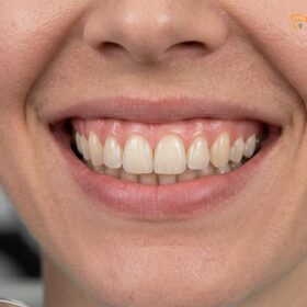
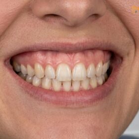
The gingival zenith point is the most apical (highest) point of the marginal gum tissue scallop surrounding a tooth. Clinically, it dictates the aesthetic emergence profile and perceived length of the clinical crown, playing a foundational role in symmetrical smile design, optimal width-to-length tooth ratios, and long-term periodontal health.
Clinical Summary:
Gingival zenith points are critical anatomical landmarks that define the aesthetic architecture of a smile. For central incisors and canines, the zenith is naturally displaced distal to the tooth’s vertical midline, whereas lateral incisors feature a centralized zenith positioned slightly more coronal. Discrepancies in these points lead to visual distortions in tooth proportion and smile symmetry. Modern periodontal plastic surgery corrects these asymmetries using advanced 810nm diode lasers, such as the AMD Picasso system, which allows for bloodless, minimally invasive contouring. Successful zenith realignment requires strict adherence to the biological width (supracrestal tissue attachment) to prevent chronic inflammation or bone resorption. In complex cases involving skeletal discrepancies, interdisciplinary approaches including orthognathic surgery or lip repositioning are utilized to achieve harmonious dentogingival aesthetics.
Key Takeaways:
- The gingival zenith is the highest point of the gum contour, crucial for determining the visual axis and length of the tooth.
- Central incisors and canines have zeniths displaced ~1mm distally, while lateral incisors have centralized zeniths.
- Asymmetrical zeniths distort the golden proportion, making teeth appear tilted, overly square, or uneven.
- AMD Picasso diode lasers (810nm) provide sutureless, bloodless zenith realignment with rapid 48-hour recovery.
- Surgical contouring must respect the biological width, maintaining a minimum 2mm distance from the alveolar bone crest.
- Severe gummy smiles may require advanced interventions like Le Fort I osteotomy or surgical lip repositioning.
Zenith Dental Meaning: Why It Matters for Your Smile
Anatomical Definitions: Gingival Zenith and Central Lines
The gingival zenith represents the peak of the gum contour, while the central line bisects the tooth vertically, serving as the primary reference for evaluating aesthetic alignment and biological width.
In the realm of periodontal aesthetics and restorative dentistry, the soft tissue framework surrounding the teeth is just as critical as the dental hard tissues. The gingival zenith is defined as the most apical point of the free gingival margin. It represents the peak of the gingival scallop and is a primary determinant of the tooth’s emergence profile. To accurately locate and evaluate the gingival zenith position, clinicians utilize the Vertical Bisected Midline (VBM), also known as the central line. The VBM is an imaginary line drawn vertically through the center of the clinical crown, bisecting the incisal edge and the cervical base equally. The relationship between the zenith and the VBM dictates the perceived axial inclination of the tooth.[1]
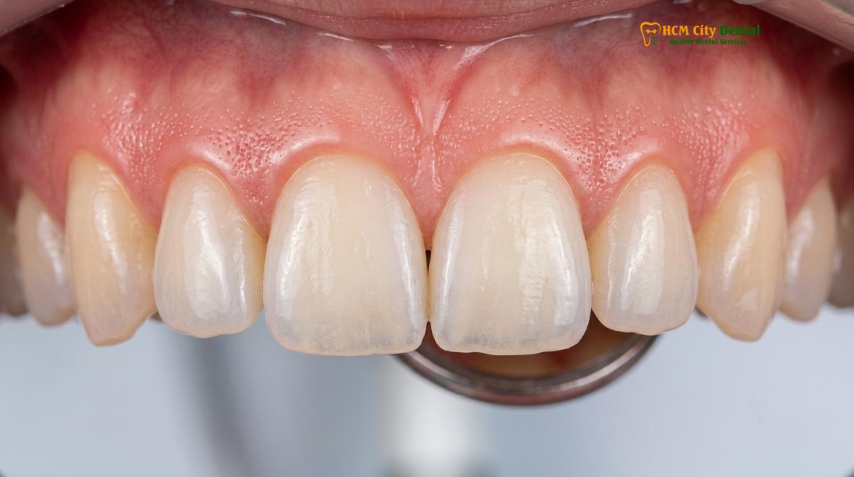
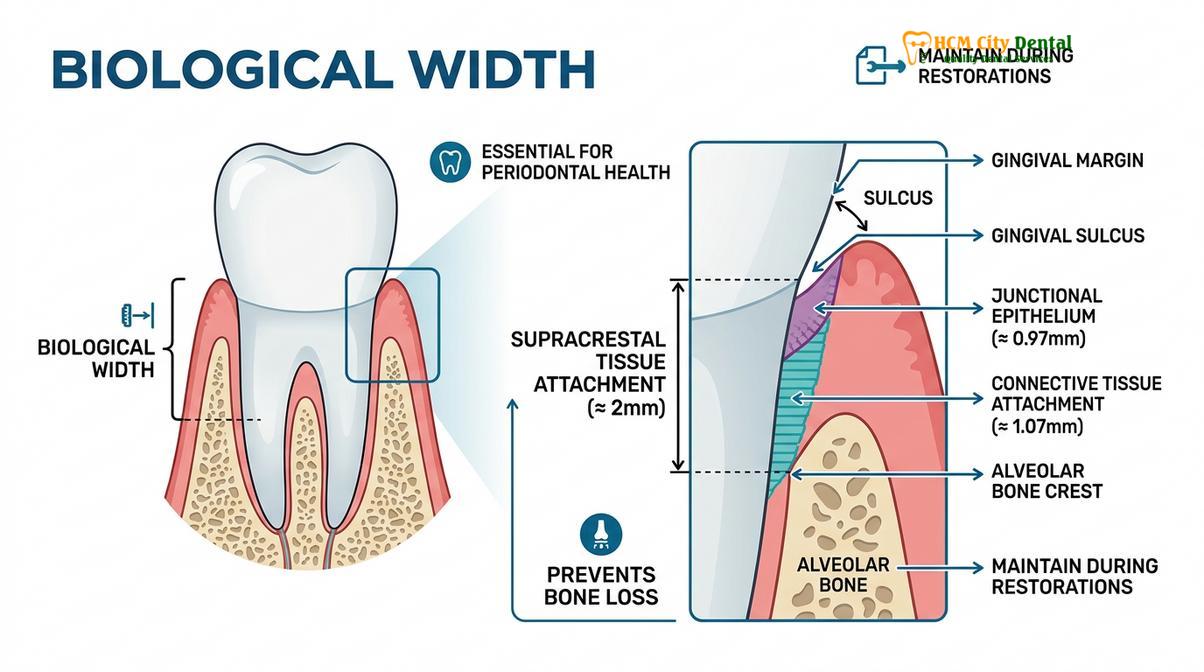
Understanding the gingival zenith requires a deep dive into the micro-anatomy of the dentogingival junction. The position of the zenith is not arbitrary; it is dictated by the underlying cementoenamel junction (CEJ) and the crest of the alveolar bone. The soft tissue that extends from the alveolar bone crest to the free gingival margin is governed by a concept historically known as the biological width, now formally termed the supracrestal tissue attachment. This attachment apparatus consists of the junctional epithelium (approximately 0.97mm) and the supracrestal connective tissue attachment (approximately 1.07mm). Together, they form a physiological barrier of roughly 2mm that protects the underlying periodontium from bacterial invasion.[2]
When evaluating the gingival zenith position, clinicians must perform bone sounding under local anesthesia to measure the distance from the free gingival margin to the bone crest. If the zenith is blunted or positioned too far coronally due to altered passive eruption (APE), the clinician must determine whether the excess tissue is purely gingival (Type I APE) or involves the alveolar bone crest being too close to the CEJ (Type II APE). Any surgical attempt to move the zenith apically must respect this 2mm supracrestal tissue attachment. If a laser or scalpel is used to raise the zenith without ensuring adequate distance to the bone, the biological width is violated, leading to chronic gingival inflammation, unpredictable tissue rebound, or pathological bone resorption.[3]
Furthermore, the gingival biotype—whether thick-flat or thin-scalloped—heavily influences the appearance and stability of the zenith. A thick-flat biotype typically exhibits a less pronounced aesthetic gums scallop and a more squared zenith, whereas a thin-scalloped biotype features a highly pronounced, delicate zenith that is highly susceptible to recession if traumatized. Comprehensive General & Preventive Dentistry protocols emphasize the maintenance of this delicate soft tissue architecture through meticulous plaque control and atraumatic restorative techniques.
Zenith Rules for Centrals, Laterals, and Canines
Zenith placement follows strict anatomical rules: distally displaced for central incisors and canines, but centrally aligned and more coronally positioned for lateral incisors.
The harmonious flow of the gingival margins across the maxillary anterior teeth is governed by specific spatial rules. The Gingival Aesthetic Line (GAL) is an imaginary line connecting the zeniths of the central incisors and the canines. In an ideal Class I gingival relationship, the zeniths of the central incisors and canines lie on this exact same horizontal plane, while the zeniths of the lateral incisors are positioned approximately 1mm coronal (lower) to this line. This step-down effect creates a dynamic, youthful appearance known as the gull-wing effect.[1]
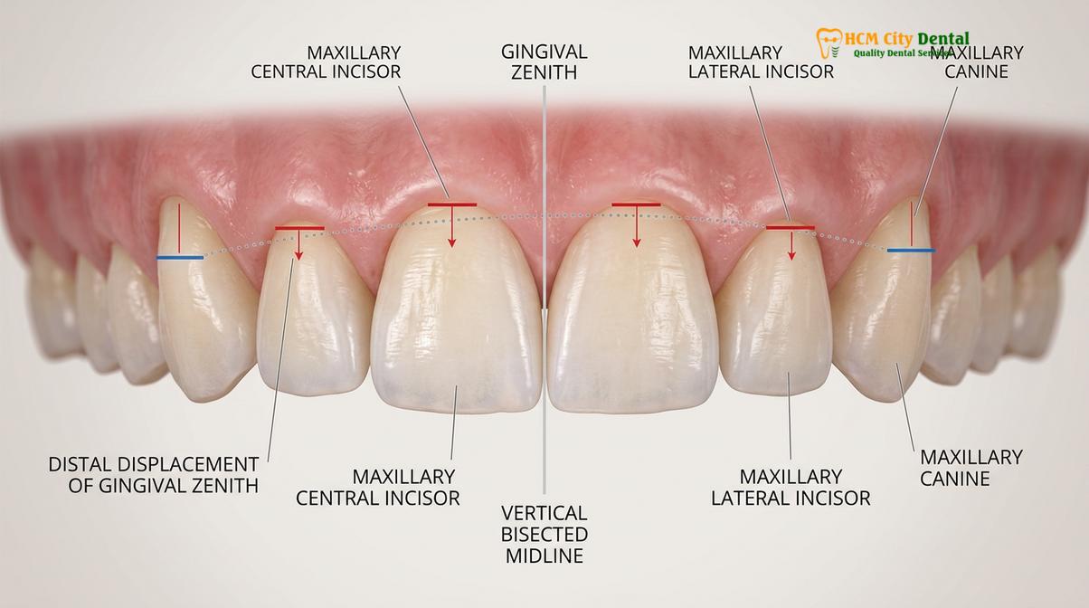
The medial-lateral positioning of the zenith relative to the Vertical Bisected Midline (VBM) is equally critical and varies by tooth type. Extensive clinical studies evaluating the tooth long axis zenith have established the following normative parameters for the maxillary anterior dentition:
- Central Incisors: The zenith is universally displaced distal to the VBM. The mean distal displacement is approximately 1.0mm. This distal shift gives the central incisor a slight mesial inclination, directing the visual flow toward the facial midline.
- Lateral Incisors: The zenith of the lateral incisor is typically coincident with the VBM (centralized) or exhibits a very minor distal deviation (mean of 0.4mm). Because the lateral incisor is narrower, a centralized zenith maintains its delicate proportions.
- Canines: The canine zenith is generally centralized along the long axis of the tooth, with minimal to no distal deviation (mean of 0mm). However, because the canine itself has a prominent cervical convexity and a slight mesial tilt, the zenith aligns perfectly with the height of contour.
To visualize these parameters clearly, the following table summarizes the ideal gingival zenith positions and levels for the maxillary anterior segment:
| Tooth Type | Zenith Position (Medial-Lateral to VBM) | Gingival Zenith Level (Apical-Coronal) | Aesthetic Function |
|---|---|---|---|
| Maxillary Central Incisor | Distally displaced (~1.0 mm) | Baseline (0 mm) – Defines the Gingival Aesthetic Line | Establishes dominance and mesial inclination |
| Maxillary Lateral Incisor | Centralized or slightly distal (~0.4 mm) | Coronal to GAL (~1.0 mm lower) | Creates the youthful gull-wing step-down effect |
| Maxillary Canine | Centralized (0 mm) | Baseline (0 mm) – Matches Central Incisor | Provides corner arch transition and robust emergence |
Deviations from these established rules result in a compromised smile arc. For instance, if the lateral incisor zenith is positioned apically (higher) than the central incisor, it creates a Class II gingival height, which visually flattens the smile and makes the lateral incisor appear disproportionately large and dominant. Conversely, if the central incisor zenith lacks its characteristic distal displacement, the tooth will appear overly square and rigid, lacking the natural dynamic flow required for high-end aesthetic rehabilitation.[3]
How Asymmetric Zenith Points Distort Teeth Proportions
Asymmetrical zeniths disrupt the golden proportion of teeth, creating visual illusions of crookedness, altered width-to-length ratios, and an unbalanced smile arc.
The human eye is highly sensitive to symmetry and proportion, particularly in the anterior aesthetic zone. When gingival zenith points are asymmetrical—whether due to uneven tooth eruption, gingival recession, periodontal disease, or iatrogenic restorative errors—the entire visual perception of the smile is distorted. The zenith acts as the upper frame of the tooth; if the frame is crooked, the picture inside appears crooked, regardless of how perfectly aligned the incisal edges might be.[1]
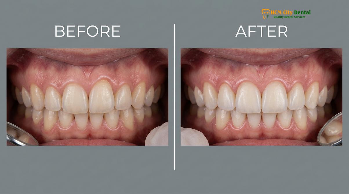
One of the most significant impacts of zenith asymmetry is the alteration of the tooth’s width-to-length ratio. In ideal dental aesthetics, the width of a maxillary central incisor should be approximately 75% to 85% of its length. If the gingival zenith is blunted or positioned too far coronally (as seen in altered passive eruption), the clinical crown appears short and square, pushing the ratio closer to 90% or 100%. Conversely, if the zenith is displaced too far apically due to gingival recession, the tooth appears excessively long and triangular, dropping the ratio below 70%. When these discrepancies occur unilaterally, one central incisor may look short and square while the adjacent one looks long and rectangular, creating severe visual dissonance.
“The gingival zenith is the silent architect of tooth proportion. A perfectly restored ceramic crown will fail aesthetically if the surrounding soft tissue zenith is asymmetric, as the eye will always be drawn to the discrepancy in the gingival margin before noticing the restorative excellence.”
Furthermore, the medial-lateral position of the zenith heavily influences the perceived axial inclination of the tooth. If the zenith of a central incisor is mistakenly positioned mesially rather than distally, the tooth will appear to tilt away from the midline, creating a divergent, unesthetic appearance. This is a common error in poorly executed direct composite veneers or traditional crown preparations where the clinician fails to map the aesthetic gums scallop prior to treatment.[3]
In modern Digital Smile Design (DSD), the evaluation of zenith points is the very first step in the diagnostic workflow. Before any hard tissue preparation is planned, the soft tissue framework must be calibrated. DSD software utilizes digital facebows and dynamic video analysis to overlay the ideal Gingival Aesthetic Line onto the patient’s face. This allows the clinician to measure the exact millimeter discrepancies in the zenith positions and plan precise surgical or orthodontic interventions to harmonize the soft tissue before addressing the teeth themselves.
Precise Surgical Realignment of Zeniths Using Soft Tissue Laser
Modern zenith realignment utilizes 810nm diode lasers for bloodless, minimally invasive contouring that preserves the biological width and accelerates healing.
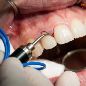
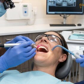
When gingival zenith discrepancies are diagnosed, surgical intervention is often required to establish symmetry. Historically, this was achieved using traditional steel scalpels, which resulted in significant bleeding, required sutures, and involved a prolonged, uncomfortable healing period. Today, the gold standard for soft tissue contouring is the use of advanced dental lasers, specifically the AMD Picasso Diode Laser operating at an 810nm wavelength. This technology has revolutionized aesthetic gum contouring procedures by offering unparalleled precision and patient comfort.[2]
The 810nm diode laser is highly absorbed by melanin and hemoglobin, making it exceptionally effective for soft tissue ablation. As the laser energy vaporizes the excess gingival tissue to relocate the zenith apically, it simultaneously cauterizes the blood vessels and nerve endings. This results in a completely bloodless surgical field, allowing the clinician to evaluate the new zenith position in real-time without the obscuration of hemorrhage. Furthermore, the sterile beam of the laser eliminates bacteria in the surgical pocket, significantly reducing the risk of postoperative infection.
According to Dr. Nguyen Van Cuong’s clinical protocols for biological width sounding and laser settings, the picasso laser zenith shaping must be executed with micro-millimeter precision. Dr. Cuong emphasizes that the laser tip should be angled to recreate the natural knife-edge bevel of the free gingival margin, rather than creating a flat, blunt cut. The procedure is typically performed under profound local anesthesia, and because there are no incisions or sutures, patients experience minimal postoperative swelling or pain. The initial coagulation layer forms instantly, and rapid re-epithelialization allows for a functional recovery within 48 hours.
Clinical Case Study: Dental Tourism at HCMC Dental Clinic
A 28-year-old international patient visited HCMC Dental Clinic in Ho Chi Minh City presenting with a gummy smile and asymmetrical central incisors. Diagnostic sounding revealed Type I altered passive eruption with adequate bone clearance. Using the AMD Picasso 810nm diode laser, the clinical team performed a bloodless zenith realignment, shifting the central zeniths 1.5mm apically and 1mm distally. The procedure was completed in 30 minutes without sutures. The patient reported zero pain post-operatively and was able to enjoy their vacation in Ho Chi Minh City the very next day, demonstrating the profound benefits of laser-assisted periodontal plastic surgery.
For patients considering these advanced aesthetic enhancements, understanding the financial investment is important. According to the latest clinic fee schedule, the pricing structure for zenith realignment and soft tissue contouring is highly accessible, particularly for international patients utilizing dental tourism pathways:
- Laser Gingivectomy (per tooth): 1,500,000 VND (~$60 USD). (Walk-in rate: 2,500,000 VND).
- Full-Arch Laser Gum Contouring (up to 10 teeth): 12,000,000 VND (~$480 USD). (Walk-in rate: 20,000,000 VND).
- Functional/Esthetic Crown Lengthening (per tooth, including bone contouring): 3,000,000 VND (~$120 USD). (Walk-in rate: 5,000,000 VND).
Note: Patients booking their consultation directly via WhatsApp are eligible for a -40% preferred international rate, reflected in the lower tier pricing above.
Advanced Interventions for Severe Gingival Disharmony
When soft tissue laser contouring is insufficient, comprehensive treatments like osseous resective surgery, orthognathic jaw surgery, or lip repositioning are required.
While diode lasers are exceptional for soft tissue zenith realignment, they are limited to cases where the underlying alveolar bone is positioned at an adequate distance from the CEJ (Type I APE). If bone sounding reveals that the alveolar crest is too close to the gingival margin (Type II APE), simply lasering the gums will violate the supracrestal tissue attachment. In these scenarios, an esthetic crown lengthening procedure is mandatory. This involves raising a full-thickness mucoperiosteal flap and performing osteotomy (removal of supporting bone) and osteoplasty (reshaping of non-supporting bone) to re-establish the 2mm biological width further apically. Once the bone is recontoured, the soft tissue flap is sutured into its new, symmetrical zenith position.[3]
In more severe cases, a gummy smile and zenith disharmony are not caused by the teeth or gums at all, but by skeletal or muscular abnormalities. Vertical Maxillary Excess (VME) is a skeletal condition where the upper jaw has grown too far downward, exposing excessive gum tissue regardless of the zenith positions. For severe skeletal VME cases, HCMC Dental Clinic works in collaboration with surgical jaw partners at the National Hospital of Odonto-Stomatology in Ho Chi Minh City (Bệnh viện Răng Hàm Mặt Trung Ương TP.HCM), the highest clinical authority in the region. Maxillofacial specialists perform a Le Fort I osteotomy, an orthognathic surgery that physically impacts and repositions the entire upper jaw upward, permanently correcting the skeletal gummy smile.[4]
“True aesthetic excellence in the anterior maxilla requires a diagnostic triad: evaluating the soft tissue zenith, the underlying osseous crest, and the dynamic mobility of the upper lip. Ignoring any one of these elements leads to compromised, unstable outcomes.”
If the skeletal foundation is normal but the patient has a hyperactive levator labii superioris muscle (the muscle that pulls the lip up), the lip will retract excessively upon smiling, exposing the zeniths and several millimeters of mucosa. For these muscular etiologies, two primary interventions are utilized:
- Botox Gummy Smile Correction: Botulinum toxin is injected into the hyperactive muscles to temporarily reduce their contractility. This lowers the lip line and conceals the excess gingiva. The cost is 4,500,000 VND (~$180 USD) for both sides (Walk-in: 7,500,000 VND). Results last 3 to 6 months.
- Surgical Lip Repositioning (Lip Lowering): A more permanent solution where a strip of mucosa is excised from the maxillary vestibule, and the lip is sutured closer to the teeth, restricting its upward mobility. The cost for this advanced procedure is 15,000,000 VND (~$600 USD) (Walk-in: 25,000,000 VND).[5]
When to See a Doctor for Gingival Zenith Evaluation
Consult a dental professional if you experience an asymmetrical smile, excessive gum display, or are planning to receive anterior cosmetic restorations like veneers.
Evaluating the gingival zenith is not merely a cosmetic endeavor; it is deeply intertwined with periodontal health. Patients should seek a clinical evaluation if they notice that their teeth appear unusually short, square, or uneven in length. Additionally, if you experience a “gummy smile” where more than 3mm of gum tissue is visible when smiling, a comprehensive diagnostic assessment is highly recommended.
Important Clinical Consideration: Never proceed with anterior cosmetic restorations (such as porcelain veneers or crowns) without first evaluating the gingival zenith points. Placing restorations over asymmetrical or biologically compromised gums will result in aesthetic failure and potential periodontal disease. Always ensure the soft tissue foundation is healthy and symmetrical before altering the hard tissues.
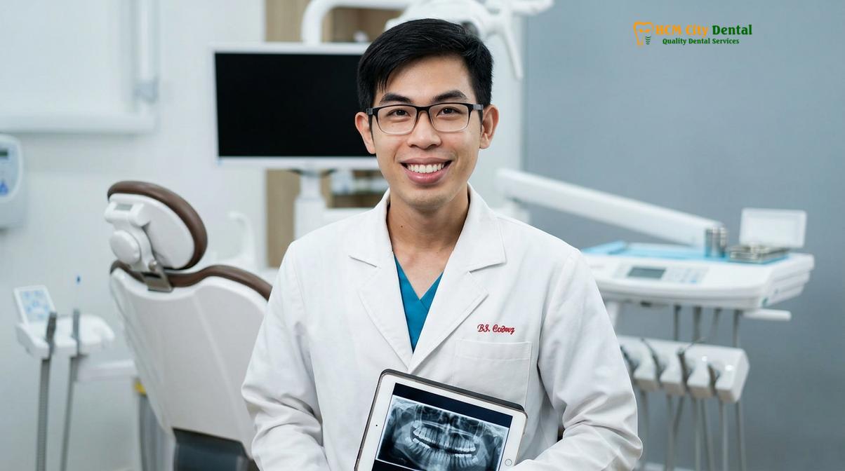
For patients planning a smile makeover, Dr. Nguyen Van Cuong advises that soft tissue calibration must be the first step in the treatment sequence. A personalized clinical examination, including digital smile design and bone sounding, is necessary to determine candidacy for laser contouring versus osseous surgery. If you are seeking expert evaluation, the team at HCMC Dental Clinic in Ho Chi Minh City is equipped with the advanced diagnostic tools and laser technology required to map and restore your ideal gingival architecture safely and predictably.
Frequently Asked Questions
Where is the zenith point located on a tooth?
The zenith point is located at the most apical (highest) position of the free gingival margin surrounding a tooth. For central incisors and canines, it is typically displaced slightly distal to the vertical midline, whereas on lateral incisors, it is centrally aligned. This specific positioning dictates the visual axis and emergence profile of the tooth.
Why are lateral incisor zenith points different?
Lateral incisor zenith points are different because of their narrower anatomical crown shape and root angulation. Clinically, their zenith is centralized along the long axis and positioned approximately 1mm more coronal than the adjacent central incisors and canines. This creates a youthful, dynamic “gull-wing” effect in the smile arc.
How does asymmetric zenith affect crown shape?
An asymmetric zenith distorts the perceived width-to-length ratio of the clinical crown, making the tooth appear tilted, overly square, or disproportionately long. This visual imbalance disrupts the harmonious flow of the gingival aesthetic line across the smile arc, drawing negative attention away from otherwise healthy teeth.
Is laser zenith realignment painful?
Laser zenith realignment is virtually painless because it utilizes a diode laser that seals nerve endings instantly during the procedure. Patients typically only require a mild local anesthetic, and postoperative discomfort is minimal compared to traditional scalpel surgery, often requiring no prescription pain medication.
How long does it take for gums to heal after zenith contouring?
Initial soft tissue healing after laser zenith contouring takes approximately 48 hours due to the laser’s cauterizing effect. Complete tissue maturation and stabilization of the new gingival margin typically require four to six weeks before final restorations, such as veneers or crowns, can be safely placed.
References
- Journal of Esthetic and Restorative Dentistry. Gingival Zenith Positions and Levels of the Maxillary Anterior Dentition. (2009).
- International Journal of Periodontics & Restorative Dentistry. Diode Laser vs. Scalpel Gingivectomy: Healing and Pain Perception. (2020).
- Journal of Clinical Periodontology. Supracrestal Tissue Attachment and Biological Width in Aesthetic Dentistry. (2021).
- Journal of Oral and Maxillofacial Surgery. Stability of Le Fort I Osteotomy in Vertical Maxillary Excess. (2022).
- Clinical Oral Investigations. Long-term Outcomes of Surgical Lip Repositioning for Gummy Smile Correction. (2018).