Esthetic crown lengthening is a precise periodontal microsurgery designed to correct excessive gingival display and expose more natural tooth structure. By carefully reshaping the gumline and underlying bone, this procedure restores biological harmony, creating a balanced, symmetrical smile without compromising long-term dental health.
Clinical Summary:
Esthetic crown lengthening addresses altered passive eruption and gummy smiles by recalibrating the dentogingival complex. Utilizing advanced diode lasers and precise osteoplasty, clinicians establish a minimum 2mm supracrestal tissue attachment to prevent relapse. The procedure harmonizes gingival zenith points, ensuring optimal tooth proportions and long-term periodontal stability while integrating seamlessly with restorative treatments like dental veneers.
Key Takeaways:
- Esthetic crown lengthening corrects short clinical crowns and excessive gingival display.
- The procedure strictly respects the biological width to prevent chronic inflammation and bone resorption.
- Advanced 810nm diode lasers offer minimally invasive tissue removal with rapid coagulation.
- Digital smile design ensures precise calibration of gingival zenith points for optimal symmetry.
- Severe skeletal cases may require orthognathic collaboration for comprehensive facial harmony.
The Biomechanics of Esthetic Crown Lengthening
Esthetic crown lengthening fundamentally relies on respecting the biological width, ensuring that the newly established gum margin maintains a healthy, physiological distance from the underlying alveolar bone crest.
The foundation of any successful periodontal surgery lies in a profound understanding of dental anatomy and biomechanics. A common aesthetic complaint among patients is the appearance of a “gummy smile” or teeth that look disproportionately small. Often, this is not due to the actual size of the teeth, but rather a condition known as altered or delayed passive eruption. In this scenario, the gingival tissue and sometimes the underlying alveolar bone fail to recede to their normal physiological levels during tooth eruption, covering a significant portion of the anatomical enamel crown.
To provide a definitive short square teeth fix, clinicians must look beyond the soft tissue. The critical anatomical concept governing this procedure is the supracrestal tissue attachment (formerly known as the biological width) [1]. This dimension represents the natural physiological space occupied by the junctional epithelium and the connective tissue attachment above the alveolar bone crest. On average, this space measures approximately 2.04 millimeters. If a clinician merely excises the excess gum tissue without evaluating the bone level, the body will naturally attempt to re-establish this biological width by regrowing the gingiva, leading to a complete relapse of the gummy smile.
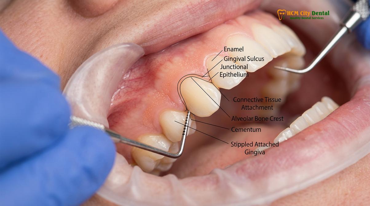
Dr. Nguyen Van Cuong, a leading expert in periodontal aesthetics, emphasizes the clinical importance of biological width sounding prior to any surgical intervention. By using a periodontal probe under local anesthesia, the clinician can accurately measure the distance from the gingival margin to the bone crest. If this distance is less than 3 millimeters, soft tissue excision alone is contraindicated. Instead, a comprehensive esthetic crown lengthening procedure involving both gingivectomy and osseous recontouring is required to permanently shift the entire dentogingival complex apically [2].
“The success of esthetic crown lengthening is not measured by the immediate removal of tissue, but by the long-term stability of the newly established supracrestal attachment. Respecting the bone crest is non-negotiable for predictable periodontal health.”
Furthermore, the biomechanics of the procedure must account for the patient’s specific gingival biotype. Patients with a thick, flat periodontal biotype are more prone to tissue rebound and require more aggressive osseous contouring compared to those with a thin, scalloped biotype. Understanding these nuanced biomechanical principles allows the surgical team to tailor the intervention, ensuring that the final restorative margins—whether for natural teeth or upcoming porcelain veneers—reside in a healthy, inflammation-free environment.
Pre-Operative Planning: Measuring Zenith Points and Bone Crest Heights
Meticulous pre-operative planning utilizes digital smile design and bone sounding to map out the exact gingival zenith points, ensuring predictable and symmetrical surgical outcomes.
The transition from a clinical diagnosis to a surgical reality requires exhaustive pre-operative planning. Modern periodontal surgery relies heavily on Digital Smile Design (DSD) to visualize the final outcome before a single incision is made. During the initial consultation at HCMC Dental Clinic in Ho Chi Minh City, comprehensive diagnostic records are gathered, including high-resolution intraoral and extraoral photography, full-arch digital impressions, and Cone Beam Computed Tomography (CBCT) scans.
A critical aspect of this digital planning is the calibration of the gingival zenith points. The zenith is the most apical (highest) point of the free gingival margin’s scallop. For optimal aesthetic harmony, the zenith of the maxillary central incisors and canines should be displaced slightly distal to the long axis of the tooth, while the zenith of the lateral incisors should be perfectly centered. To reshape gums zenith accurately, the clinician uses digital overlays to create a surgical guide. This guide dictates exactly where the new gingival margin should lie, ensuring that the central incisors and canines are level, with the lateral incisors positioned approximately 0.5 to 1.0 millimeter more coronally.
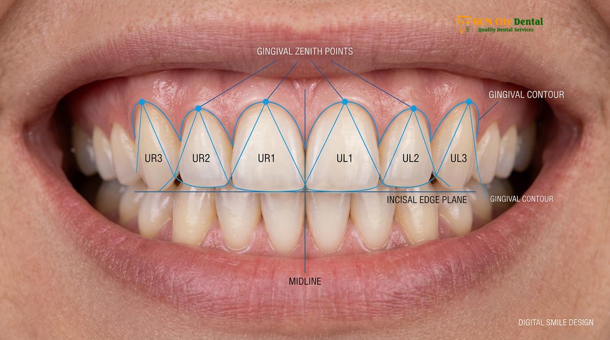
Once the soft tissue parameters are digitally established, the clinician must plan the hard tissue modifications. The CBCT scan provides a three-dimensional view of the alveolar bone crest in relation to the cementoenamel junction (CEJ). If the bone crest is situated directly at the CEJ—a common finding in altered passive eruption—the surgical plan must include osteoplasty to lower the bone crest by at least 2 to 3 millimeters. This meticulous mapping prevents the violation of the supracrestal tissue attachment and guarantees that the newly exposed crown length remains stable over the patient’s lifetime.
To provide transparency in treatment planning, the clinic utilizes a structured fee schedule for these advanced interventions. According to the latest clinic fee schedule, patients can benefit from a highly competitive pricing model, particularly when utilizing the -40% WhatsApp booking discount.
| Procedure Type | Standard Walk-In Price | Discounted Price (-40%) | Clinical Scope |
|---|---|---|---|
| Laser Gingivectomy (Per Tooth) | 2,500,000 VND | 1,500,000 VND (~$60 USD) | Soft tissue removal only using 810nm diode laser. |
| Full-Arch Laser Gum Contouring | 20,000,000 VND | 12,000,000 VND (~$480 USD) | Up to 10 teeth in the aesthetic zone. |
| Esthetic Crown Lengthening (Per Tooth) | 5,000,000 VND | 3,000,000 VND (~$120 USD) | Includes flap elevation and cosmetic bone shaping. |
| Surgical Lip Repositioning | 25,000,000 VND | 15,000,000 VND (~$600 USD) | Mucosal excision to restrict hypermobile lip elevation. |
| Botox Gummy Smile Correction | 7,500,000 VND | 4,500,000 VND (~$180 USD) | Neuromodulator injection for hyperactive levator muscle. |
This transparent approach to pre-operative planning ensures that patients are fully informed about both the clinical workflow and the financial investment required to achieve their ideal smile.
The Surgical Phase: Flap Elevation and Osteoplasty
The surgical phase involves precise soft tissue excision followed by flap elevation and osteoplasty to recontour the alveolar bone, permanently establishing a new biological width.
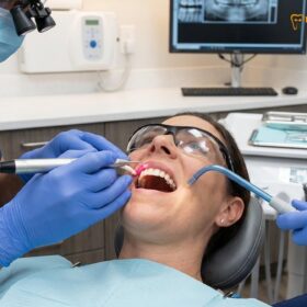
The execution of esthetic crown lengthening is a delicate interplay between soft tissue management and hard tissue sculpting. The procedure begins with the administration of profound local anesthesia to ensure complete patient comfort. The initial step involves an internal bevel incision, guided by the pre-fabricated surgical stent. This incision outlines the new gingival margin, effectively removing the excess collar of tissue.
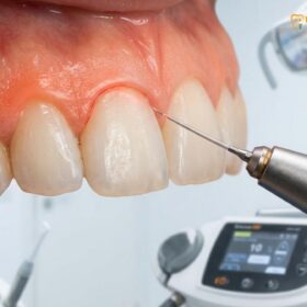
At this stage, the integration of advanced technology becomes paramount. The clinic utilizes AMD Picasso Lasers, a state-of-the-art diode soft tissue laser operating at an 810nm wavelength. The diode laser is highly absorbed by melanin and hemoglobin, making it exceptionally effective for soft tissue surgery. It provides bloodless, sutureless cuts, instant coagulation, and cauterization. This minimally invasive approach significantly reduces postoperative pain and swelling, allowing for a rapid 48-hour initial recovery. Furthermore, the sterile beam of the laser minimizes the risk of bacterial infection in the surgical field [3].
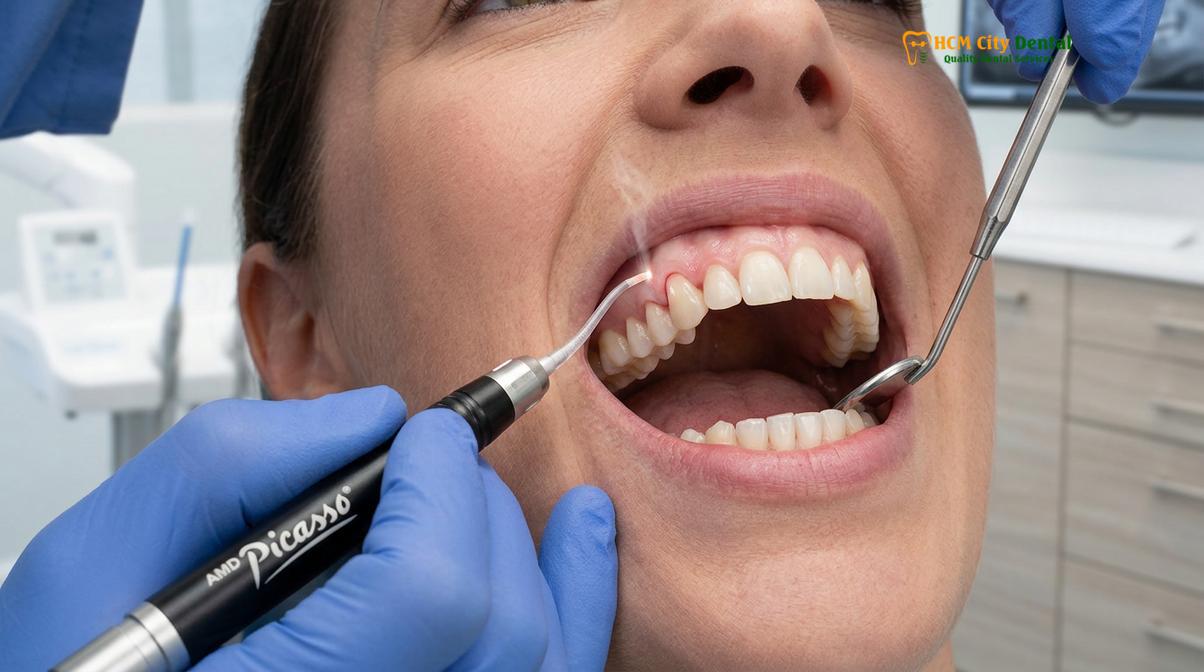
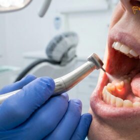
Following the initial gingivectomy, if bone removal is indicated, a sulcular incision is made, and a full-thickness mucoperiosteal flap is carefully elevated. This exposes the underlying alveolar bone and the root surfaces. The clinician then performs an osteotomy (the removal of supporting bone attached to the tooth) and osteoplasty (the reshaping of non-supporting bone to create a physiological contour). This cosmetic bone shaping is executed using high-speed surgical burs under copious sterile saline irrigation to prevent thermal necrosis of the bone cells [4].
Clinical Contraindications: Esthetic crown lengthening with osteoplasty is contraindicated in patients with inadequate root length (poor crown-to-root ratio), active untreated periodontitis, or severe systemic conditions that impair bone healing. A thorough radiographic evaluation is mandatory to ensure sufficient bone support remains after the procedure.
The goal of the osseous resection is to recreate the natural, scalloped architecture of the bone, positioning it exactly 2.5 to 3.0 millimeters apical to the newly established gingival margin. Once the ideal bone contour is achieved, the surgical site is thoroughly debrided, and the roots are planed to remove any residual tissue tags. The mucoperiosteal flap is then repositioned and secured using fine, monofilament sutures. These sutures stabilize the tissue during the critical early phases of healing, ensuring that the papillae are tightly adapted to the interproximal spaces.
Achieving Symmetric Gumline Harmony Across the Aesthetic Zone
Creating a harmonious smile requires calibrating the gingival margins across the entire anterior sextant, ensuring the central incisors, laterals, and canines follow a natural, aesthetically pleasing scallop.
The aesthetic zone, typically defined as the visible area from the right maxillary canine to the left maxillary canine, demands the highest level of surgical precision. A successful Gum Contouring & Gummy Smile procedure does not merely expose more tooth structure; it creates a cohesive, flowing architecture that complements the patient’s facial features. This concept, known as gingival scallop restoration, is vital for achieving a natural-looking result.
In a naturally beautiful smile, the gingival margins follow a specific “high-low-high” pattern. The central incisors and canines share a similar apical height, while the lateral incisors sit slightly lower (more coronal). When performing full-arch laser gum contouring, the clinician must meticulously recreate this undulating pattern. Failure to respect this relationship can result in a flat, artificial-looking gumline that detracts from the overall aesthetics of the face.
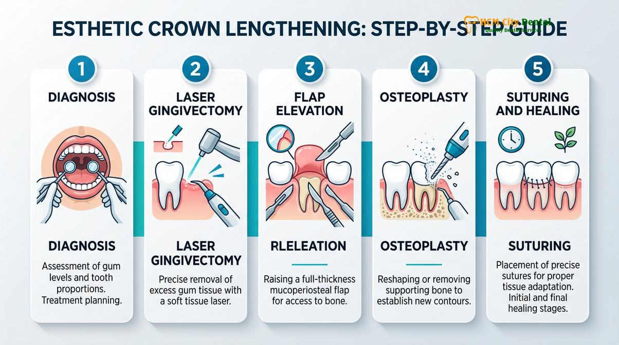
Furthermore, the interdental papillae—the small, triangular peaks of gum tissue between the teeth—must be carefully preserved and managed during the surgery. The papillae frame the teeth and eliminate dark triangles in the interproximal spaces. During flap elevation and suturing, the surgeon uses specialized microsurgical techniques to ensure the papillae are not blunted or lost. The tension-free closure of the flap is critical to maintaining the blood supply to these delicate tissues, promoting optimal healing and regeneration.
Clinical Case Review: A 28-year-old patient presented to HCMC Dental Clinic in Ho Chi Minh City with a chief complaint of short, square teeth and excessive gingival display. Clinical and radiographic examination revealed delayed passive eruption with thick, fibrotic gingiva. A comprehensive esthetic crown lengthening procedure, including laser gingivectomy and osseous recontouring, was performed across the upper six anterior teeth. Following a 3-month healing period, the patient achieved a harmonious, symmetrical gumline with ideal tooth proportions, completely resolving the gummy smile without the need for restorative veneers.
Achieving this level of symmetry requires a deep understanding of macro-aesthetics (the relationship of the teeth to the face) and micro-aesthetics (the intricate details of the teeth and gums themselves). By combining digital planning with precise surgical execution, clinicians can predictably deliver results that profoundly enhance the patient’s confidence and facial harmony.
Advanced Interventions: Jaw Surgery and Lip Repositioning
For patients with severe vertical maxillary excess or hypermobile lips, crown lengthening is often combined with orthognathic surgery or lip repositioning to achieve comprehensive facial balance.
While esthetic crown lengthening is highly effective for dentoalveolar discrepancies, some gummy smiles are skeletal or muscular in origin. When a patient presents with Vertical Maxillary Excess (VME)—a condition where the entire upper jaw has grown too far downward—periodontal surgery alone is insufficient. In these severe skeletal cases, the clinic works in close collaboration with Surgical Jaw Partners. Specifically, complex orthognathic procedures, such as the Le Fort I osteotomy, are coordinated with maxillofacial specialists at the National Hospital of Odonto-Stomatology in Ho Chi Minh City (Bệnh viện Răng Hàm Mặt Trung Ương TP.HCM), recognized as the highest clinical authority in the region [5].
The Le Fort I osteotomy involves surgically detaching the maxilla, impacting it upward to reduce the vertical height, and securing it with titanium plates and screws. This dramatically reduces the amount of gum tissue displayed during a full smile. Once the skeletal discrepancy is corrected, esthetic crown lengthening can be performed as a secondary procedure to fine-tune the gingival margins and optimize tooth proportions.
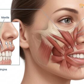
Another common etiology for a gummy smile is a hyperactive levator muscle. In these patients, the upper lip is of normal length at rest, but elevates excessively upon smiling, exposing a wide band of gingiva. For moderate cases, a surgical lip repositioning (or lip lowering) procedure is highly effective. This involves removing a continuous strip of mucosa from the maxillary vestibule and suturing the lip mucosa to the mucogingival junction. This physically restricts the upward pull of the elevator muscles, resulting in a narrower, more aesthetically pleasing smile line.
For patients seeking a non-surgical alternative for a hypermobile lip, neuromodulator injections offer a temporary but effective solution. Botox gummy smile correction involves injecting small amounts of botulinum toxin into the levator labii superioris alaeque nasi muscle. This partially paralyzes the muscle, preventing the lip from rising too high. While the results typically last 3 to 4 months, it serves as an excellent diagnostic tool or a maintenance therapy for patients who decline surgical lip repositioning.
Post-Operative Care and Dental Tourism in Vietnam
Proper post-operative care, including gentle oral hygiene and routine dental scaling, ensures rapid healing and long-term stability of the newly contoured gingival margins.
The success of any periodontal surgery extends well beyond the operating chair; it relies heavily on meticulous post-operative care and patient compliance. Immediately following the procedure, patients are instructed to apply ice packs to the face to minimize swelling and to adhere to a soft diet for the first few days. While the AMD Picasso diode laser significantly reduces postoperative discomfort, mild analgesics may be prescribed to manage any residual soreness.
Oral hygiene is critical during the healing phase. Patients must avoid aggressive brushing around the surgical site to prevent disrupting the sutures or the delicate healing tissues. Instead, an antimicrobial mouthwash containing chlorhexidine gluconate is often prescribed to maintain chemical plaque control. After the initial healing phase of 10 to 14 days, the sutures are removed, and patients can gradually resume normal brushing techniques using an ultra-soft toothbrush.
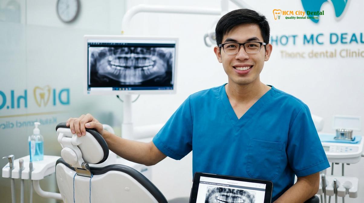
Dr. Nguyen Van Cuong advises that while the initial soft tissue healing occurs within a few weeks, the complete maturation of the supracrestal tissue attachment and the underlying bone remodeling can take up to six months. During this period, routine dental scaling and professional prophylaxis are essential to prevent plaque accumulation, which could trigger inflammation and compromise the aesthetic result. If the crown lengthening was performed as a preparatory step for restorative work, such as porcelain veneers, the clinician will wait until this maturation phase is complete before taking final impressions, ensuring the margins remain perfectly stable.
“Healing is a dynamic biological process. We provide the precise surgical foundation, but the patient’s commitment to post-operative hygiene dictates the ultimate longevity and beauty of the result.”
The high standard of care, combined with advanced technology and competitive pricing, has made Ho Chi Minh City a premier destination for dental tourism. International patients frequently travel to Vietnam to receive world-class periodontal and restorative treatments at a fraction of the cost in their home countries. Clinics equipped with digital workflows and English-speaking specialists ensure a seamless, comfortable experience for global patients seeking comprehensive smile transformations.
When to See a Doctor
If you are self-conscious about a gummy smile, uneven gumlines, or teeth that appear unusually short, a professional clinical evaluation is highly recommended. You should schedule a consultation with a periodontal specialist if you experience chronic gingival inflammation around short clinical crowns, or if your restorative dentist has indicated that a tooth lacks sufficient structure to support a crown or veneer. Early diagnostic imaging and bone sounding can determine whether you are a suitable candidate for esthetic crown lengthening, ensuring that any subsequent cosmetic or restorative treatments are built upon a healthy, stable biological foundation. Always seek a personalized assessment to rule out underlying skeletal or muscular etiologies before proceeding with soft tissue surgery.
Frequently Asked Questions
Are stitches required for esthetic crown lengthening?
Yes, sutures are typically required if a mucoperiosteal flap is elevated for bone contouring. However, if only a soft-tissue laser gingivectomy is performed using an advanced diode laser, stitches are generally unnecessary due to the laser’s instant cauterization. The decision depends entirely on whether osteoplasty is clinically indicated to respect the biological width.
Does the bone contouring weaken my teeth?
When performed correctly by a periodontal specialist, bone contouring does not weaken the teeth. The procedure carefully removes only a minimal amount of crestal bone to re-establish the biological width, leaving ample root support for long-term stability. Radiographic planning ensures that the crown-to-root ratio remains highly favorable.
How long does the procedure take?
A full-arch esthetic crown lengthening procedure typically takes between 60 to 90 minutes. Single-tooth adjustments may take as little as 20 minutes, while complex cases involving extensive osteoplasty or lip repositioning may require up to two hours. The use of digital surgical guides significantly streamlines the operative time.
Will my gums grow back after the contouring procedure?
If the underlying alveolar bone is properly recontoured to respect the supracrestal tissue attachment, the gums will not grow back. Relapse only occurs if soft tissue is removed without addressing the underlying bone crest level, prompting the body to regenerate tissue to protect the bone.
How soon can I receive dental veneers after crown lengthening?
Clinicians generally recommend waiting 3 to 6 months after surgical crown lengthening before taking final impressions for veneers. This healing period ensures the gingival margins have fully stabilized and matured, preventing aesthetic discrepancies or margin exposure that could occur if the tissue recedes further during the remodeling phase.
References
- Journal of Periodontology. Dimensions and relations of the dentogingival junction in humans. (1961).
- International Journal of Periodontics & Restorative Dentistry. Biologic rationale of esthetic crown lengthening. (2011).
- Journal of Clinical Periodontology. Healing of the supracrestal tissue attachment after surgical crown lengthening. (2020).
- American Academy of Periodontology. Parameters of Care: Periodontal Flap Surgery and Osteoplasty. (2019).
- Journal of Oral and Maxillofacial Surgery. Stability of Le Fort I osteotomy for vertical maxillary excess. (2022).