Crown lengthening and gingivectomy are distinct periodontal procedures used to correct gummy smiles or expose tooth structure. While a gingivectomy solely removes excess soft tissue, crown lengthening involves both tissue trimming and bone contouring to preserve the biological width and ensure long-term restorative stability.
Clinical Summary:
Gingivectomy is a minimally invasive soft-tissue procedure ideal for treating gingival overgrowth or minor esthetic concerns, often performed with an advanced diode laser for rapid, sutureless healing. Conversely, crown lengthening is a comprehensive surgical intervention requiring osteotomy (bone reduction) to establish a healthy supracrestal tissue attachment. The clinical choice depends heavily on bone sounding diagnostics; if the alveolar crest is too close to the cementoenamel junction, bone contouring is mandatory to prevent tissue rebound and chronic periodontal inflammation.
Key Takeaways:
- Gingivectomy removes only soft tissue, while crown lengthening reshapes both gums and underlying bone.
- Respecting the biological width (minimum 2mm) is critical to prevent postoperative bone resorption and tissue rebound.
- AMD Picasso Lasers (810nm) enable bloodless, sutureless soft tissue cuts with rapid 48-hour recovery.
- Severe skeletal gummy smiles may require orthognathic surgery (Le Fort I) rather than simple gum contouring.
- Diagnostic bone sounding determines whether gums vs bone trimming is clinically appropriate for the patient.
Gingivectomy: Exposing Gums Only (Soft Tissue Correction)
A gingivectomy is a soft-tissue excision procedure designed to remove excess gingiva, eliminate pseudopockets, and correct minor esthetic discrepancies without altering the underlying alveolar bone.
In the realm of periodontal therapy and cosmetic dentistry, a gingivectomy serves as the primary surgical intervention for patients presenting with excess gingival tissue that is not complicated by underlying osseous (bone) excess. This procedure focuses entirely on the soft tissue architecture. Historically performed with a surgical scalpel, modern clinical protocols now heavily favor the use of advanced laser technology to achieve superior hemostasis and patient comfort. The primary objective is to excise hyperplastic tissue, thereby revealing the natural anatomical crown of the tooth that has been obscured by overgrown gums.
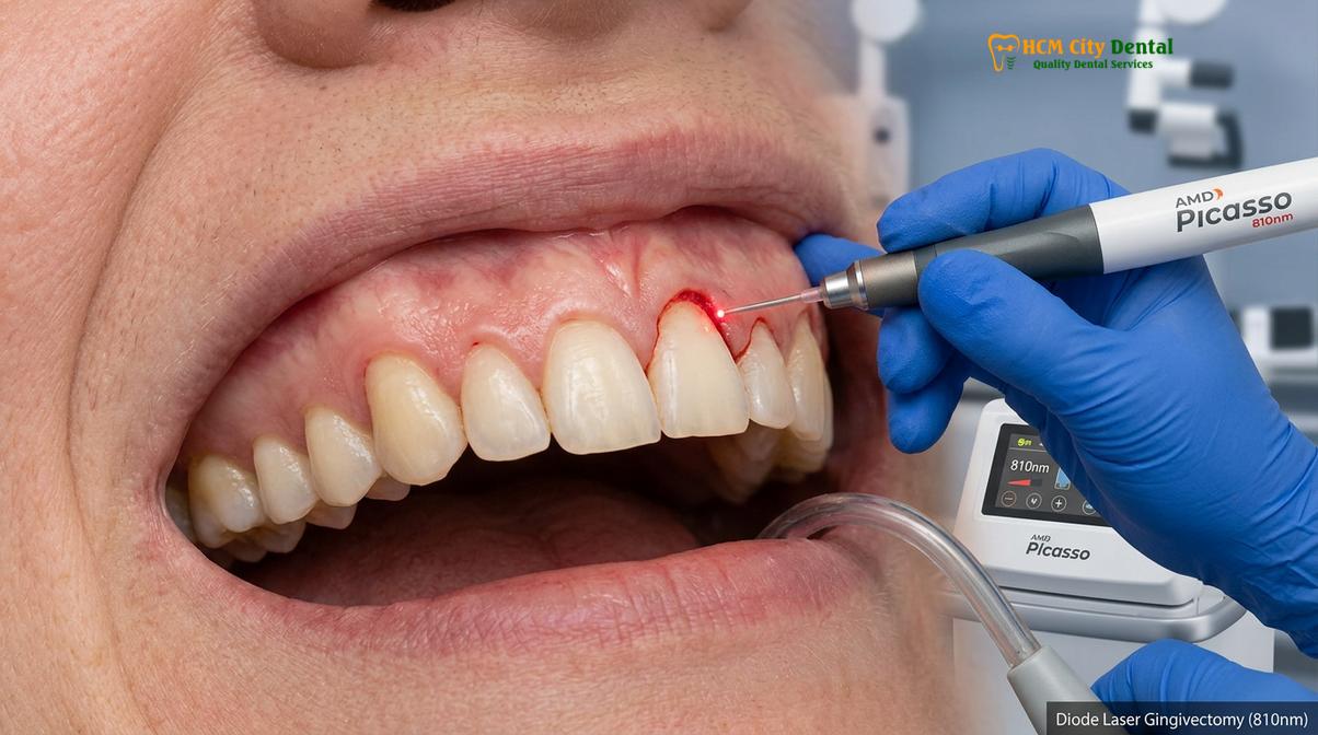
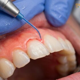
At the forefront of this soft-tissue management is the utilization of AMD Picasso Lasers. Operating at an 810nm wavelength, this diode laser is specifically absorbed by melanin and hemoglobin, making it exceptionally effective for soft tissue applications. The laser provides a bloodless, sutureless cut by instantly coagulating and cauterizing the blood vessels as it vaporizes the targeted tissue. This photothermal interaction not only ensures a clear, dry surgical field for the clinician but also sterilizes the surgical pocket, significantly minimizing the risk of postoperative infection [1]. Patients undergoing laser gingivectomy typically experience drastically reduced postoperative pain and swelling, often achieving a rapid 48-hour recovery.
Before any surgical excision begins, a thorough professional dental scaling and root planing is often required to ensure the gingival tissues are free from local irritants such as calculus and plaque. Operating on inflamed, edematous tissue can lead to unpredictable healing and compromised esthetic outcomes. Once the tissue is healthy and firm, the clinician can precisely sculpt the gingival margins. A critical aspect of this sculpting is the calibration of the zenith points—the most apical point of the gingival scallop, which should ideally be displaced slightly distal to the long axis of the tooth for maxillary centrals and canines.
“The transition from traditional scalpel gingivectomy to diode laser applications has revolutionized soft tissue management, offering unparalleled precision while virtually eliminating the need for sutures and prolonged healing phases.”
Dr. Nguyen Van Cuong, a leading specialist in periodontal esthetics, emphasizes that while a gingivectomy is highly effective, its application must be strictly limited to cases where the underlying bone level is anatomically appropriate. If a clinician attempts to perform a simple gingivectomy when the bone is too close to the desired new gum line, the tissue will inevitably grow back—a phenomenon known as tissue rebound. Therefore, precise periodontal probing and bone sounding under local anesthesia are mandatory diagnostic steps prior to initiating any soft tissue excision.
Crown Lengthening: Bone Contouring (Osteotomy) and Tissue Trimming
Crown lengthening is a dual-tissue surgery that involves reflecting a mucoperiosteal flap to perform an osteotomy and osteoplasty, ensuring the new gum line maintains a healthy distance from the bone crest.
When diagnostic bone sounding reveals that the alveolar bone crest is situated too close to the cementoenamel junction (CEJ) or the planned restorative margin, a simple gingivectomy is contraindicated. In these scenarios, surgical crown lengthening becomes the mandatory clinical protocol. This procedure is significantly more complex than a gingivectomy because it involves the surgical modification of the supporting alveolar bone in addition to the soft tissue excision. The fundamental biological principle driving this requirement is the preservation of the supracrestal tissue attachment, historically referred to as the biological width.
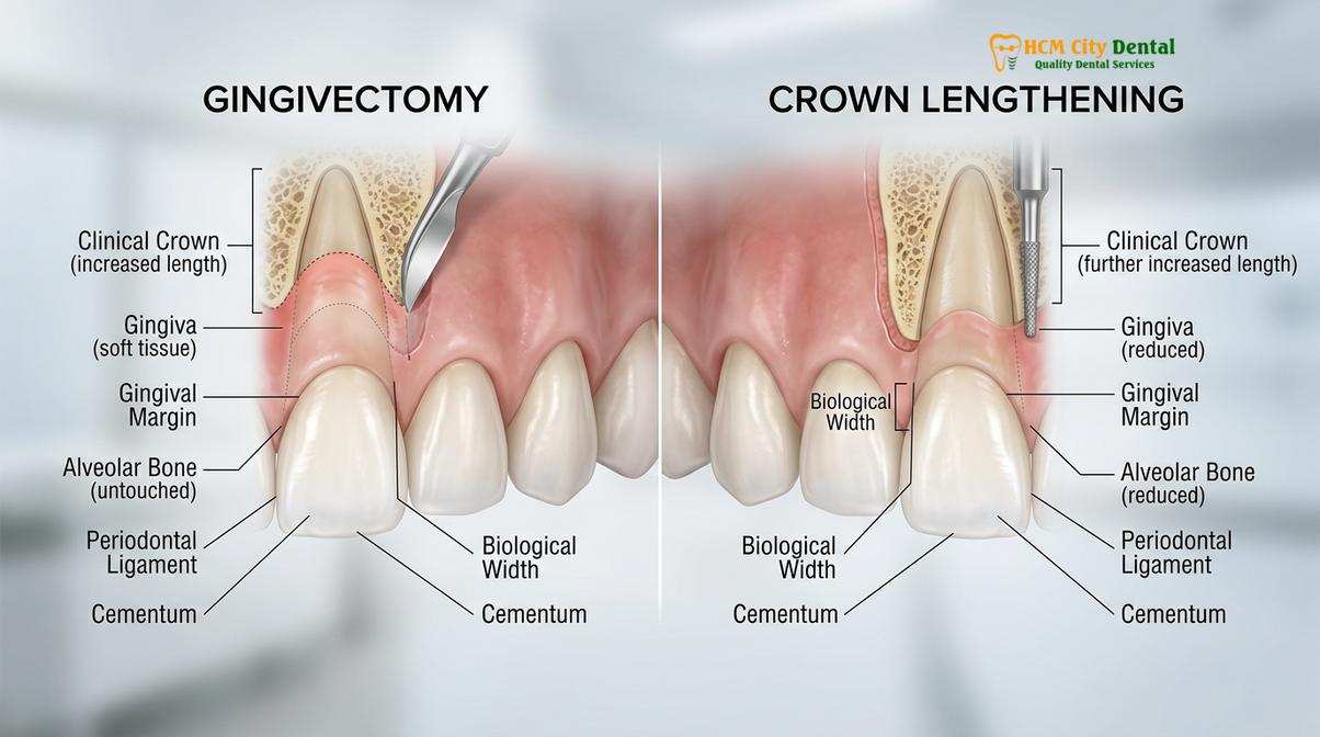
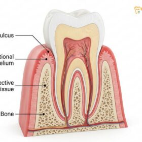
The biological width is a natural physiological dimension comprising the junctional epithelium and the supracrestal connective tissue attachment, which together occupy approximately 2.04 millimeters of space above the alveolar bone crest. If a dental restoration or a newly sculpted gum line encroaches upon this sacred space, the body responds with chronic, localized inflammation. This violation leads to unpredictable bone resorption, persistent bleeding, and the formation of deep periodontal pockets [2]. To prevent this, crown lengthening surgically moves the bone crest apically (downward), re-establishing the necessary space for the soft tissue to attach healthily to the root surface.
The surgical workflow for crown lengthening begins with the administration of profound local anesthesia, followed by precise inverse bevel incisions to remove the excess marginal gingiva. A full-thickness mucoperiosteal flap is then carefully reflected to expose the underlying alveolar bone. At this stage, the clinician performs an osteotomy in dentistry (the removal of tooth-supporting bone) and an osteoplasty (the reshaping of non-supporting bone to achieve a physiological, tapered contour). This is typically accomplished using high-speed rotary instruments under copious sterile saline irrigation, refined with specialized hand instruments like Ochsenbein chisels.
Once the bone has been meticulously recontoured to follow the desired scalloped architecture of the new gum line—ensuring at least 3 millimeters of clearance from the future restorative margin—the surgical flap is apically positioned and secured with sutures. The integration of biological width attachment principles ensures that as the tissue heals, it forms a stable, healthy seal around the tooth. This procedure is not only vital for esthetic corrections but is frequently utilized functionally to expose healthy tooth structure when a tooth has fractured at or below the gum line, or when deep subgingival caries threaten the viability of a restorative crown.
Clinical Indications: When Do You Need Bone Reduction?
Bone reduction is clinically mandatory when the alveolar crest is less than 3 millimeters from the planned restorative margin or when correcting altered passive eruption for esthetic harmony.
Understanding the etiology of a gummy smile or a short clinical crown is paramount in determining whether a patient requires a simple gingivectomy or comprehensive bone reduction. The diagnostic phase is the most critical component of treatment planning. Esthetic crown lengthening is primarily indicated for patients diagnosed with altered passive eruption (APE). APE occurs when the gingival margin fails to recede to its normal physiological position at the cervical convexity of the tooth during adulthood, leaving the anatomical crown hidden beneath thick fibrotic tissue and, frequently, a thick ledge of alveolar bone [3].
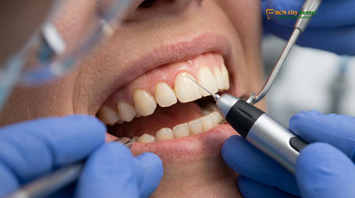
In cases of APE Type I Subgroup B, where there is an adequate band of keratinized tissue but the bone crest is situated directly at the CEJ, bone reduction is absolutely necessary. Attempting to resolve this with a laser gingivectomy alone will result in rapid tissue rebound and failure of the esthetic outcome. By performing an osteotomy, the clinician permanently alters the hard tissue foundation, allowing the gums to drape naturally at a higher, more esthetic level. This meticulous approach to gums vs bone trimming is what separates temporary cosmetic fixes from permanent, biologically sound periodontal therapy.
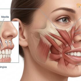
However, not all gummy smiles are caused by dental or periodontal factors. Some patients present with a hyperactive levator muscle of the upper lip. In these cases, the teeth and bone are perfectly proportioned, but the lip retracts excessively upon smiling, exposing a wide band of gingiva. For these muscular etiologies, periodontal surgery is ineffective. Instead, treatments such as Botox injections to relax the hyperactive muscle, or a surgical lip repositioning procedure, are the indicated therapies. Botox offers a temporary but highly effective solution, while lip repositioning provides a more permanent restriction of the lip’s upward mobility.
Furthermore, severe skeletal discrepancies require multidisciplinary intervention. Patients presenting with Vertical Maxillary Excess (VME)—a condition where the upper jaw bone itself has grown too far downward—cannot be treated with gum contouring alone. For these complex skeletal cases, HCMC Dental Clinic collaborates with Surgical Jaw Partners. Specifically, patients are referred for orthognathic surgery, such as the Le Fort I osteotomy, performed in conjunction with maxillofacial specialists at the National Hospital of Odonto-Stomatology in Ho Chi Minh City (Bệnh viện Răng Hàm Mặt Trung Ương TP.HCM), the highest clinical authority in the region [4]. This ensures that the root cause of the skeletal deformity is addressed safely and effectively.
“The preservation of a healthy periodontium is critical for the long-term success of a restored tooth. Dentists must constantly balance the restorative and esthetic needs of their patients with periodontal health, ensuring the biological width is never compromised.”
Comparing Pain Levels, Surgical Scope, and Costs
While gingivectomy is a quick, minimally invasive laser treatment with lower costs, crown lengthening is a more complex surgical procedure requiring bone modification, longer healing, and higher investment.
When evaluating the two procedures, patients must understand that the surgical scope directly dictates the recovery timeline, the level of postoperative discomfort, and the financial investment required. A laser gingivectomy is highly localized to the soft tissue. Because the AMD Picasso diode laser seals nerve endings and lymphatic vessels as it cuts, intraoperative pain is non-existent under local anesthesia, and postoperative discomfort is typically limited to a mild soreness that resolves within 24 to 48 hours. Patients can usually return to work the very next day.
In contrast, crown lengthening is a true minor oral surgery. The reflection of a mucoperiosteal flap and the removal of alveolar bone induce a more significant inflammatory response. While the procedure itself is painless due to profound local anesthesia, patients can expect moderate swelling and discomfort for 3 to 5 days postoperatively. Sutures are usually removed after 7 to 14 days, but complete tissue maturation—especially critical if the patient is waiting for permanent anterior veneers or crowns—can take anywhere from 3 to 6 months. Rushing the restorative phase before the tissue has fully stabilized can lead to aesthetic failures, such as visible crown margins if the tissue recedes further during the final healing phase.
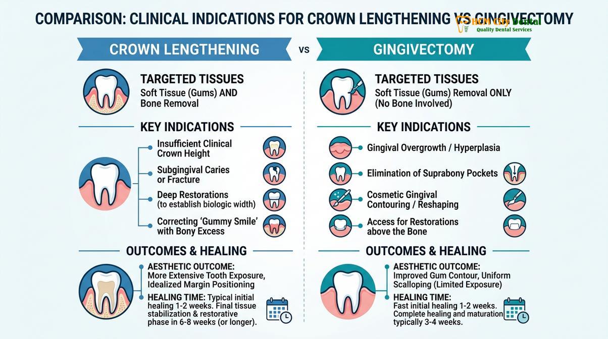
From a financial perspective, the complexity of the procedure is reflected in the pricing structure. For patients exploring comprehensive gum contouring procedures through dental tourism in Vietnam, the cost savings are substantial without compromising on world-class clinical standards. According to the latest clinic fee schedule at HCMC Dental Clinic, the pricing parameters are transparently structured:
| Procedure Type | Target Tissue | Estimated Healing | Standard Price (VND) |
|---|---|---|---|
| Laser Gingivectomy (per tooth) | Soft Tissue Only | 2 – 3 Days | 1,500,000 VND (~$60 USD) |
| Full-Arch Laser Gum Contouring (up to 10 teeth) | Soft Tissue Only | 3 – 5 Days | 12,000,000 VND (~$480 USD) |
| Functional/Esthetic Crown Lengthening (per tooth) | Bone & Soft Tissue | 3 – 6 Months (Maturation) | 3,000,000 VND (~$120 USD) |
| Surgical Lip Repositioning | Mucosal Tissue | 2 – 3 Weeks | 15,000,000 VND (~$600 USD) |
| Botox Gummy Smile Correction (both sides) | Levator Muscle | No downtime | 4,500,000 VND (~$180 USD) |
Note: Walk-in rates are higher. Patients booking in advance via WhatsApp can secure a -40% discount on the walk-in rates, bringing the costs down to the standard prices listed above.
Clinical Case Review: Esthetic Harmony
A 28-year-old patient traveled to Ho Chi Minh City seeking treatment for a severe gummy smile that affected her confidence. During her consultation at HCMC Dental Clinic, Dr. Nguyen Van Cuong utilized Digital Smile Design (DSD) and 3D CBCT imaging to diagnose altered passive eruption combined with a hyperactive lip. The treatment plan involved esthetic crown lengthening on the maxillary anterior sextant to establish proper biological width, followed by a surgical lip repositioning procedure [5]. The integration of AMD Picasso lasers ensured minimal bleeding, and the patient achieved a perfectly balanced, natural smile within a single trip.
When to See a Doctor
Determining the exact cause of a gummy smile or short clinical crowns requires professional diagnostic imaging and clinical probing. You should schedule a consultation with a periodontal specialist if you experience any of the following:
- Your teeth appear disproportionately short or square, and a large band of gum tissue is visible when you smile.
- You have been told you need a dental crown, but the tooth is broken off at or below the gum line, making it impossible to attach the restoration securely.
- You experience chronic gingival inflammation, bleeding, or deep pockets around existing dental crowns, which may indicate a biological width violation.
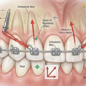
- You have overgrown gum tissue (gingival hyperplasia) resulting from orthodontic treatment (braces) or certain medications.
Important Clinical Consideration
Do not attempt to self-diagnose the etiology of your gummy smile. Only a qualified dental professional can perform the necessary bone sounding and radiographic analysis to determine whether your condition requires a simple soft-tissue laser gingivectomy, complex osseous crown lengthening, or orthognathic jaw surgery. Incorrect treatment selection can lead to tissue rebound or irreversible bone loss.
For a definitive diagnosis and a personalized treatment plan, we encourage patients to book a comprehensive clinical evaluation at HCMC Dental Clinic in Ho Chi Minh City, where our specialists utilize advanced digital diagnostics to ensure predictable, esthetic outcomes.
Frequently Asked Questions
Why does crown lengthening cost more than a simple gingivectomy?
Crown lengthening costs more because it is a complex surgical procedure that requires flap reflection, precise bone contouring (osteotomy), and suturing. Unlike a simple soft-tissue laser excision, it involves modifying the underlying alveolar bone to establish a new biological width, demanding advanced surgical expertise, specialized instrumentation, and longer clinical chair time to ensure the periodontium remains healthy and stable.
Can gums grow back after a simple gingivectomy?
Yes, gums can grow back after a simple gingivectomy if the underlying bone is too close to the new gum line, violating the biological width. The gingival tissue will naturally rebound coronally in an attempt to re-establish the required 2-3 millimeters of space between the bone crest and the gingival margin. This is why bone sounding is a mandatory diagnostic step before any soft tissue is removed.
Is crown lengthening painful?
Crown lengthening is performed under profound local anesthesia, making the procedure itself virtually painless. Postoperative discomfort is generally mild to moderate and can be effectively managed with prescribed non-steroidal anti-inflammatory drugs (NSAIDs) and cold compresses during the first 48 to 72 hours of the healing phase. Most patients find the recovery highly manageable when following strict postoperative care instructions.
How long does it take to heal from laser gum contouring?
Initial healing from laser gum contouring typically takes 48 to 72 hours due to the minimally invasive nature of diode lasers. Because the laser cauterizes nerve endings and blood vessels simultaneously, patients experience minimal swelling, no bleeding, and can usually resume normal dietary habits and daily activities within a few days, making it an excellent option for rapid esthetic enhancement.
Can I get veneers immediately after crown lengthening?
No, you cannot get permanent veneers immediately after crown lengthening; a healing period of 3 to 6 months is required. This maturation phase ensures that the gingival margins have completely stabilized and settled into their final position. Placing permanent restorations too early risks aesthetic failures, such as margin exposure, if the tissue continues to recede slightly during the final stages of healing.
References
- International Journal of Periodontics & Restorative Dentistry. Diode laser vs scalpel gingivectomy. (2020).
- Journal of Aesthetic and Restorative Dentistry. Biological width around restorations. (2019).
- Journal of Clinical Periodontology. Esthetic crown lengthening outcomes. (2021).
- Journal of Oral and Maxillofacial Surgery. Le Fort I osteotomy stability. (2022).
- Clinical Oral Investigations. Surgical lip repositioning longevity. (2018).