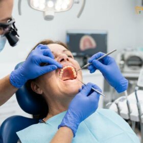
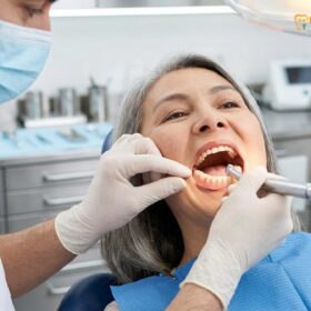
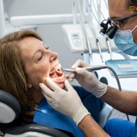
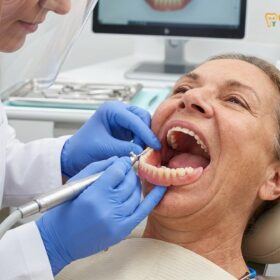
Denture stomatitis treatment focuses on eradicating fungal biofilms from prosthetic surfaces and healing the inflamed oral mucosa. Clinical management requires a synchronized combination of mechanical prosthesis disinfection, targeted topical antifungal therapies, and correcting ill-fitting acrylic bases to restore optimal tissue health and prevent chronic recurrence.
Clinical Summary:
Denture stomatitis, primarily driven by the opportunistic pathogen Candida albicans, is a prevalent inflammatory condition affecting the mucosal tissues beneath removable dental prosthetics. Effective clinical resolution demands a rigorous, dual-pronged approach: treating the infected oral mucosa with specific antifungal agents while simultaneously decontaminating the porous acrylic denture base. Addressing underlying systemic and local risk factors—such as nocturnal denture wear, poor glycemic control, inadequate salivary flow, and mechanical tissue trauma—is critical for long-term success. Prosthodontic interventions, including professional relining or rebasing, are frequently necessary to eliminate microscopic biofilm reservoirs, improve tissue adaptation, and ensure the sustained health of the oral cavity.
Key Takeaways:
- Affects up to 65% of complete and partial denture wearers, often presenting asymptomatically in its early stages.
- Candida albicans biofilm accumulation within the microscopic pores of acrylic resin is the primary etiological driver.
- Successful treatment mandates the simultaneous disinfection of both the oral mucosal tissues and the prosthetic appliance.
- Continuous nocturnal denture wear exponentially increases the risk of fungal proliferation by creating an anaerobic microenvironment.
- Topical antifungal medications, combined with professional prosthodontic adjustments, form the cornerstone of clinical management.
What is Denture Stomatitis? The Fungal Invasion of Gums mucosa
Denture stomatitis is a chronic inflammatory condition characterized by localized erythema and edema of the oral mucosa directly beneath a removable prosthesis, primarily caused by fungal infection and mechanical trauma.
Denture stomatitis, frequently referred to in clinical literature as chronic atrophic candidiasis, represents one of the most common mucosal pathologies encountered in prosthodontic practice. It predominantly affects the palatal mucosa of patients wearing maxillary complete or partial dentures, though it can also manifest beneath mandibular prostheses. The condition is fundamentally a biofilm-mediated opportunistic infection, heavily influenced by the microenvironment created by the physical presence of the denture. When a prosthesis covers the oral tissues, it alters the local microbiome, reduces the natural cleansing action of saliva, and creates a protected, anaerobic niche that favors the proliferation of pathogenic microorganisms [1].
The pathogenesis of this condition is multifactorial, involving a complex interplay between microbiological, mechanical, and systemic factors. While the presence of a prosthesis is the prerequisite, the primary biological driver is the overgrowth of fungal species. However, mechanical trauma from an ill-fitting denture plays a crucial synergistic role. A prosthesis that lacks stability or exhibits poor tissue adaptation will continuously rub against the delicate mucosal surface during mastication and speech. This chronic micro-trauma compromises the epithelial barrier, inducing a localized inflammatory response that makes the tissue highly susceptible to microbial invasion. Consequently, effective General Dentistry protocols must address both the biological infection and the mechanical discrepancies of the appliance.
To standardize diagnosis and guide treatment workflows, clinicians universally rely on Newton’s Classification system, which categorizes the severity of the inflammation based on visual clinical presentation. Understanding these stages is vital for determining the appropriate aggressiveness of the therapeutic intervention.
| Newton’s Classification | Clinical Presentation | Pathophysiological Characteristics | Primary Clinical Focus |
|---|---|---|---|
| Type I (Localized) | Pinpoint hyperemic lesions; localized redness. | Early-stage inflammation, often directly correlated with minor mechanical trauma from specific pressure points on the denture intaglio surface. | Relieving pressure spots; improving basic prosthesis hygiene. |
| Type II (Generalized) | Diffuse erythema involving the entire denture-bearing area. | Widespread chronic atrophic candidiasis; established fungal biofilm on the prosthesis interacting with the mucosal immune system. | Topical antifungal therapy; rigorous chemical denture disinfection. |
| Type III (Granular) | Inflammatory papillary hyperplasia; nodular mucosal overgrowth. | Chronic, long-standing infection and trauma leading to fibrotic tissue changes and deep fungal infiltration into the hyperplastic nodules. | Surgical excision of hyperplastic tissue; fabrication of a new prosthesis. |
In its early stages (Type I and II), the palate tissue inflammation is entirely reversible through conservative management. However, if left untreated, the chronic inflammatory cascade stimulates abnormal cellular proliferation, leading to Type III granular stomatitis. At this advanced stage, the mucosal architecture is permanently altered, and conservative pharmacological treatments alone are insufficient to restore normal tissue contours, often necessitating surgical intervention prior to the fabrication of new prosthetics.
Candida albicans: The Microscopic Culprit in Porous Acrylic Gaps
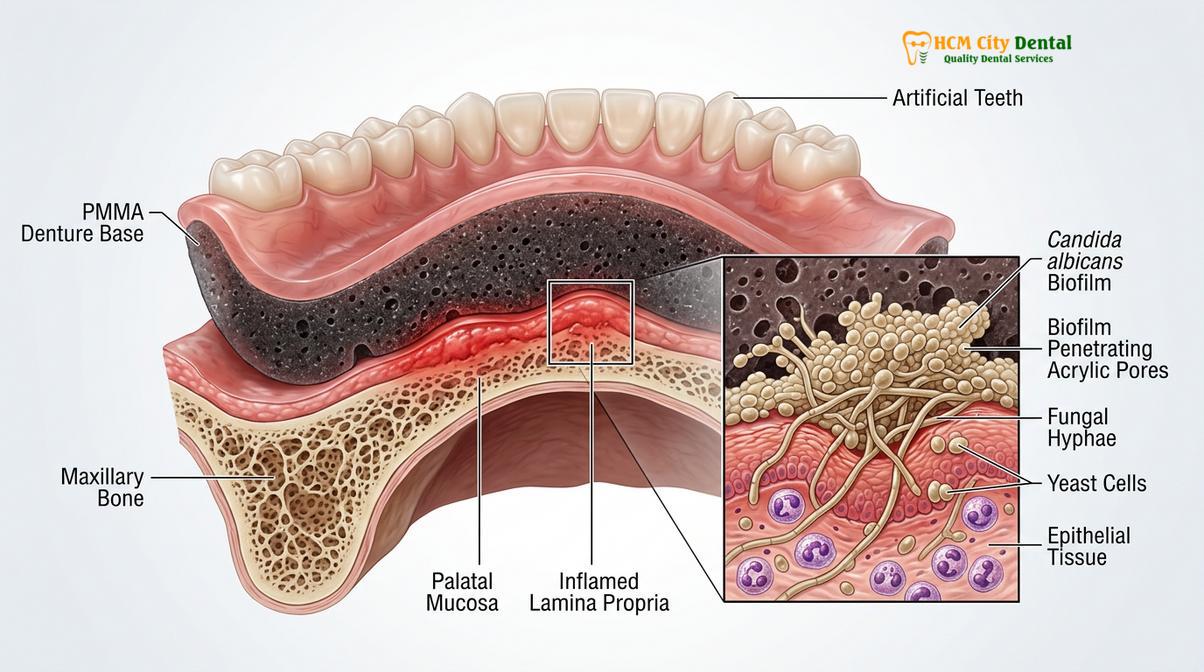
Candida albicans exploits the microscopic porosities of polymethyl methacrylate (PMMA) denture bases to establish highly resistant biofilms, transitioning from benign yeast to invasive hyphal forms.
The primary etiological agent responsible for denture stomatitis is Candida albicans, a dimorphic fungus that exists as a normal commensal organism in the oral cavity of up to 70% of the healthy human population. In a balanced oral microbiome, Candida exists primarily in its benign yeast form, kept in check by competitive bacterial flora and the host’s immune defenses. However, the introduction of a removable prosthesis drastically alters this ecological balance. The intaglio (tissue-facing) surface of traditional dentures is fabricated from polymethyl methacrylate (PMMA), a material that, despite appearing smooth to the naked eye, is inherently porous at the microscopic level [2].
These microscopic acrylic porosities serve as ideal sanctuaries for microbial colonization. When a denture is placed in the mouth, salivary proteins—specifically mucins and statherins—rapidly coat the acrylic surface, forming an acquired pellicle. Candida albicans possesses specialized surface receptors that bind aggressively to these specific salivary proteins. Once attached, the fungal cells begin to secrete an extracellular polymeric substance (EPS), a sticky, protective matrix that encapsulates the colony, forming a robust biofilm. This biofilm is highly resistant to mechanical disruption and significantly impedes the penetration of standard antimicrobial agents.
Within the protective environment of the biofilm, and stimulated by the dark, moist, and low-oxygen conditions beneath the denture, Candida albicans undergoes a critical morphological transition. It shifts from its benign yeast form into its pathogenic hyphal form. These elongated hyphae possess the mechanical strength and enzymatic capability to penetrate the superficial layers of the oral epithelium. As the hyphae invade the mucosal tissues, they release hydrolytic enzymes, such as secreted aspartyl proteinases (SAPs) and phospholipases, which degrade host cell membranes and trigger a robust localized inflammatory response. This cellular damage and subsequent immune reaction manifest clinically as the characteristic redness and swelling of a Candida albicans infection.
Furthermore, the physical properties of the denture material itself can exacerbate this process. Dentures that have been adjusted chairside with coarse acrylic burs, or those that have degraded over years of use, exhibit increased surface roughness. This increased surface area exponentially enhances the capacity for biofilm retention. Therefore, maintaining the highly polished surface of the prosthesis through proper comprehensive denture cleaning is not merely an aesthetic concern, but a fundamental requirement for preventing fungal colonization and subsequent mucosal disease.
Clinical Symptoms: Redness, Burning Sensation, and Swollen Palates
While frequently asymptomatic, clinical signs include diffuse mucosal redness, tissue edema, and occasionally a burning sensation, often accompanied by secondary lesions like angular cheilitis.
One of the most challenging aspects of managing denture stomatitis is its frequently asymptomatic nature. A significant percentage of patients are entirely unaware of the severe inflammation occurring beneath their prostheses until it is incidentally discovered by a clinician during a routine dental examination. Because the condition develops gradually, the sensory nerves in the palate may adapt to the chronic low-grade irritation, resulting in a lack of acute pain. However, the absence of pain does not equate to an absence of pathology; the underlying tissue destruction and fungal proliferation continue unabated.
When symptoms do manifest, they typically present as a generalized burning sensation across the hard palate, which may be exacerbated by consuming hot, spicy, or highly acidic foods. Patients may also report a feeling of dryness, altered taste perception (dysgeusia), or persistent oral malodor (halitosis) resulting from the metabolic byproducts of the fungal biofilm. Visually, the clinician will observe a stark demarcation of erythema (redness) that perfectly outlines the borders of the denture base. The affected mucosa often appears swollen, edematous, and smooth, having lost its normal stippled texture [3].
“The deceptive nature of denture stomatitis lies in its silence. Patients often present to the clinic with severely inflamed, fiery-red palatal tissues yet report zero discomfort. This underscores the absolute necessity of annual prosthodontic evaluations; we must lift the prosthesis to visualize the tissue, as the patient cannot feel the fungal invasion occurring beneath it.”
— Dr. Nguyen Van Cuong, Prosthodontic Specialist
In addition to the primary palatal lesions, chronic atrophic candidiasis is frequently associated with secondary oral manifestations. The most common of these is angular cheilitis, characterized by painful, cracking, and bleeding fissures at the commissures (corners) of the lips. This occurs because the fungal load from the denture acts as a continuous reservoir, seeding the infection to the lips. Angular cheilitis is further exacerbated if the patient’s dentures are heavily worn, leading to a loss of vertical dimension of occlusion (VDO), which causes the corners of the mouth to fold inward and trap moisture. Another associated condition is median rhomboid glossitis, a red, depapillated patch in the center of the tongue, resulting from the tongue resting against the infected palatal denture surface.
Why Keeping Your Dentures in at Night Invites Infection
Continuous nocturnal denture wear deprives the mucosa of oxygen and salivary cleansing, creating an ideal anaerobic microenvironment that exponentially accelerates fungal biofilm proliferation.
The single most significant behavioral risk factor for the development and perpetuation of denture stomatitis is the continuous, 24-hour wearing of removable prostheses. The oral mucosa is not biologically designed to be permanently covered by a rigid, non-porous material. Under normal physiological conditions, the epithelial tissues of the palate and alveolar ridges undergo continuous cellular turnover and are constantly bathed in saliva. Saliva is a complex biological fluid containing critical antimicrobial components, including lysozymes, lactoferrin, histatins, and secretory immunoglobulin A (sIgA), which actively suppress microbial overgrowth.
When a denture is left in place overnight, it acts as a physical barrier, completely shielding the underlying mucosa from these protective salivary components. Furthermore, salivary flow naturally decreases during sleep due to reduced parasympathetic nervous system activity. This combination of physical shielding and reduced salivary volume creates a stagnant, dark, and moist microenvironment beneath the denture base. The temperature in this confined space remains consistently warm, providing the exact optimal conditions required for Candida albicans to rapidly multiply and transition into its invasive hyphal state.
Clinical Warning: Overnight Denture Soaking vs. Wearing
Never sleep with your dentures in your mouth. Removing the prosthesis for at least 8 hours every night is a non-negotiable clinical requirement to allow the mucosal tissues to oxygenate and recover from mechanical compression. During this rest period, practice proper overnight denture soaking in a designated cleansing solution to actively dissolve the daily accumulation of fungal biofilms.
Beyond the microbiological implications, continuous wear subjects the underlying bone and soft tissue to unrelenting mechanical pressure. The mucosal tissues require a period of rest to recover from the compressive forces of mastication. Without this recovery period, the localized blood supply is chronically compromised, leading to tissue ischemia (lack of oxygen). Ischemic tissues exhibit a significantly reduced immune response, making them even more susceptible to fungal invasion. Therefore, instructing patients to remove their prostheses at night is the first and most critical step in any treatment protocol, often resulting in a dramatic reduction of clinical symptoms even before pharmacological intervention begins.
Antifungal Protocols: Nystatin, Palate Hygiene, and Denture Disinfection
Eradicating the infection requires a synchronized protocol of topical antifungal medications applied to the mucosa and rigorous chemical and mechanical decontamination of the prosthesis.
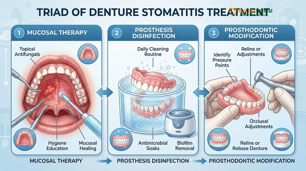
The clinical management of denture stomatitis requires a comprehensive, multi-targeted approach. Treating the oral mucosa while neglecting the prosthesis, or vice versa, will inevitably result in rapid reinfection. The therapeutic protocol is built upon a triad of interventions: mechanical debridement, chemical disinfection, and targeted pharmacological therapy. This comprehensive strategy ensures that the fungal load is reduced to levels that the host’s immune system can effectively manage [4].
1. Mechanical and Chemical Prosthesis Disinfection
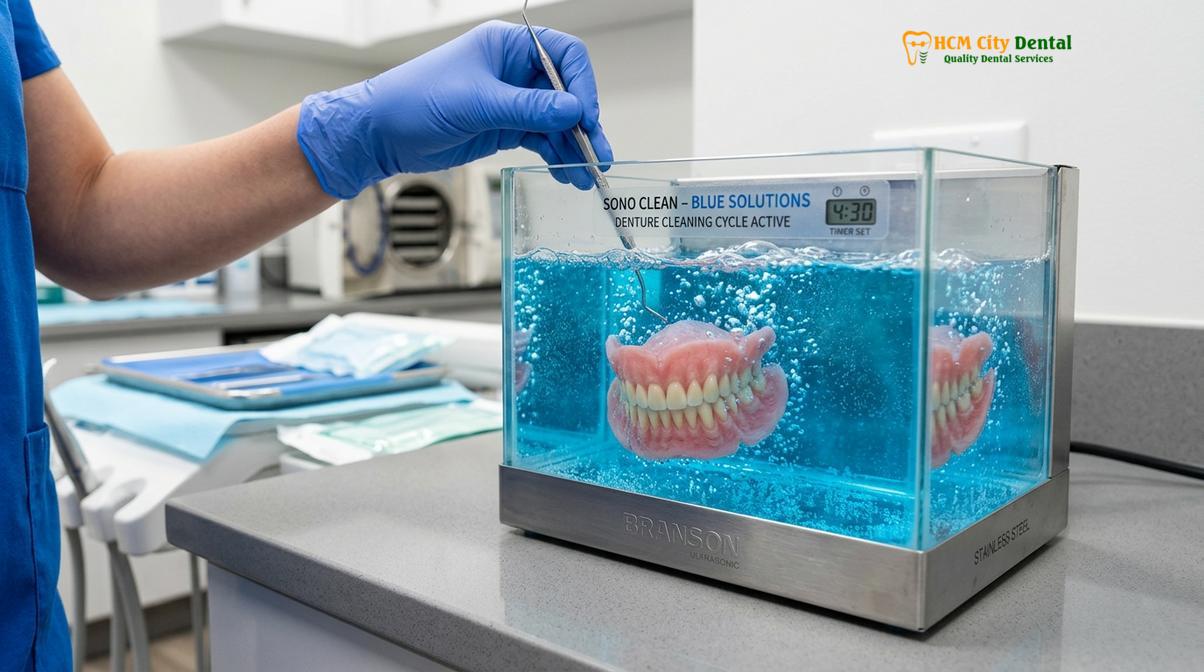
The first line of defense is the rigorous decontamination of the denture itself. Mechanical brushing is essential to disrupt the extracellular matrix of the fungal biofilm. Patients must be instructed to use a dedicated, soft-bristled denture brush and a non-abrasive cleansing agent. Standard toothpaste should never be used, as its abrasive silica particles will create microscopic scratches in the PMMA, further increasing surface roughness and future biofilm retention. Following mechanical disruption, chemical disinfection is required to eliminate the residual fungal spores embedded deep within the acrylic pores.
For oral thrush disinfection, soaking the prosthesis in a dilute sodium hypochlorite solution (bleach) is highly effective against Candida species. However, this must be carefully monitored, as prolonged exposure or high concentrations can degrade the acrylic resin and tarnish metal components of partial dentures. Alternatively, prescription chlorhexidine gluconate (0.12%) solutions can be utilized as a daily soak. It is imperative that the denture is thoroughly rinsed with water before being reinserted into the oral cavity to prevent chemical burns to the mucosa.
2. Pharmacological Intervention: Topical Antifungals
While prosthesis hygiene is paramount, the infected mucosal tissues must also be treated medically. Topical antifungal agents are the standard of care for localized infections. Polyene antifungals, such as Nystatin, are frequently prescribed. Nystatin works by binding to ergosterol, a vital component of the fungal cell membrane, creating pores that lead to cell death. It is typically administered as an oral suspension, which the patient is instructed to swish and hold in the mouth for several minutes before swallowing. Alternatively, Nystatin or Miconazole can be prescribed in an ointment or gel formulation, which is applied directly to the intaglio surface of the denture before insertion, ensuring prolonged contact time with the infected palate.
For patients utilizing an antifungal mouthwash, it is crucial to understand that the rinse must contact the bare tissues; the denture must be removed during application. In cases where the infection is refractory to topical treatments, or if the patient is severely immunocompromised, systemic azole antifungals (such as Fluconazole) may be prescribed. However, systemic therapy is generally reserved for severe cases due to the potential for hepatotoxicity and complex drug interactions.
Clinical Case Review: Refractory Stomatitis Management
A 68-year-old patient presented to HCMC Dental Clinic in Ho Chi Minh City with severe, burning Type II denture stomatitis that had not responded to standard Nystatin therapy. Clinical evaluation revealed a heavily worn, 10-year-old maxillary denture with significant acrylic porosity. The treatment protocol was immediately shifted: the patient was prescribed a localized Miconazole gel, instructed on strict nocturnal removal, and the clinic performed a professional ultrasonic decontamination of the prosthesis. To address the mechanical trauma and eliminate the porous biofilm reservoir, a denture reline for loose dentures was performed using a high-density, non-porous hard acrylic. Complete mucosal healing was achieved within three weeks.
3. Prosthodontic Modification and Tissue Conditioning
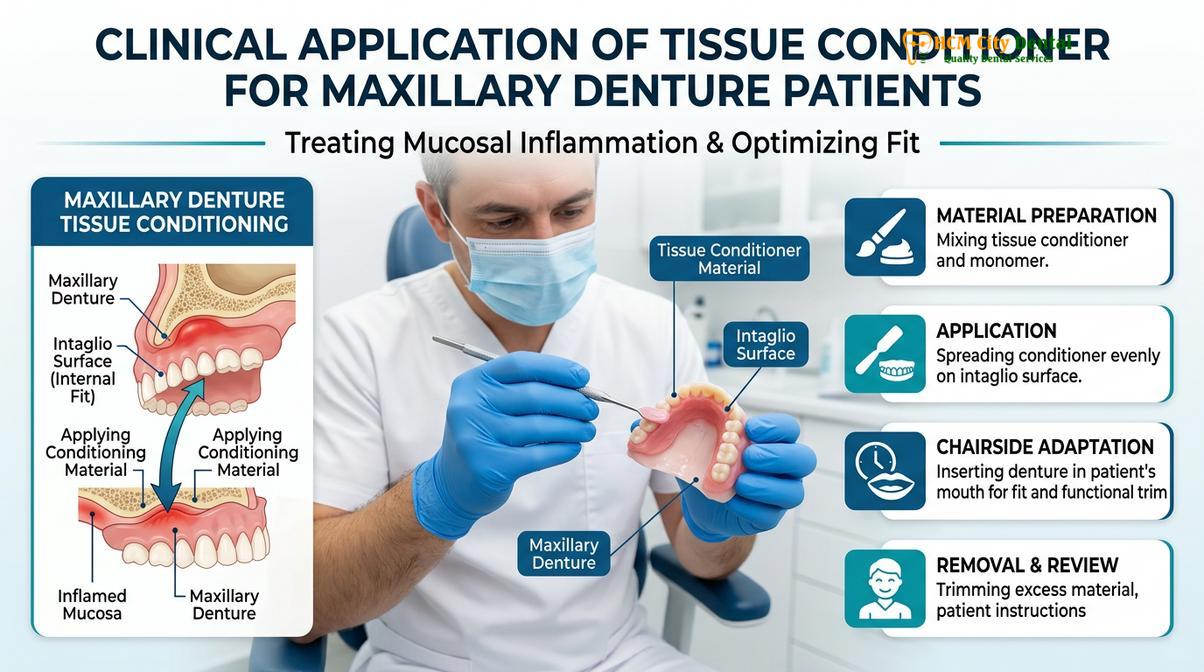
Pharmacological treatment will fail if the underlying mechanical trauma is not addressed. An ill-fitting denture will continue to abrade the tissues, perpetuating the inflammatory cycle. During the active infection phase, clinicians often apply a soft tissue conditioner to the intaglio surface of the denture. This temporary, resilient material acts as a cushion, absorbing masticatory forces and allowing the traumatized mucosa to heal. Once the tissues have returned to a state of health, a permanent modification, such as a hard reline or the fabrication of a new prosthesis, is required to ensure long-term stability. Patients must also be educated on proper denture adhesive application, as excessive use of adhesives can create a sticky matrix that further traps fungal organisms if not cleaned meticulously.
Systemic Risk Factors: Diabetes, Dry Mouth, and Immunocompromised Gums
Systemic conditions such as uncontrolled diabetes, medication-induced xerostomia, and immune suppression severely compromise oral defenses, acting as major catalysts for chronic fungal infections.
While local factors like denture hygiene and fit are the primary drivers of stomatitis, the patient’s systemic health plays a profound role in their susceptibility to infection and their ability to heal. The oral cavity is not an isolated system; it is deeply interconnected with overall physiological well-being. When systemic health declines, the oral mucosal barrier is often one of the first lines of defense to fail, allowing opportunistic pathogens like Candida albicans to thrive [5].
Diabetes Mellitus: Poorly controlled diabetes is a massive risk factor for all oral fungal infections. Hyperglycemia (high blood sugar) directly correlates with elevated glucose levels in the saliva and gingival crevicular fluid. This glucose-rich environment provides an abundant food source that fuels rapid fungal proliferation. Furthermore, chronic hyperglycemia impairs the function of neutrophils and macrophages—the white blood cells responsible for hunting and destroying fungal pathogens. Patients with uncontrolled diabetes also experience delayed wound healing, meaning the micro-traumas caused by the denture take significantly longer to repair, providing a prolonged window for infection.
Xerostomia (Dry Mouth): Adequate salivary flow is the mouth’s primary self-cleaning mechanism. Saliva physically washes away unattached microbes and delivers a constant stream of antimicrobial proteins. Xerostomia, often induced by polypharmacy (the use of multiple medications such as antihypertensives, antidepressants, and antihistamines), radiation therapy to the head and neck, or autoimmune conditions like Sjögren’s syndrome, devastatingly alters the oral environment. Without sufficient saliva, the friction between the denture and the mucosa increases dramatically, causing severe mechanical trauma. Simultaneously, the lack of salivary lysozymes and immunoglobulins removes the biological brakes on fungal growth.
“We cannot treat the mouth in isolation. When a patient presents with recurrent, aggressive denture stomatitis despite excellent prosthesis hygiene, we must look beyond the acrylic. Often, this oral presentation is the first clinical indicator of undiagnosed diabetes or severe medication-induced xerostomia that requires immediate medical co-management.”
— Dr. Nguyen Van Cuong, Prosthodontic Specialist
Immunosuppression: Patients with compromised immune systems—whether due to advanced age, nutritional deficiencies (such as iron, folate, or vitamin B12 deficiency), HIV/AIDS, or immunosuppressive therapies for cancer or organ transplantation—are at an exceptionally high risk. In these individuals, the immune system lacks the capacity to mount an effective localized response against the invading hyphae. What might be a mild, localized Type I stomatitis in a healthy individual can rapidly progress to a severe, generalized infection in an immunocompromised patient, highlighting the need for aggressive and proactive clinical management.
When to See a Doctor for Denture-Related Lesions
While mild redness beneath a new prosthesis may occasionally be attributed to the initial adaptation period, persistent or worsening mucosal changes require immediate professional evaluation. Patients should not attempt to self-diagnose or rely solely on over-the-counter remedies if symptoms persist beyond a few days. Delaying professional care allows the fungal biofilm to penetrate deeper into the tissues, complicating the eventual treatment protocol and increasing the risk of permanent tissue hyperplasia.
You should schedule a clinical examination at HCMC Dental Clinic if you experience any of the following red-flag symptoms:
- Persistent Erythema: Redness beneath the denture that does not resolve after leaving the prosthesis out for 24-48 hours.
- Pain or Burning: Any significant discomfort, burning sensation, or pain when consuming normal foods or wearing the appliance.
- Tissue Overgrowth: The development of bumps, nodules, or a granular texture on the roof of the mouth (indicating potential Type III hyperplasia).
- Bleeding or Ulceration: Any areas of the mucosa that bleed easily upon brushing or exhibit open sores.
- Difficulty Functioning: If the inflammation is interfering with your ability to practice speaking with new dentures or eating with dentures comfortably.
A comprehensive prosthodontic evaluation will involve a thorough visual inspection, palpation of the tissues, and a detailed assessment of the denture’s fit, occlusion, and material integrity. In rare cases where lesions do not respond to standard antifungal protocols, a tissue biopsy may be indicated to rule out dysplastic changes or other underlying mucosal pathologies. Early intervention is the key to restoring comfort and ensuring the longevity of both your oral health and your prosthetic investment.
Frequently Asked Questions
Can denture stomatitis spread to other parts of the body?
Yes, if left untreated in immunocompromised individuals, the localized fungal infection can spread to the throat, esophagus, or systemically. In healthy patients, it typically remains confined to the oral mucosa beneath the prosthesis, though it may cause secondary issues like angular cheilitis at the corners of the mouth. Maintaining strict hygiene prevents this localized biofilm from becoming a broader systemic threat.
How do I disinfect my dentures if I have oral thrush?
You must use a targeted chemical soak, such as a diluted sodium hypochlorite solution or a prescription chlorhexidine rinse, combined with rigorous mechanical brushing. Never use hot water, as it warps the acrylic, and always consult your dentist for the correct dilution ratios to prevent material degradation. Daily decontamination is essential to break the cycle of reinfection.
How long does it take for denture stomatitis to clear up?
With strict adherence to antifungal protocols and proper prosthesis hygiene, mild to moderate denture stomatitis typically resolves within two to four weeks. However, if the underlying cause—such as an ill-fitting denture or uncontrolled diabetes—is not addressed, the inflammation will rapidly recur. Continuous monitoring by a dental professional is required to confirm complete eradication.
Should I buy a new denture if I have chronic stomatitis?
Not necessarily, but your current prosthesis will likely require professional modification, such as a hard reline or rebase, to eliminate microscopic porosities and improve tissue adaptation. If the acrylic is severely degraded, heavily colonized by deep fungal biofilms, or if the bite alignment is fundamentally flawed, fabricating a new prosthesis becomes clinically necessary to protect your oral health.
Is antifungal mouthwash enough to cure denture stomatitis?
No, an antifungal mouthwash alone is insufficient because it only treats the oral tissues, leaving the fungal biofilm intact on the denture surface. Successful eradication requires a dual approach: medicating the oral mucosa while simultaneously executing rigorous chemical and mechanical disinfection of the prosthetic appliance. Treating one without the other guarantees rapid reinfection.
References
- Journal of Prosthodontic Research. Epidemiology and etiology of denture stomatitis: A comprehensive review. (2021).
- International Journal of Dentistry. The role of Candida albicans biofilm in chronic atrophic candidiasis. (2020).
- Journal of Oral Pathology & Medicine. Newton’s classification and clinical management of denture-induced stomatitis. (2019).
- Clinical Oral Investigations. Efficacy of topical antifungals and denture disinfection protocols. (2022).
- Journal of the American Dental Association. Systemic risk factors and glycemic control in oral candidiasis. (2018).
For pricing, booking, and a free clinical assessment, visit our Dentures & Removable Prosthetics service page at HCMC Dental Clinic in Ho Chi Minh City.