Ultimate Guide to Wisdom Teeth Removal & Care

Wisdom teeth removal at HCMC Dental Clinic. Dr. Cuong explains the procedure, recovery steps, and cost details. Get your free consultation today!
Wisdom teeth, clinically termed third molars, represent a unique anatomical intersection of human evolution and contemporary oral health challenges. These vestigial structures, once essential for masticating the coarse diets of our ancestors, now frequently pose significant clinical issues due to the evolutionary reduction in modern jaw size. Despite this diminished functional need, the genetic blueprint for third molars persists, often leading to their problematic eruption into an already crowded dental arch.
Understanding the development and potential complications of these final molars is paramount for proactive oral health management. Typically erupting between the ages of 17 and 25, their trajectory can be complex. Early diagnostic evaluation, often through panoramic X-rays as early as age 15, allows dental professionals to accurately assess the angulation and position of developing wisdom teeth. This foresight is crucial for predicting potential impaction, evaluating risks to adjacent dentition, and formulating an individualized, preventive treatment strategy long before symptoms manifest. This guide aims to thoroughly explore these aspects, from their evolutionary history to advanced management techniques.
2. Impaction Classifications & Clinical Risks
When a wisdom tooth fails to fully emerge through the gums, it is classified as “impacted.” Impactions are categorized based on their orientation within the jawbone and their relationship to the adjacent second molar. Understanding the specific type of impaction is crucial, as it dictates the complexity of the surgical extraction.

Fig 2. Clinical classification of third molar impactions and their pressure vectors on adjacent teeth.
2.1. Mesioangular Impaction
This is the most common type of impaction, accounting for approximately 44% of cases. In a mesioangular impaction, the wisdom tooth is tilted forward, aggressively pushing against the back of the second molar. This angulation not only makes the tooth prone to partial eruption but also creates a significant plaque trap between the two teeth, severely increasing the risk of deep dental caries (decay) on the vital second molar. Extraction often involves sectioning the tooth to prevent damage to the adjacent structures.
2.2. Horizontal Impaction
Horizontal impaction is considered one of the most surgically complex orientations. The wisdom tooth grows completely sideways, running parallel to the jawbone and directly into the root system of the adjacent second molar. Because the tooth is deeply embedded within the alveolar bone and often in close proximity to the inferior alveolar nerve, extracting a horizontally impacted wisdom tooth requires advanced surgical skill, careful bone guttering, and precise sectioning. If left untreated, it can cause severe root resorption of the neighboring healthy teeth.
2.3. Vertical & Distoangular Positions
A vertical impaction means the tooth is in a relatively normal, upright position but remains trapped beneath the bone or soft tissue due to lack of space. While seemingly straightforward, they can still cause recurrent infections if a tissue flap (operculum) forms. Distoangular impactions, conversely, are angled backward, away from the second molar and toward the ramus of the mandible (the ascending back portion of the jaw). These are challenging because they are deeply anchored in dense cortical bone, requiring extensive osseous removal for safe extraction.
2.4. Pathological Risks of Non-Extraction
Ignoring impacted wisdom teeth is a clinical risk that extends beyond mere discomfort. The most frequent complication is Pericoronitis, an acute and painful localized infection of the gum tissue overlying a partially erupted tooth. Because this tissue flap is nearly impossible to clean, bacteria thrive, leading to swelling, purulent discharge, and restricted jaw movement (trismus). Furthermore, impacted teeth can develop dentigerous cysts—fluid-filled sacs that expand within the jaw, silently destroying surrounding bone and neighboring tooth roots. Over time, these cysts can compromise the structural integrity of the mandible itself.
3. Surgical Extraction Procedure: Step-by-Step
At HCMC Dental, wisdom tooth extraction is a meticulously planned surgical procedure, not just a routine pull. Our protocol ensures maximum safety, minimal trauma, and an accelerated recovery.

Fig 3. Precision diagnostic planning using 3D Cone Beam Computed Tomography (CBCT) to map nerve pathways.
3.1. Pre-operative Planning & 3D CBCT Scanning
Standard 2D X-rays are insufficient for complex lower impactions. We utilize state-of-the-art 3D Cone Beam Computed Tomography (CBCT) to capture a highly detailed, three-dimensional view of your craniofacial anatomy. This is critical for mapping the exact trajectory of the inferior alveolar nerve—the major sensory nerve of the lower jaw. By knowing the precise millimeter distance between the nerve canal and the tooth roots, Dr. Cuong can plan a surgical approach that strictly avoids nerve injury, virtually eliminating the risk of postoperative paresthesia (numbness).
3.2. Anesthesia & Patient Comfort Options
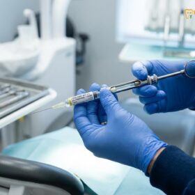
Your comfort is our priority. We employ advanced computer-controlled local anesthesia delivery systems (like The Wand) that provide profound numbing without the pain of traditional injections. For patients experiencing dental anxiety or undergoing multiple complex extractions, we offer conscious IV sedation. Administered by a certified anesthesiologist, IV sedation places you in a deeply relaxed, twilight state where you remain breathing on your own but will have little to no memory of the procedure itself.
3.3. Surgical Technique
Once profound anesthesia is achieved, the surgical area is isolated. A precise mucoperiosteal flap is created to gently retract the gum tissue. If the tooth is encased in bone, a conservative amount of bone is removed (guttering) to expose the crown. Rather than applying forceful leverage—which causes trauma and swelling—our surgeons use a technique called odontisection. The tooth is strategically divided into smaller, manageable fragments using specialized surgical burs, allowing each piece to be gently lifted from the socket. The site is then thoroughly irrigated with sterile saline and closed using resorbable sutures.
3.4. Coronectomy: A Safe Alternative for High-Risk Cases
When lower wisdom teeth roots are deeply entangled with the inferior alveolar nerve canal, conventional surgical extraction carries a 2% to 5% risk of nerve injury. To mitigate this risk, Dr. Cuong performs a highly specialized procedure called a Coronectomy (or intentional partial odontectomy).
During a coronectomy, the clinical crown of the wisdom tooth is surgically cut and removed at the cementoenamel junction, exposing the pulp. Specifically, the surgeon uses a fissure bur to cut through the crown of the tooth at an angle of 45 degrees, stopping just short of the lingual cortex to prevent injury to the lingual nerve. The crown is then gently fractured off, and the remaining root surfaces are planed using a round bur to ensure they are at least 2mm below the surrounding bone crest. This prevents the root from hindering primary gum closure. The root pulp must remain completely untouched to preserve its vitality. Non-vital roots will undergo inflammatory resorption, leading to localized infections that require secondary removal. Once root reduction is complete, the vital root pulp tissue is left intact to prevent inflammatory bone resorption. The gum tissue is then closed over the roots. Clinical studies show that in over 85% of cases, the remaining roots remain stable and are slowly covered by new jawbone. In roughly 15% of cases, the roots may migrate slightly upward, away from the nerve canal, making any future extraction (if necessary) extremely safe and simple. By avoiding root extraction near the nerve canal, the risk of permanent lip and chin numbness (paresthesia) is reduced to practically zero.
Clinical Indications and Contraindications: A coronectomy is indicated only when there is radiographic evidence (via 3D CBCT) of close proximity, contact, or wrapping of the roots around the inferior alveolar nerve canal, and when the tooth pulp is vital and free of infection. Conversely, the procedure is strictly contraindicated in cases of active tooth decay extending into the root canal system, tooth mobility, local or systemic immunological compromise, or pre-existing root infection (periapical pathology). In these contraindicated scenarios, retaining the roots would lead to chronic focal infection, requiring secondary surgical intervention to extract the retained root fragments.
Root Migration Dynamics: Following a successful coronectomy, the retained roots undergo a natural biological phenomenon known as coronal migration. Due to the remodeling of the periodontal ligament (PDL) and bone deposition at the apex, the root fragments slowly travel upward, away from the inferior alveolar nerve canal. This migration typically occurs during the first 3 to 6 months post-surgery, with an average movement of 1mm to 4mm. Once the roots migrate sufficiently, they settle and are capped by new cortical bone formation. In rare instances where migration continues until the roots emerge through the gums, they are then located far from the nerve canal, allowing for a trivial and completely safe extraction without any neurological risk.
4. Advanced Clinical Technologies Used at HCMC Dental
We leverage cutting-edge medical technology to transition wisdom tooth extraction from a traumatic experience to a refined, tissue-preserving procedure.

Fig 4. The Piezotome Solo: Ultrasonic technology that cuts bone without damaging soft tissue or nerves.
4.1. Piezotome Ultrasonic Surgery
Traditional bone removal relies on rotary drills, which can generate heat and inadvertently damage adjacent soft tissues. At HCMC Dental, we utilize Piezosurgery (Piezotome). This revolutionary technology uses modulated ultrasonic micro-vibrations specifically tuned to cut only mineralized tissue (bone). It is entirely inactive on soft tissue, meaning it will not cut blood vessels, membranes, or nerves, even upon direct contact. This selective cutting capability drastically reduces surgical trauma, minimizes postoperative swelling and bruising, and significantly accelerates the healing process.
The biophysics of Piezosurgery is based on the inverse piezoelectric effect, where high-frequency electrical currents are converted into micro-vibrations of 28 to 36 kHz. This frequency range is specifically calibrated to act selectively on mineralized bone, while tissues like the inferior alveolar nerve, buccal mucosa, and periodontal ligament remain entirely unharmed. When conventional rotary burs contact bone, they generate high frictional heat, which can lead to thermal osteonecrosis (localized bone cell death) if irrigation is even slightly compromised. In contrast, Piezotome micro-vibrations preserve the osteocytes (bone cells) at the cut margin, which promotes immediate osteogenesis (new bone formation).
| Clinical Parameter | Traditional Rotary Drills | Piezotome Ultrasonic Surgery | Surgical Benefit |
|---|---|---|---|
| Cutting Action | Macro-rotation and friction | Ultrasonic micro-vibrations (28-36 kHz) | Selective cutting of bone only |
| Soft Tissue Safety | High risk of soft tissue tears/nerves | Zero tissue cut upon contact | Protects Inferior Alveolar Nerve |
| Thermal Bio-Effect | Risks thermal osteonecrosis | Minimal friction, cold cavitation spray | Preserves bone cells, less post-op swelling |
| Bleeding & Visibility | Drill blocks site, high blood pooling | Ultrasonic cavitation flushes area | Excellent visual field for surgeon |
| Healing Speed | Standard recovery (7-10 days) | Accelerated bone remodeling | Immediate trigger of bone healing |

Fig 5. PRF Therapy: Utilizing the patient’s own concentrated growth factors to supercharge socket healing.
4.2. Platelet-Rich Fibrin (PRF) Therapy
To further enhance recovery, we offer Platelet-Rich Fibrin (PRF) therapy. Immediately prior to surgery, a small sample of your blood is drawn and spun in a specialized centrifuge. This process separates the red blood cells, isolating a highly concentrated matrix of platelets, white blood cells, and natural growth factors. This autologous PRF clot is then placed directly into the empty tooth socket before suturing. PRF acts as a bioactive bandage, drastically accelerating hard and soft tissue regeneration, reducing pain, and virtually eliminating the risk of dry socket by protecting the underlying bone.
The isolation of PRF is a delicate clinical procedure. We collect a small volume of the patient’s blood (approximately 10ml per socket) in sterile, additive-free tubes. These are spun immediately in our specialized centrifuge at a precise speed of 2,700 RPM for 12 minutes (L-PRF protocol). This slow centrifugation separates the blood into three distinct layers: a bottom layer of packed red blood cells (RBCs), a top layer of acellular Platelet-Poor Plasma (PPP), and a dense, gelatinous Platelet-Rich Fibrin (PRF) clot in the middle. The PRF clot is extracted and compressed into a resilient membrane. This membrane is a three-dimensional autologous network containing concentrated platelets, leucocytes, and cytokines trapped within a polymerized fibrin matrix.
Biologically, the PRF matrix serves as a sustained-release reservoir of essential growth factors that are slowly released over a 7 to 14-day healing period, which aligns with the critical early phases of socket epithelialization and osteogenesis:
- Platelet-Derived Growth Factor (PDGF): Stimulates cell replication and chemotaxis of osteoblasts (bone-forming cells) and fibroblasts (tissue-forming cells).
- Transforming Growth Factor-Beta (TGF-β): Promotes collagen synthesis and fibronectin deposition, forming the structural scaffold for new bone.
- Vascular Endothelial Growth Factor (VEGF): Directs angiogenesis, stimulating the formation of new capillary networks to deliver oxygen and nutrients to the healing socket.
- Epidermal Growth Factor (EGF): Accelerates re-epithelialization, closing the open gum wound over the socket.
| Post-Op Stage | Standard Socket Healing | PRF-Accelerated Socket Healing | Clinical Advantage |
|---|---|---|---|
| Day 1 (Clot Formation) | Fragile red blood cell clot forms | Dense PRF fibrin membrane seals socket | 99% reduction in immediate bleeding and pain |
| Day 3 (Tissue Growth) | Early granular tissue starts forming | Active release of PDGF & VEGF spurs capillaries | Prevents dry socket by keeping bone insulated |
| Day 7 (Epithelial Cover) | Gum margins starting to close | EGF triggers complete epithelial closure | No food trapping in socket |
| Day 14 (Bone Matrix) | Soft osteoid tissue forms slowly | TGF-β initiates calcium mineralization | Accelerated jawbone recovery |
5. Cost Comparison & Global Price Index
Ho Chi Minh City has emerged as a premier destination for world-class dental care at a fraction of the cost found in Western nations. By combining elite surgical expertise with lower regional operational costs, HCMC Dental provides unparalleled value.

Fig 6. Experience premium, stress-free dental care while enjoying the vibrant city of Ho Chi Minh.
| Procedure Type | USA / Australia Average | HCMC Dental (Vietnam) | Savings Margin |
|---|---|---|---|
| Simple Extraction (Erupted) | $250 – $400 USD | $50 – $80 USD | ~80% Savings |
| Complex Surgical Extraction (Impacted) | $600 – $1,200 USD | $150 – $250 USD | ~75% Savings |
| 3D CBCT Scan | $300 – $500 USD | $50 USD (Often complimentary with surgery) | ~90% Savings |
| IV Sedation (Per Hour) | $500 – $800 USD | $150 – $250 USD | ~70% Savings |
*Note: Prices are estimates and may vary based on anatomical complexity. Even factoring in flights and premium hotel accommodations, international patients routinely save thousands of dollars on multiple extractions.
6. Post-Operative Recovery & Risk Management
The success of your surgery extends into your home care. Following our strict recovery protocols ensures a smooth, complication-free healing phase.

Fig 7. Proper aftercare, including cold compress therapy, is vital for a smooth recovery.
6.1. The Critical First 24 Hours
Immediately after surgery, bite firmly on the provided sterile gauze for 45 minutes to establish a stable blood clot. Bleeding control is paramount; minor oozing is normal, but active bleeding requires replacing the gauze and applying steady pressure. Ice packs should be applied externally to the jaw (20 minutes on, 20 minutes off) for the first 24-48 hours to restrict blood flow and aggressively control inflammatory swelling. Stick to a strict liquid and soft-food diet (smoothies, mashed potatoes, lukewarm soups) and avoid chewing near the surgical sites.
6.2. Preventing Dry Socket (Alveolar Osteitis)
Dry socket occurs when the protective blood clot in the socket is dislodged or dissolves prematurely, exposing the highly sensitive bone and nerves underneath. This is an intensely painful condition. To prevent it, you must absolutely avoid creating negative pressure in your mouth. Do not use straws, do not spit forcefully, do not smoke, and do not aggressively rinse your mouth for at least 72 hours. Gentle head rolls with warm salt water can begin after 24 hours.
6.3. Flight Guidelines for Dental Tourists
For our international patients, we strongly advise against flying within 48 to 72 hours of complex surgical extractions. Cabin pressure changes at high altitudes can cause pain, increase swelling, and potentially dislodge delicate blood clots. Plan your itinerary to allow for adequate rest in Ho Chi Minh City post-surgery. Our team provides comprehensive travel documentation and fit-to-fly certificates when appropriate.
6.4. Long-Term Recovery & Surgical Risk Management
Minor postoperative complications like mild swelling, stiffness in the jaw joint (trismus), and minor bruising are normal physiological reactions that peak at 48 hours and resolve by day 5 to 7. However, recognizing abnormal healing signals is crucial for timely risk management:
- Dry Socket Indicators: If a sudden, throbbing pain develops 3 to 4 days post-extraction and radiates to your ear or temple, accompanied by a foul odor and taste, the protective clot has likely dislodged. Contact us immediately for a gentle cleaning of the socket and placement of a protective eugenol-based sedative dressing (Alvogyl).
- Infection Signals: Worsening pain or swelling after day 4, persistent fever, or difficulty swallowing require immediate clinical attention and prescription of targeted antibiotic therapy (such as Amoxicillin-Clavulanic acid).
- IAN Neurological Recovery: If temporary nerve paresthesia occurs (numbness in the lip, chin, or tongue due to proximity to the inferior alveolar nerve), we prescribe supportive neurotropic B-vitamins (B1, B6, B12) to accelerate axon regeneration. Paresthesia is almost always transient and resolves within weeks to months.
6.5. Post-Surgical Dietary Timelines
To avoid trauma to the blood clot and surgical sutures, follow this dietary progression:
- Day 1: Strictly cold or cool liquids. Lukewarm clear broths, fruit smoothies (no seeds, eaten with a spoon, no straw), protein shakes, and plain Greek yogurt. Do not consume anything hot, carbonated, or alcoholic.
- Days 2 to 3: Transition to soft, non-chewing foods. Mashed potatoes, soft scrambled eggs, applesauce, oatmeal, well-cooked pasta, and soft tofu. Restrict chewing to the opposite side of the mouth.
- Days 4 to 7: Introduce soft chewable foods. Flaky fish, steamed vegetables, soft-cooked chicken, pancakes, and soft bread. Avoid small, hard elements like rice, popcorn, chips, or nuts, which can get ketted in the healing socket.
- Day 8+: Gradually resume a normal diet as tolerated, ensuring the extraction sites are gently rinsed with warm saline after meals to prevent food impaction.
Nutritional Biology for Accelerated Wound Healing: Post-surgical recovery is heavily dependent on specific micronutrients and macronutrients. We advise patients to optimize their diet with foods rich in Vitamin C (essential for collagen synthesis and tissue cross-linking) and Zinc (a cofactor in cellular replication and re-epithelialization). Adequate intake of high-quality protein (such as bone broths, Greek yogurt, and protein shakes) is necessary to provide the amino acids required for soft tissue reconstruction. Incorporating a recovery smoothie made with avocado, spinach, banana, and a scoop of high-quality whey or plant-based protein powder offers a nutrient-dense, easy-to-consume option during the initial days.
The Physics of Negative Intraoral Pressure: It is critically important to understand why drinking straws must be avoided. Sucking through a straw creates a vacuum inside the oral cavity, generating significant negative pressure (measured up to -100 mmHg). This pressure differential exerts a physical suction force directly on the freshly formed fibrin blood clot inside the extraction socket. Because the clot is only anchored by early fibrin strands during the first 72 hours, this suction force can easily dislodge the clot, exposing the raw alveolar bone and leading to an agonizing dry socket. Patients must consume all liquids by drinking directly from a cup or using a spoon.
7. Medical Case Review: Complex Horizontal Lower Left Third Molar

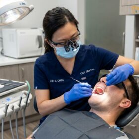
Fig 8. Our expert surgical team executing a complex horizontal impaction removal in our sterile operating theater.
Patient Profile: 24-year-old male presenting with acute, localized pain in the lower left quadrant and restricted mouth opening.
Diagnosis: Severe pericoronitis associated with a deeply impacted horizontal left mandibular third molar (Tooth #38). 3D CBCT revealed the apical roots were entangled with the superior border of the inferior alveolar nerve canal.
Clinical Approach: Under IV sedation, a full mucoperiosteal flap was raised. Utilizing the Piezotome, Dr. Cuong selectively removed overlying cortical bone without thermal injury. The tooth was carefully sectioned into three fragments using a high-speed surgical handpiece. The fragments were elevated individually, ensuring zero pressure was applied toward the nerve canal. The socket was completely debrided, and autologous PRF membranes were inserted before securing the flap with Vicryl sutures.
Outcome: The patient reported zero numbness post-operatively, indicating successful nerve preservation. Swelling peaked at 48 hours and subsided entirely by day 5. The patient returned to a normal diet within 7 days, highlighting the effectiveness of combining Piezosurgery with PRF therapy for rapid healing in complex cases.
8. Frequently Asked Questions (FAQ)
Do all wisdom teeth need to be extracted?
No. If wisdom teeth erupt fully, are properly aligned, and can be easily cleaned, they do not require extraction. However, if they are impacted, causing pain, damaging adjacent teeth, or prone to recurrent infection, surgical removal is medically necessary.
Will the extraction be painful?
The procedure itself is entirely painless due to profound local anesthesia and, if chosen, IV sedation. Post-operative discomfort is expected but is effectively managed with prescribed anti-inflammatory medications and analgesics.
How long does the surgery take?
A simple extraction can take as little as 10-15 minutes. Complex, deeply impacted surgical extractions typically take 30-45 minutes per tooth. For all four teeth, the procedure generally takes about an hour under sedation.
When can I eat solid foods again?
Stick to a liquid and very soft diet for the first 48 hours. You can gradually introduce semi-solid foods (like pasta or scrambled eggs) as tolerated. Avoid hard, crunchy, or spicy foods for at least 7-10 days.
What is the risk of nerve damage?
While there is a slight risk of altered sensation if the tooth is near the inferior alveolar nerve, our use of mandatory 3D CBCT scanning allows us to map the nerve accurately and alter our surgical approach to practically eliminate this risk.
Can I go back to work the next day?
For simple extractions, many patients return to work the following day. For surgical impactions, we recommend resting for 2 to 3 days, as swelling typically peaks on the second or third day.
Is IV sedation safe?
Yes, IV sedation is exceedingly safe when administered by our certified medical anesthesiologists. Your vital signs are continuously monitored throughout the procedure, ensuring a highly controlled and safe environment.
Why should I travel to Vietnam for wisdom teeth removal?
Vietnam, specifically HCMC Dental, offers a combination of highly trained maxillo-facial surgeons, state-of-the-art technology (CBCT, Piezotome, PRF), and stringent international sterilization protocols, all at a cost that is 70-80% lower than Western clinics.
What should patients know about one day dental care?
In clinical practice, one day dental care is an essential factor in maintaining long-term oral health and preventing decay. Regular scaling, routine examinations, and personalized treatment plans are key to managing this aspect effectively.
What should patients know about contemporary dental care?
In clinical practice, contemporary dental care is an essential factor in maintaining long-term oral health and preventing decay. Regular scaling, routine examinations, and personalized treatment plans are key to managing this aspect effectively.