An oroantral communication (OAC) is a pathological, unnatural opening connecting the sterile environment of the maxillary sinus cavity to the bacteria-rich oral cavity. This complication most frequently occurs following the extraction of maxillary posterior teeth—specifically the first and second molars—where the apices of the roots often protrude into or sit intimately adjacent to the Schneiderian membrane of the sinus floor. When an acute communication is left untreated, or if spontaneous healing fails due to infection or excessive defect size, the tract rapidly epithelializes, transforming into a chronic oroantral fistula (OAF). For international patients seeking advanced oral and maxillofacial sinus fistula surgery in Ho Chi Minh City, achieving a hermetic, tension-free closure is paramount to preventing chronic sinusitis and preserving the alveolar ridge for future dental implant rehabilitation. At HCMC Dental Clinic, our clinical protocols for oroantral communication fistula closure buccal fat pad prf sinus repair integrate cutting-edge autogenous biomaterials and advanced flap designs to ensure predictable, long-term success.
Under the clinical direction of Dr. Nguyen Van Cuong (Dr. Cuong), DDS, our facility in District 1 employs a highly specialized, evidence-based approach to sinus complications. Whether managing an acute sinus perforation after upper molar extraction treatment or executing a complex oroantral fistula repair buccal fat pad flap in Vietnam, our surgical decision-making is guided by strict anatomical parameters. By combining the regenerative power of Leukocyte-Platelet Rich Fibrin (L-PRF) with the robust vascularity of the pedicled buccal fat pad (Bichat’s ball), we provide a double-layer closure that not only seals the sinus but also meticulously preserves the vestibular depth required for future implant prosthetics.
Clinical Summary:
The surgical management of an oroantral communication (OAC) or chronic fistula (OAF) requires precise anatomical evaluation and technique selection based on defect size. Small perforations (<2mm) may heal spontaneously via blood clot formation, while moderate defects (2mm–5mm) are effectively managed using autogenous L-PRF plugs and figure-eight suturing. For large defects (>5mm) or chronic epithelialized fistulas, HCMC Dental Clinic utilizes a double-layer hermetic closure protocol. This involves the complete excision of the fistulous tract, placement of an L-PRF membrane, and the mobilization of a pedicled Buccal Fat Pad (Bichat’s flap) or a Rehrmann buccal advancement flap. The buccal fat pad is highly preferred when future dental implants are planned, as it provides exceptional vascularity without compromising vestibular depth. Strict 14-day postoperative sinus precautions are mandatory for successful integration.
Key Takeaways:
- Acute vs. Chronic: An OAC is an acute perforation usually occurring immediately post-extraction, whereas an OAF is a chronic, epithelialized tract requiring surgical excision.
- Size-Based Protocols: Defects >5mm mandate advanced surgical flap closure to prevent chronic maxillary sinusitis and bone loss.
- Vestibular Preservation: The pedicled Buccal Fat Pad (BFP) flap is superior to the Rehrmann sliding flap for preserving the anatomical architecture necessary for future dental implants.
- Autogenous Healing: L-PRF (Leukocyte-Platelet Rich Fibrin) acts as a bioactive membrane, accelerating soft tissue healing and providing a secondary barrier against sinus infection.
- Strict Post-Op Regimen: Success relies heavily on patient compliance with a 14-day regimen of decongestants, antibiotics, and the strict avoidance of nose-blowing or pressure-inducing activities.
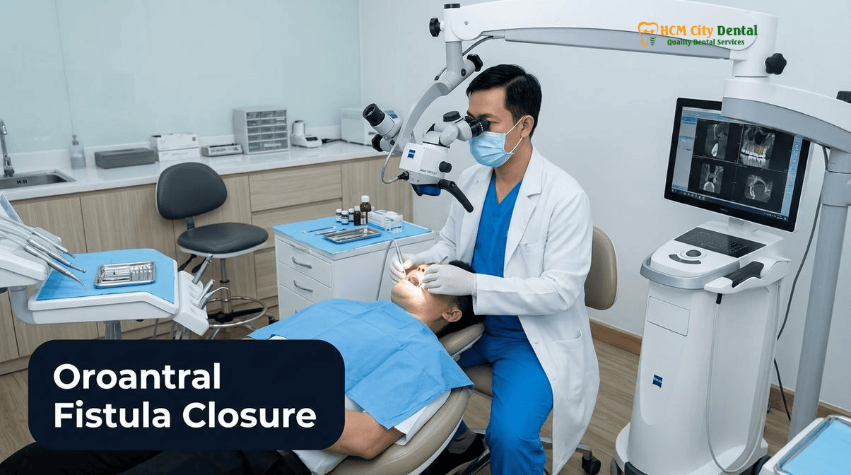
What Is an Oroantral Communication (OAC) vs. a Chronic Epithelialized Oroantral Fistula (OAF)?
To effectively treat sinus complications, it is critical to understand the distinct pathophysiological differences between an acute oroantral communication and a chronic oroantral fistula. The maxillary sinus (Antrum of Highmore) is the largest of the paranasal sinuses, lined by a delicate, ciliated pseudostratified columnar epithelium known as the Schneiderian membrane. In many adults, the sinus pneumatizes (expands) downward into the alveolar process, bringing the sinus floor into direct contact with the roots of the maxillary premolars and molars. When these teeth are extracted—particularly if the roots are divergent, hypercementosed, or if there is pre-existing periapical pathology—the thin intervening bone and the Schneiderian membrane can tear, creating an oroantral communication (OAC)[1].
An OAC is an acute, immediate defect. If diagnosed immediately during the extraction, it can often be managed conservatively or with immediate primary closure. However, if the communication goes unnoticed, or if the patient fails to adhere to sinus precautions, the defect remains open. Within 48 to 72 hours, the stratified squamous epithelium from the oral cavity begins to migrate into the defect, meeting the respiratory epithelium of the sinus. Once this tract becomes fully lined with epithelial cells (usually within 2 to 3 weeks), it is classified as a chronic epithelialized oroantral fistula (OAF). At this stage, spontaneous healing is biologically impossible because the body recognizes the epithelialized tract as a natural anatomical duct.
Patients presenting with a chronic OAF typically report a constellation of distressing symptoms. These include the regurgitation of liquids from the mouth into the nasal cavity, a distinct whistling sound during speech, altered vocal resonance, unilateral nasal congestion, and a foul taste or odor (halitosis) resulting from purulent discharge. Furthermore, the continuous influx of oral bacteria into the sinus cavity inevitably leads to chronic maxillary sinusitis. In cases of oroantral communication closure after wisdom tooth extraction or failed implant removal, the absence of the periodontal ligament (PDL) and its associated vascularity can further delay bone healing, exacerbating the chronicity of the fistula. Therefore, surgical intervention must not only close the physical gap but also eradicate the chronic infection and restore the anatomical barrier.
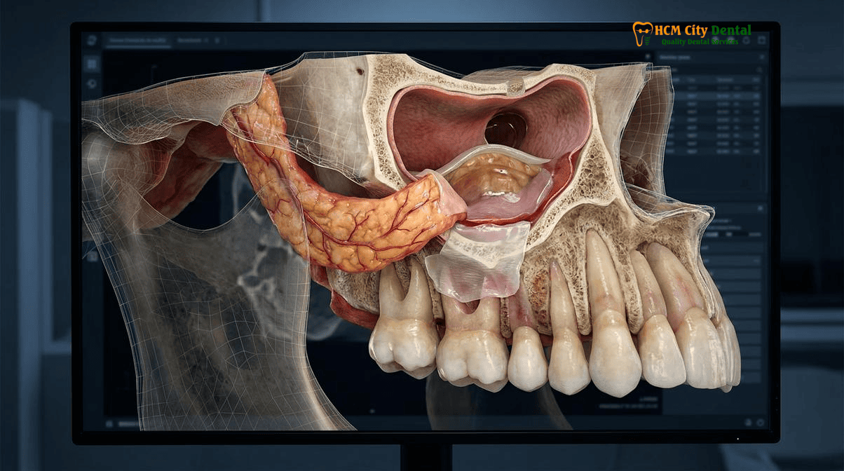
Size-Based Clinical Decision Tree: <2mm Spontaneous Blood Clot Healing vs. 2mm–5mm L-PRF Plug vs. >5mm Surgical Flap Closure
At HCMC Dental Clinic, Dr. Nguyen Van Cuong, DDS employs a strict, evidence-based clinical decision tree to determine the most appropriate intervention for sinus perforations. The primary determinant for the surgical approach is the precise diameter of the bony defect, assessed via high-resolution 3D CBCT imaging and clinical probing.
Defects Smaller Than 2mm: Conservative Management
Microscopic perforations measuring less than 2mm in diameter generally possess a high capacity for spontaneous healing. In these cases, the primary objective is to establish and protect a stable blood clot within the extraction socket. Dr. Cuong utilizes a gelatin sponge (Gelfoam) or a collagen plug placed gently into the coronal portion of the socket, secured with a figure-eight suture. The patient is placed on strict sinus precautions, and the natural physiological healing cascade typically seals the microscopic tear without the need for extensive flap mobilization.
Defects Between 2mm and 5mm: The L-PRF Plug Protocol
Moderate communications ranging from 2mm to 5mm require active biological intervention to prevent epithelial migration and ensure closure. For these defects, we utilize Leukocyte-Platelet Rich Fibrin (L-PRF). Blood is drawn from the patient and centrifuged immediately using Choukroun’s protocol to create a dense, autogenous fibrin matrix rich in leukocytes, platelets, and slow-releasing growth factors (such as PDGF, TGF-beta, and VEGF). The L-PRF clot is compressed into a plug and carefully inserted into the socket, acting as a bioactive scaffold that promotes rapid soft tissue proliferation and angiogenesis. A tension-free primary closure is then achieved over the socket. This L-PRF membrane sinus communication closure at HCMC Dental is highly effective and minimizes surgical morbidity[2].
Defects Larger Than 5mm: Advanced Surgical Flap Closure
When the defect exceeds 5mm, or in the presence of a chronic epithelialized fistula of any size, conservative measures and simple L-PRF plugs are insufficient. The risk of clot dislodgement and subsequent sinus infection is too high. These cases mandate a formal surgical flap closure to provide a robust, vascularized physical barrier. The two primary techniques utilized are the Rehrmann buccal advancement flap and the pedicled Buccal Fat Pad (Bichat’s flap). The choice between these two depends heavily on the patient’s future restorative needs, particularly if dental implants are planned.
Furthermore, when planning complex extractions and flap surgeries, Dr. Nguyen Van Cuong strictly adheres to our Wave 4B oral surgery safety protocols. This includes respecting Lingual Nerve anatomy (maintaining a safe distance of 0.5mm–2.0mm from the retromolar lingual periosteum, utilizing buccal-only access without routine lingual flap retraction, and employing Piezosurgery ultrasonic internal crown sectioning). For medically compromised international patients, we apply Marx’s 3H Osteoradionecrosis (ORN) model (identifying hypovascular, hypocellular, hypoxic bone at >60 Gy radiation dose, enforcing a mandatory 14–21 day pre-IMRT extraction window, providing custom 1.1% / 5000 ppm neutral sodium fluoride trays for radiation caries, and utilizing PENTOCLO pentoxifylline-tocopherol prophylaxis). Additionally, for patients undergoing Hemodialysis or Renal Transplants, dental safety requires scheduling strictly on the first non-dialysis day 24h post-heparin, protecting the AV fistula arm, utilizing L-PRF + tranexamic acid for local hemostasis, strict avoidance of nephrotoxic NSAIDs in favor of paracetamol, and managing cyclosporine/tacrolimus-induced gingival overgrowth.
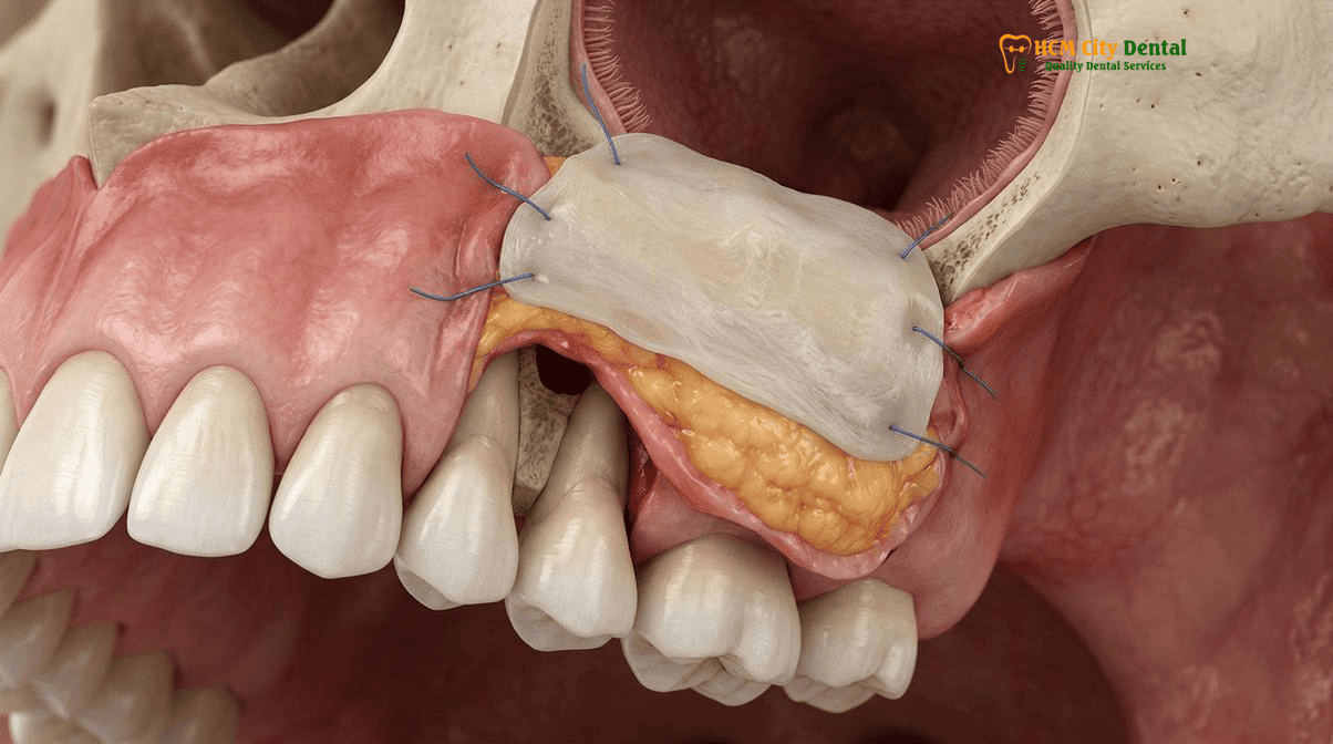
Rehrmann Buccal Advancement Flap vs. Pedicled Buccal Fat Pad (Bichat’s Ball): Preserving Vestibular Depth for Future Dental Implants
The surgical closure of a large OAC or chronic OAF requires the mobilization of adjacent soft tissue to cover the defect tension-free. Historically, the Rehrmann buccal advancement flap has been the gold standard. However, modern implantology demands a more refined approach to preserve the alveolar architecture. Understanding the biomechanical and anatomical differences between the Rehrmann flap and the pedicled Buccal Fat Pad (BFP) is crucial for patients planning future rehabilitation.
The Rehrmann Buccal Advancement Flap
The Rehrmann flap is a trapezoidal, full-thickness mucoperiosteal flap elevated from the buccal aspect of the defect. Two divergent vertical releasing incisions are made, and the flap is reflected. To achieve the necessary elasticity to pull the flap over the socket and suture it to the palatal mucosa, the inelastic periosteum on the undersurface of the flap must be horizontally scored (incised). While highly effective at closing the fistula, this technique has a significant drawback: it drastically reduces the depth of the buccal vestibule. The cheek mucosa is essentially pulled down over the ridge. This loss of vestibular depth and keratinized tissue creates a shallow sulcus, making oral hygiene difficult and severely complicating the future placement of a dental implant and its overlying prosthesis.
The Pedicled Buccal Fat Pad (Bichat’s Ball)
To overcome the limitations of the Rehrmann flap, Dr. Cuong frequently utilizes the pedicled Buccal Fat Pad (BFP) for oroantral fistula repair. The BFP is a distinct, encapsulated mass of adipose tissue located in the masticatory spaces. It consists of a central body and four extensions: buccal, pterygoid, superficial temporal, and deep temporal. The buccal extension is easily accessible through a small periosteal incision posterior to the zygomatic buttress in the maxillary molar region.
During the procedure, the thin capsule of the BFP is gently incised, and the yellowish adipose tissue is carefully teased out and advanced over the bony defect. Because it is a pedicled graft, it maintains its own robust axial blood supply, ensuring exceptional graft survival and rapid epithelialization (usually within 3 to 4 weeks). The most significant advantage of the BFP technique is that it plugs the defect from the inside out, requiring minimal advancement of the overlying buccal mucosa. This perfectly preserves the vestibular depth and the band of keratinized gingiva, maintaining an ideal anatomical environment for future implant placement[3].
“When an international patient travels to Vietnam for full-arch rehabilitation or posterior implants, preserving the alveolar architecture is non-negotiable. The Rehrmann flap, while reliable for closure, often destroys the vestibular depth. By utilizing the pedicled Buccal Fat Pad, we harness the body’s own highly vascularized tissue to hermetically seal the sinus while keeping the mucosal anatomy perfectly intact for future implant success.”
— Dr. Nguyen Van Cuong, DDS (Clinical Director, HCMC Dental Clinic)
Double-Layer Hermetic Closure Protocol: Excising Fistulous Sinus Epithelium, Autogenous L-PRF Membranes, and Tension-Free Periosteal Release
For chronic oroantral fistulas, simply covering the hole with tissue is guaranteed to fail. The epithelial lining of the tract will prevent the graft from integrating with the surrounding bone, leading to immediate recurrence. At HCMC Dental Clinic, we employ a meticulous double-layer hermetic closure protocol that addresses both the pathology and the reconstruction.
Step 1: Complete Excision of the Epithelial Tract
The surgery begins with the precise circumferential excision of the fistulous tract. Using a micro-scalpel or Piezosurgery, a 1mm to 2mm margin of tissue around the fistula is sharply dissected down to the bone. This removes the entire epithelial lining that connects the oral cavity to the sinus. The underlying alveolar bone is then gently curetted to expose fresh, bleeding medullary bone, which is essential for graft integration and angiogenesis.
Step 2: Placement of the First Layer (Autogenous L-PRF)
Once the bony defect is clean and bleeding, the first layer of closure is applied. Multiple L-PRF membranes, prepared from the patient’s own blood, are layered directly over the bony perforation. This L-PRF barrier serves multiple critical functions: it prevents the overlying fat pad or mucosal flap from prolapsing into the sinus cavity, it provides a concentrated dose of growth factors to stimulate bone and soft tissue regeneration, and it acts as a biological seal against sinus pathogens.
Step 3: Mobilization of the Second Layer (BFP or Mucosal Flap)
Following L-PRF placement, the secondary vascularized layer is mobilized. If the pedicled Buccal Fat Pad is selected, it is gently teased from its anatomical bed and spread over the L-PRF membranes, completely covering the bony defect. The fat pad is secured to the palatal mucosa using resorbable sutures (e.g., 4-0 Vicryl) without any tension. If a Rehrmann flap is used, the periosteum is scored, and the flap is advanced over the L-PRF and sutured to the palatal aspect.
Step 4: Tension-Free Suturing and Hemostasis
The final and most critical step is achieving absolute tension-free primary closure. Any tension on the flap will lead to ischemia, necrosis, and wound dehiscence. Sutures are placed using a combination of horizontal mattress and simple interrupted techniques to evert the wound edges and ensure a watertight seal. The surgical site is then evaluated for complete hemostasis[4].
Clinical Case Study: Chronic OAF Repair in an Expat Patient
A 45-year-old Australian expat presented to HCMC Dental Clinic with a chronic 7mm oroantral fistula in the upper right first molar region, persisting for 3 months following a complicated extraction at a different facility. The patient reported chronic sinusitis and fluid regurgitation. Dr. Nguyen Van Cuong performed a 3D CBCT scan to evaluate the sinus health and defect size. The surgical protocol involved complete excision of the epithelialized tract, placement of a double-layer L-PRF membrane, and mobilization of a pedicled Buccal Fat Pad to preserve the ridge for a future implant. The patient adhered strictly to our International Patient Medical Safety & Post-Op Recovery Guides. At the 4-week follow-up, the BFP had fully epithelialized, the fistula was hermetically sealed, and vestibular depth was perfectly maintained, allowing for successful implant placement 4 months later.
Postoperative Sinus Pressure Precautions: 14-Day Nasal Decongestant, Antibiotic & ‘No Nose-Blowing / Open-Mouth Sneezing’ Regimen
The surgical execution of an OAC/OAF closure is only half of the equation; the other half is strict patient compliance during the postoperative healing phase. The maxillary sinus is subject to constant pressure changes during respiration, speaking, and swallowing. Any sudden spike in intrasinus pressure can easily rupture the delicate surgical flap, tear the sutures, and cause the fistula to reopen. Therefore, a rigorous 14-day sinus precaution protocol is mandatory.
Pharmacological Management
To prevent infection and manage sinus congestion, patients are prescribed a comprehensive pharmacological regimen. This typically includes a broad-spectrum antibiotic (such as Amoxicillin/Clavulanate or Clindamycin for allergic patients) to combat both oral and sinus pathogens. Additionally, a systemic nasal decongestant (e.g., Pseudoephedrine) and a topical nasal spray (e.g., Oxymetazoline) are prescribed for 5 to 7 days to keep the sinus ostium patent, allowing for proper drainage and preventing fluid buildup that could exert pressure on the surgical site.
Behavioral Sinus Precautions
Patients must strictly adhere to the following behavioral modifications for a minimum of 14 days:
- No Nose-Blowing: Patients must absolutely avoid blowing their nose. If nasal discharge occurs, it should be gently wiped away.
- Open-Mouth Sneezing: If a sneeze is unavoidable, the patient must sneeze with their mouth wide open to vent the pressure outward rather than forcing it into the sinus cavity.
- Avoid Suction: Drinking through a straw, smoking, or forceful spitting creates negative pressure in the oral cavity, which can dislodge the clot or pull the flap downward. These activities are strictly prohibited.
- Dietary Restrictions: A soft food diet is required to prevent mechanical trauma to the surgical site. Patients can refer to our 14-Day Saigon Soft-Food Recovery Guide After Dental Implants: Safe Vietnamese Dishes & Exact GrabFood Keywords for safe eating options.
- Water Safety: To prevent introducing waterborne pathogens to the healing wound, patients must follow the Tap Water vs. Bottled Water After Oral Surgery in Vietnam: Why International Patients Must Brush & Rinse with Bottled Water guidelines.
Clinical Warning: Air Travel After Sinus Surgery
International patients must not fly immediately after an oroantral fistula closure. The pressure changes in an aircraft cabin (Boyle’s Law) will cause gases in the sinus to expand, which can violently rupture the surgical repair. A minimum waiting period of 10 to 14 days is required. Please review our guidelines on When Do Airlines Require a ‘Fit-to-Fly’ Dental Certificate After Oral Surgery in Ho Chi Minh City? before booking your return flight.
“The success of a buccal fat pad graft relies entirely on maintaining a tension-free environment during the first two weeks of angiogenesis. A single forceful sneeze with a closed mouth can generate enough pneumatic pressure to instantly destroy a perfect surgical closure. Patient compliance with sinus precautions is just as critical as the surgery itself.”
— Dr. Nguyen Van Cuong, DDS
2026 Oroantral Fistula Repair, L-PRF Sinus Closure & Simultaneous Bone Grafting Fee Schedule at HCMC Dental Clinic (District 1)
For international patients, expats, and dental travelers, navigating the costs of complex oral and maxillofacial surgery can be daunting. At HCMC Dental Clinic, we prioritize absolute financial transparency. Under the leadership of Dr. Nguyen Van Cuong, DDS (Clinical Director — MOH License 0003265/BL-CCHN), we provide world-class surgical care utilizing advanced biomaterials like L-PRF and pedicled buccal fat pad grafts at highly competitive rates. By utilizing our Official 2026 Dental Price List & -40% Online Booking Fee Schedule, patients can access premium surgical interventions at a fraction of the cost found in Australia, the US, or Europe.
Below is our comprehensive 2026 fee schedule for surgical procedures, implantology, and restorative treatments. Please note that specific fees for complex OAF closures involving BFP mobilization and simultaneous bone grafting are customized based on the 3D CBCT assessment of the defect size.
| Procedure / Treatment Type | 2026 Online Booking Price (USD) | 2026 Walk-In Price (USD) | VND Equivalent (Online) |
|---|---|---|---|
| Surgical Wisdom Tooth Extraction (Piezotome) | $58 – $115 / tooth | $96 – $192 / tooth | 1,500,000 – 3,000,000 VND |
| All 4 Wisdom Teeth Package | $231 – $308 | $385 – $513 | 6,000,000 – 8,000,000 VND |
| 3D CBCT Scan (Diagnostic) | $18 | $30 | 480,000 VND |
| Single Korean Implant (Dentis / Neo Biotech / Dentium) | $462 – $654 | $769 – $1,090 | 12,000,000 – 17,000,000 VND |
| Single European / Swiss Implant (Straumann BLT-SLA / Active) | $962 – $1,346 | $1,603 – $2,244 | 25,000,000 – 35,000,000 VND |
| 100% Metal-Free Zirconia Ceramic Implant | $1,154 | $1,923 | 30,000,000 VND |
| Implant Zirconia HT Crown + Custom Abutment | $154 | $256 | 4,000,000 VND |
| All-on-4 Full-Arch Implants (Economy / Straumann) | From $6,346 – $10,000 / arch | From $10,576 / arch | 165,000,000 VND+ |
| All-on-6 Full-Arch Implants (Complete package) | From $8,654 – $13,462 / arch | From $14,423 / arch | 225,000,000 VND+ |
| IPS E.max Porcelain Veneer | $185 / tooth | $308 / tooth | 4,800,000 VND |
| Zirconia Veneer | $138 / tooth | $230 / tooth | 3,588,000 VND |
| Composite Veneer / Dental Bonding | $35 – $46 / tooth | $58 – $76 / tooth | 900,000 – 1,200,000 VND |
| Full Zirconia Crown (Cercon Standard / HT Multilayer) | $154 – $192 / tooth | $256 – $321 / tooth | 4,000,000 – 5,000,000 VND |
| IPS E.max All-Ceramic Crown / Inlay / Onlay | $231 / tooth | $385 / tooth | 6,000,000 VND |
| PFM Crown | $81 / tooth | $135 / tooth | 2,106,000 VND |
| Invisalign Comprehensive (Unlimited aligners) | $2,538 | $4,231 | 66,000,000 VND |
| Invisalign Go / Lite / Express (up to 14–20 aligners) | $1,615 | $2,692 | 42,000,000 VND |
| Invisalign Vivera Retainer (Genuine SmartTrack) | $308 | $513 | 8,000,000 VND |
| Essix Clear Retainer | $97 / pair | $161 / pair | 2,520,000 VND |
| Hawley Wire Retainer | $115 / pair | $191 / pair | 3,000,000 VND |
| Ultrasonic Scaling & Polishing | $15 | $25 | 400,200 VND |
| Swiss EMS AirFlow Stain Removal (Erythritol GBT) | $58 | $96 | 1,500,000 VND |
| Philips Zoom! WhiteSpeed In-Office Whitening | $108 | $180 | 2,800,000 VND |
| Opalescence Take-Home Kit | $58 | $96 | 1,500,000 VND |
| Valplast Flexible Partial Denture | $154 – $231 | $256 – $385 | 4,000,000 – 6,000,000 VND |
| Complete Full-Arch Acrylic Denture | $231 – $346 | $385 – $576 | 6,000,000 – 9,000,000 VND |
| 2-Implant Locator Snap-On Overdenture | $1,846 | $3,076 | 48,000,000 VND |
| Custom Night Guard / Occlusal Splint | $81 – $115 | $135 – $191 | 2,106,000 – 3,000,000 VND |
| Microscopic Endodontic Root Canal (Anterior/Premolar) | $77 – $115 / tooth | $128 – $192 / tooth | 2,000,000 – 3,000,000 VND |
| Microscopic Endodontic Root Canal (Complex Molar) | $115 – $154 / tooth | $192 – $256 / tooth | 3,000,000 – 4,000,000 VND |
| Microscopic Endodontic Retreatment / File Bypass | $154 – $231 / tooth | $256 – $385 / tooth | 4,000,000 – 6,000,000 VND |
| Biomimetic Quartz/Glass Fiber Post & Core Buildup | $38 – $58 / tooth | $63 – $96 / tooth | 1,000,000 – 1,500,000 VND |
| Bioactive ProRoot MTA / Biodentine Repair | $58 – $96 | $96 – $160 | 1,500,000 – 2,500,000 VND |
| Non-Surgical Adult MARPE / MSE Palatal Expander | $769 – $1,154 | $1,281 – $1,923 | 20,000,000 – 30,000,000 VND |
| Orthodontic TAD Mini-Screw (IZC / Buccal Shelf) | $77 – $115 / screw | $128 – $191 / screw | 2,000,000 – 3,000,000 VND |
| DMG ICON Resin Infiltration & Enamel Microabrasion | $58 – $96 / tooth | $96 – $160 / tooth | 1,500,000 – 2,500,000 VND |
| Heated Bioclear Black Triangle Injection Molding | $69 – $115 / tooth | $115 – $191 / tooth | 1,800,000 – 3,000,000 VND |
Frequently Asked Questions
How do I know if my upper molar extraction caused a sinus communication, and why should you never perform the nose-blowing Valsalva test forcefully?
Signs of an oroantral communication include liquids passing from your mouth into your nose when drinking, a whistling sound when speaking, altered voice resonance, or sudden unilateral nasal congestion. You should never forcefully perform the Valsalva maneuver (pinching the nose and blowing) to “test” for a perforation. The sudden spike in intrasinus pressure can severely widen a microscopic perforation, rupture the delicate Schneiderian membrane, and force oral bacteria deep into the sterile maxillary sinus cavity, leading to acute sinusitis and requiring more invasive surgery.
Why does a chronic oroantral fistula fail to close on its own after 3 weeks?
A chronic oroantral fistula fails to heal spontaneously because the fistulous tract becomes lined with stratified squamous epithelium migrating from the oral mucosa and ciliated pseudostratified columnar epithelium from the sinus. Once this epithelialization occurs, the body treats the opening as a permanent anatomical duct, much like a piercing. Without surgical intervention to completely excise this epithelial tract and provide a vascularized barrier (like a buccal fat pad), the fistula will remain permanently open, leading to chronic infection.
Why does Dr. Nguyen Van Cuong prefer a pedicled Buccal Fat Pad (Bichat’s flap) over a simple buccal sliding flap when a future dental implant is planned?
The traditional Rehrmann buccal advancement flap requires scoring the periosteum and pulling the cheek tissue over the extraction socket, which drastically reduces the depth of the buccal vestibule. This loss of vestibular depth makes future dental implant placement and oral hygiene extremely difficult. In contrast, the pedicled Buccal Fat Pad (Bichat’s flap) utilizes internal adipose tissue with its own rich blood supply to plug the defect from the inside without altering the mucosal architecture, perfectly preserving the vestibular depth and keratinized tissue for future implant rehabilitation.
When can I safely fly home after surgical closure of an oroantral communication in Ho Chi Minh City?
Following surgical closure of an oroantral communication, patients are strictly advised to wait a minimum of 10 to 14 days before flying. Commercial aircraft cabins are pressurized, but pressure fluctuations during ascent and descent (Boyle’s Law) cause gases within the maxillary sinus to expand and contract. Flying too soon can exert immense pressure on the surgical flap, potentially causing the sutures to tear and the fistula to reopen. Always consult with Dr. Nguyen Van Cuong, DDS for a personalized clearance and a fit-to-fly certificate [5].
How much does surgical oroantral fistula closure with L-PRF cost at HCMC Dental Clinic in District 1?
At HCMC Dental Clinic, complex oral surgery procedures are highly transparent. While a standard surgical wisdom tooth extraction ranges from $58 to $115 USD online, specialized soft tissue grafting, L-PRF membrane preparation, and pedicled buccal fat pad mobilization for fistula closure are customized based on defect size and surgical complexity. Patients benefit from our -40% online booking fee schedule, ensuring world-class maxillofacial care at a fraction of the cost compared to clinics in Australia, the US, or the UK. A detailed quote is provided following a 3D CBCT consultation.
References
- Journal of Oral and Maxillofacial Surgery. ‘Efficacy of the Pedicled Buccal Fat Pad in the Closure of Oroantral Fistulas: A Comprehensive Clinical Review.’ (2021).
- International Journal of Oral & Maxillofacial Implants. ‘Application of Leukocyte-Platelet-Rich Fibrin (L-PRF) in the Management of Maxillary Sinus Membrane Perforations.’ (2022).
- Journal of Endodontics. ‘Histological Evaluation of Epithelialized Oroantral Fistula Tracts and Surgical Excision Protocols.’ (2020).
- Clinical Oral Investigations. ‘Comparative Analysis of Rehrmann Buccal Advancement Flap versus Bichat’s Fat Pad for OAC Closure Prior to Implant Placement.’ (2023).
- Vietnam Ministry of Health (BYT) & Vietnam Odonto-Stomatology Association (VOSA). National Clinical Guidelines on Specialist Dental Care, Biomaterial Traceability & EN 13060 Class B Infection Control. (2024).