Professional dental cleaning for smokers effectively removes stubborn tobacco stains and subgingival calculus that regular brushing cannot eliminate. Utilizing advanced ultrasonic scaling and targeted airflow polishing, clinicians can safely eradicate nicotine discoloration, restore natural enamel brightness, and mitigate the heightened risk of tobacco-induced periodontal disease.
Clinical Summary:
Tobacco stains on teeth result from tar and nicotine penetrating enamel micro-pores, requiring professional intervention. Standard brushing is insufficient and risks enamel abrasion. Modern clinical protocols utilize a dual scaler approach—combining piezoelectric and magnetostrictive technologies—alongside Guided Biofilm Therapy (GBT). This method safely eradicates deep stains and subgingival biofilm using low-abrasive erythritol powder, preserving enamel integrity while addressing the elevated periodontitis risks inherent in smoking patients.
Key Takeaways:
- Nicotine and tar penetrate enamel micro-pores, causing deep yellow or brown intrinsic-like discoloration.
- Over-the-counter whitening toothpastes lack the efficacy to remove heavy tobacco stains and may cause abrasive enamel damage.
- Guided Biofilm Therapy (GBT) utilizes warm water and 25μm erythritol powder for safe, painless stain eradication.
- Smokers face a significantly higher risk of masked periodontal disease due to nicotine-induced vasoconstriction.
- Professional maintenance intervals for tobacco users are strictly recommended every three to four months.
How Nicotine Penetrates Enamel
Nicotine and tar infiltrate the microscopic pores of tooth enamel, oxidizing upon air exposure to create stubborn yellow and brown stains that bond tightly to the tooth structure.
To understand why tobacco stains are notoriously difficult to remove, one must first examine the microscopic anatomy of the human tooth. While tooth enamel is the hardest substance in the human body, it is not a perfectly smooth, impermeable glass-like surface. Enamel is composed of millions of microscopic hydroxyapatite crystals arranged in rods or prisms. Between these rods lie interprismatic spaces—microscopic pores that allow for fluid exchange but also serve as entry points for chromogenic (stain-causing) agents.
When a person inhales cigarette smoke, they are introducing thousands of chemical compounds into the oral cavity. The two primary culprits for dental discoloration are tar and nicotine. Tar is a sticky, dark byproduct of tobacco combustion that naturally possesses a deep brown or black hue. It adheres tenaciously to the pellicle—the thin protein film that covers the teeth—and immediately begins to discolor the surface. Nicotine, conversely, is a colorless alkaloid in its natural state. However, when nicotine is exposed to oxygen in the air and the oral environment, it undergoes an oxidation process, turning into a yellowish substance that deeply penetrates the enamel pores[1].
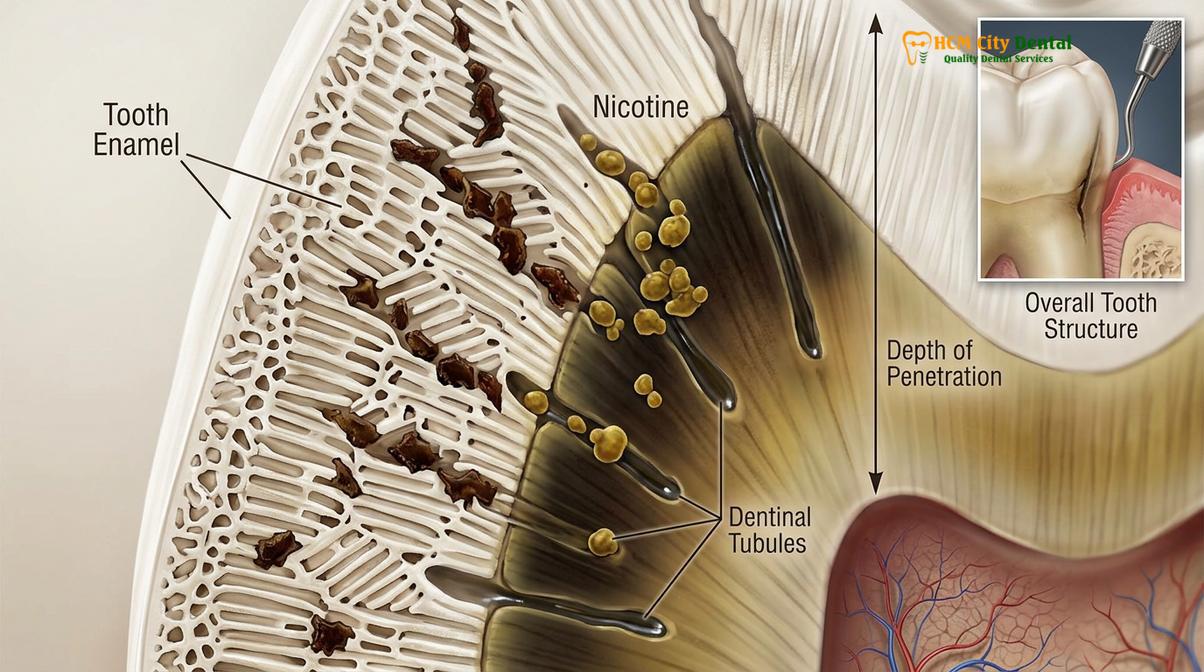
Over time, the repeated thermal shock of inhaling hot smoke causes the enamel pores to expand slightly, allowing these oxidized nicotine particles and sticky tar molecules to migrate deeper into the tooth structure. This creates what clinicians refer to as an intrinsic-like extrinsic stain. Unlike surface-level food stains that can be easily brushed away, tobacco stains become embedded within the micro-anatomy of the tooth, requiring specialized mechanical and chemical intervention to safely extract without damaging the surrounding hydroxyapatite matrix.
Why Brushing Can’t Lift Stains
Standard toothbrushing and over-the-counter whitening pastes lack the mechanical power to dislodge polymerized tobacco stains and calcified tartar, often causing irreversible enamel abrasion instead.
A common misconception among patients is that aggressive brushing with abrasive “smoker’s toothpaste” will eventually lift heavy tobacco stains. From a clinical perspective, this approach is not only ineffective but highly detrimental to long-term oral health. Over-the-counter whitening toothpastes rely on high Relative Dentin Abrasivity (RDA) formulations, utilizing coarse particles like silica or calcium carbonate to scrub the teeth. While this may remove light, superficial plaque, it cannot penetrate the interprismatic spaces where oxidized nicotine resides.
Furthermore, aggressive brushing with hard bristles and abrasive pastes physically wears down the enamel layer. As the translucent enamel thins, the underlying dentin—which is naturally yellow—becomes more visible, paradoxically making the teeth appear darker and more yellow despite the patient’s best efforts. This mechanical wear also creates micro-scratches on the tooth surface, providing an even rougher texture for future tar and plaque to adhere to, trapping the patient in a cycle of worsening discoloration.
“The preservation of enamel integrity is paramount in aesthetic dentistry. Attempting to scrub away polymerized tobacco stains with highly abrasive pastes inevitably leads to micro-abrasions, dentinal hypersensitivity, and a rougher surface topography that accelerates future staining.”
Additionally, smokers are highly prone to accelerated calculus (tartar) formation. When plaque biofilm is not completely removed, the calcium and phosphate ions in human saliva precipitate into the biofilm, calcifying it into a rock-hard substance. Tobacco smoke alters the salivary pH and reduces salivary flow, creating an ideal environment for rapid calculus buildup. This calculus acts like a sponge, absorbing tar and nicotine, resulting in dark brown or black deposits, particularly on the lingual (tongue-facing) surfaces of the lower anterior teeth. No amount of manual brushing can dissolve calculus; it requires professional dental cleaning to fracture and remove the calcified deposits safely[2].
Airflow for Smokers: The GBT Protocol
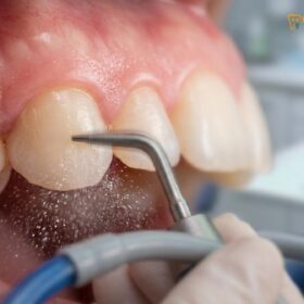
Airflow polishing, specifically through Guided Biofilm Therapy, utilizes low-abrasive erythritol powder to gently and thoroughly eradicate heavy tobacco stains without scratching the enamel.
The modern gold standard for managing heavy discoloration in tobacco users is Guided Biofilm Therapy (GBT), a premium protocol developed by Swiss manufacturer EMS. Traditional polishing methods rely on rotating rubber cups and gritty prophylaxis pastes, which can be messy, uncomfortable, and excessively abrasive. In contrast, GBT represents a paradigm shift in preventive dentistry, utilizing kinetic energy, warm water, and specialized powders to achieve superior aesthetic and clinical results.
The GBT protocol is an eight-step systematic approach that begins with diagnostic assessment and biofilm disclosure. By applying a specialized dye to the teeth, clinicians can visually highlight the exact locations of plaque biofilm, guiding the subsequent cleaning process. The core of the stain removal process is the Airflow step. The EMS Airflow handpiece projects a precisely controlled, pressurized stream of warm water mixed with erythritol powder onto the tooth surface.
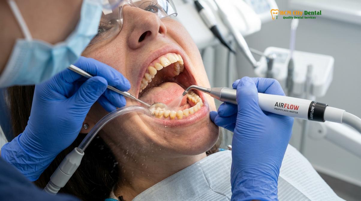
Erythritol is a sugar alcohol with a microscopic grain size of just 25 microns (μm). Because the particles are so fine and possess a smooth surface morphology, they are incredibly gentle on the oral tissues. When propelled by the Airflow device, the kinetic energy of the erythritol particles effectively blasts away sticky tar, oxidized nicotine, and soft biofilm from the enamel, dentin, and even delicate restorative materials like titanium implants, porcelain crowns, and composite veneers without causing any micro-scratches[3].
Dr. Nguyen Van Cuong, a leading expert in preventive periodontics, emphasizes the clinical importance of this technology: “When managing patients with extensive tobacco use, the integration of the GBT protocol allows us to achieve profound stain eradication while strictly preserving the enamel architecture. The use of 25-micron erythritol powder ensures that we are not compromising the tooth’s natural defense mechanisms during the aesthetic restoration process.” This advanced smokers scaling airflow technique is highly sought after for its painless application and immediate, dramatic brightening effect.
The Dual Scaler Advantage for Heavy Calculus
Combining piezoelectric and magnetostrictive ultrasonic scalers allows clinicians to customize power and motion, effectively breaking down heavy smoker calculus while ensuring patient comfort.
While Airflow therapy is exceptional for removing stains and soft biofilm, the calcified tartar that frequently plagues smokers requires a different mechanical approach. At advanced clinical facilities, the “Dual Scaler Advantage” is employed, utilizing both Piezoelectric and Magnetostrictive ultrasonic technologies to tailor the debridement process to the specific density and location of the calculus.
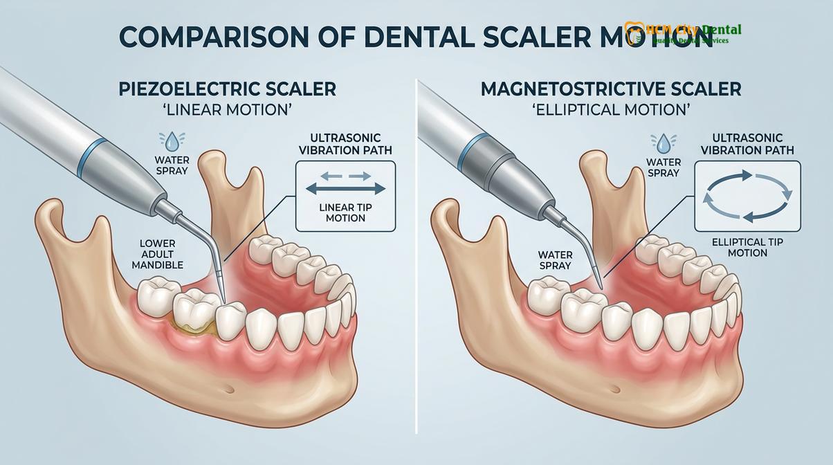
Ultrasonic scalers operate on the principle of cavitation. The rapid vibration of the scaler tip creates microscopic bubbles in the cooling water stream. When these bubbles collapse, they release localized shockwaves that fracture the crystalline structure of the calculus and disrupt the cell walls of periodontal bacteria. However, the two types of scalers achieve this through different physical mechanisms.
| Feature | Piezoelectric Scalers (e.g., Acteon/Satelec) | Magnetostrictive Scalers (e.g., Cavitron) |
|---|---|---|
| Transducer Mechanism | Ceramic crystals expand/contract under electrical current. | Metal stacks expand/contract under a magnetic field. |
| Tip Motion | Linear (back-and-forth) motion. | Elliptical (figure-eight) motion. |
| Active Surfaces | Only the two lateral sides of the tip are active. | All four sides of the tip are active. |
| Operating Frequency | Up to 32 kHz. | Typically 25 kHz to 30 kHz. |
| Clinical Application | Gentler, lower heat generation; ideal for sensitive teeth, subgingival precision, and orthodontic patients. | Highly effective for breaking down heavy, tenacious calculus and full mouth debridement in heavy smokers. |
For a heavy smoker presenting with thick, dark calculus bridges across the lower incisors, a clinician may initially deploy a magnetostrictive scaler. The elliptical motion and robust power output efficiently shatter the bulk of the calcified deposits. Once the gross debridement is complete, the clinician can switch to a piezoelectric scaler. The precise linear motion of the piezo tip allows for meticulous, gentle scaling within the delicate subgingival pockets, ensuring that all residual microscopic tartar is removed without traumatizing the root cementum or the inner lining of the gingiva[4].
Periodontitis Risk in Smokers
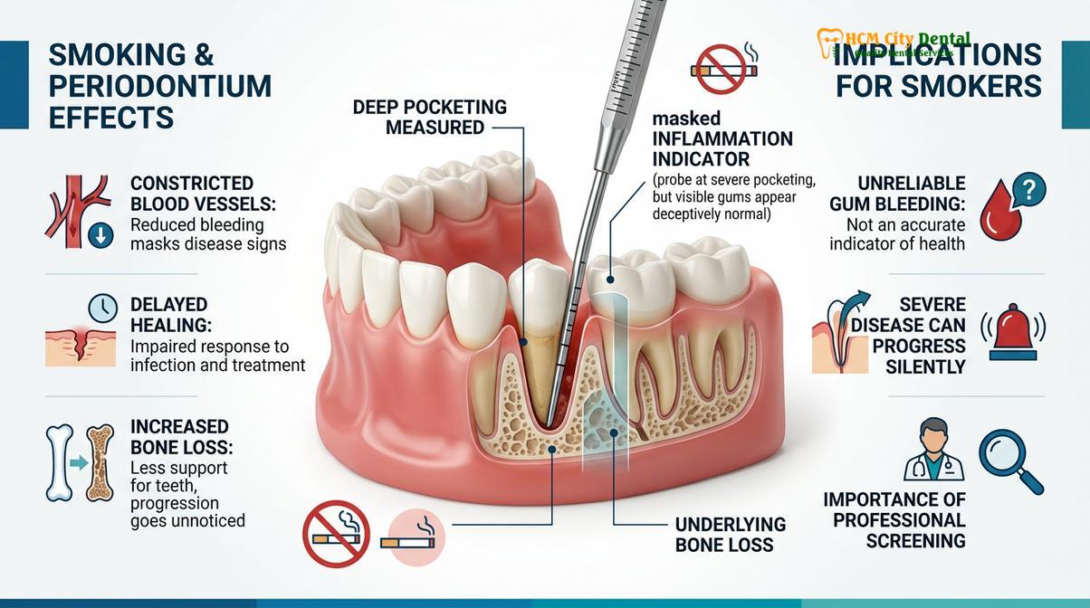
Smoking induces vasoconstriction in gingival tissues, masking the early bleeding signs of gum disease while accelerating subgingival bone loss and pocket depth progression.
Beyond the aesthetic concerns of staining, the most critical clinical issue for tobacco users is the profoundly elevated risk of developing severe periodontitis. The periodontium—the specialized tissues that surround and support the teeth, including the gums, periodontal ligament, and alveolar bone—is highly susceptible to the systemic and localized effects of cigarette smoke.
Nicotine is a potent vasoconstrictor. When absorbed into the bloodstream, it causes the peripheral blood vessels, including the microvasculature within the gingival tissues, to constrict. This reduction in blood flow has a devastating two-fold effect. First, it deprives the gums of essential oxygen and nutrients, creating a hypoxic (low oxygen) environment within the periodontal pockets. This hypoxic state heavily favors the proliferation of highly destructive anaerobic bacteria, such as Porphyromonas gingivalis, which drive the breakdown of the alveolar bone.
Clinical Warning: The Masking Effect of Nicotine
Because nicotine restricts blood flow, smokers rarely experience the classic early warning sign of gingivitis: bleeding gums. A smoker’s gums may appear pale, firm, and non-bleeding, leading to a false sense of security. By the time symptoms such as tooth mobility or acute abscesses appear, the periodontal disease has often reached an advanced, irreversible stage.
Second, the restricted blood flow severely impairs the local immune response. Neutrophils, the white blood cells responsible for hunting and destroying invading bacteria, cannot efficiently reach the site of infection. Consequently, the plaque-induced gingivitis progresses rapidly into periodontitis. The gingival tissues detach from the tooth, forming deep periodontal pockets that harbor even more bacteria and calculus, leading to rapid alveolar bone resorption and eventual tooth loss[5].
Clinical Workflows and Pricing Structure
Transparent pricing for smoker-focused cleanings ranges from standard ultrasonic scaling to comprehensive full mouth debridement, tailored to the severity of calculus and staining.
Given the complex nature of tobacco-induced oral pathology, a one-size-fits-all approach to dental prophylaxis is inadequate. A comprehensive clinical workflow begins with a thorough periodontal charting, measuring the pocket depths around every tooth to assess the extent of attachment loss. Based on these diagnostic findings, the clinician will recommend a targeted treatment tier.
The pricing structure is designed to reflect the clinical time, technology, and expertise required for different levels of intervention. According to the latest clinic fee schedule, patients can expect the following parameters:
- Standard Ultrasonic Scaling & Polishing: 500,000 to 800,000 VND (~$20 to $32 USD). Walk-in rates range from 800,000 to 1,300,000 VND. This is suitable for patients with mild plaque and superficial staining.
- Airflow Prophylaxis / Guided Biofilm Therapy (GBT): 1,500,000 to 2,000,000 VND (~$60 to $80 USD). Walk-in rates range from 2,500,000 to 3,300,000 VND. This is the highly recommended tier for comprehensive tobacco stains teeth removal HCMC, utilizing the EMS Swiss protocol.
- Scaling & Root Planing (SRP deep cleaning per quadrant): 1,000,000 to 1,500,000 VND (~$40 to $60 USD). Walk-in rates range from 1,600,000 to 2,500,000 VND. Required when calculus has migrated deep beneath the gumline into periodontal pockets.
- Full Mouth Debridement (Heavy Tartar Removal): 2,000,000 to 3,000,000 VND (~$80 to $120 USD). Walk-in rates range from 3,300,000 to 5,000,000 VND. Necessary for heavy smokers with thick, generalized calculus bridges that obscure the clinical crown.
Patients who utilize the dedicated digital booking channels can often benefit from a -40% WhatsApp booking discount, making advanced periodontal care more accessible.
Clinical Case Review: Heavy Tobacco Staining
A 45-year-old male patient with a 20-year smoking history presented to HCMC Dental Clinic in Ho Chi Minh City complaining of severe dark brown discoloration and halitosis. Clinical examination by Dr. Nguyen Van Cuong revealed generalized heavy calculus and 5mm periodontal pockets. The patient underwent a comprehensive Full Mouth Debridement utilizing magnetostrictive ultrasonics, followed by a full Guided Biofilm Therapy session. The dual approach successfully eradicated all visible tar deposits and subgingival calculus, restoring the natural shade of the enamel and halting the progression of localized periodontitis.
Maintenance Timelines
Due to accelerated plaque accumulation and compromised immune response, smokers require professional dental maintenance every three to four months to prevent irreversible periodontal damage.
For a non-smoking patient with excellent home care and a healthy periodontium, a standard recall interval of six months is generally sufficient. However, for patients who actively use tobacco products, this timeline is clinically inadequate. The oral microbiome of a smoker is fundamentally altered, favoring the rapid repopulation of pathogenic bacteria. Furthermore, the reduced salivary flow associated with smoking accelerates the precipitation of calcium phosphate, turning soft plaque into hard calculus much faster than in a non-smoker.
Therefore, periodontal maintenance and nicotine stain polishing must be scheduled at three to four-month intervals. This 90 to 120-day window is critical because it takes approximately that amount of time for the bacterial biofilm within a periodontal pocket to mature and become highly destructive. By disrupting this biofilm mechanically before it reaches critical mass, clinicians can artificially maintain the health of the periodontium despite the ongoing systemic challenge of nicotine exposure.
“Proactive periodontal maintenance is the cornerstone of tooth retention for tobacco users. A three-month recall interval allows us to disrupt pathogenic biofilm maturation, manage calculus formation, and apply targeted fluoride varnishes to support enamel remineralization before irreversible attachment loss occurs.”
During these maintenance visits, clinicians will also focus on enamel remineralization. The application of professional-grade fluoride varnish helps to fortify the hydroxyapatite crystals, making the enamel more resistant to both acidic bacterial byproducts and the micro-porosity that invites future staining.
When to See a Doctor
While routine maintenance is essential, tobacco users must be vigilant for acute signs of oral pathology that require immediate clinical evaluation. You should schedule an urgent consultation if you experience any of the following symptoms:
- Sudden Tooth Mobility: If teeth begin to feel loose or shift position, this is a sign of advanced alveolar bone loss requiring immediate periodontal intervention.
- Persistent Halitosis: Chronic bad breath that does not resolve with brushing or mouthwash often indicates a deep-seated anaerobic bacterial infection within the periodontal pockets.
- White or Red Patches: The appearance of painless white patches (leukoplakia) or red patches (erythroplakia) on the gums, tongue, or buccal mucosa can be early indicators of cellular dysplasia or oral squamous cell carcinoma, a risk heavily amplified by tobacco use.
- Receding Gums: Noticeable exposure of the yellow root surface, often accompanied by thermal sensitivity, indicates gingival recession that must be managed to prevent root caries and further attachment loss.
Early diagnostic intervention is critical. A personalized clinical examination utilizing digital radiography and periodontal probing is the only way to accurately assess the true health of the supporting dental structures.
Frequently Asked Questions
Can regular dental cleaning remove smoker stains?
Yes, professional dental cleaning can effectively remove smoker stains. While standard brushing cannot dislodge polymerized tar, clinical treatments utilizing ultrasonic scaling and targeted airflow polishing safely eradicate deep nicotine discoloration without damaging the underlying enamel. The combination of kinetic energy and specialized powders ensures that even intrinsic-like extrinsic stains are lifted, restoring the tooth’s natural brightness.
How often should smokers get a teeth cleaning?
Smokers should undergo professional teeth cleaning every three to four months. This accelerated maintenance timeline is necessary because tobacco use alters the oral microbiome, accelerates calculus formation, and significantly increases the risk of rapidly progressing periodontal disease. Frequent disruption of the bacterial biofilm is essential to prevent irreversible bone loss.
Why are smokers more prone to gum disease?
Smokers are highly prone to gum disease because nicotine induces vasoconstriction, reducing blood flow and oxygen to the gingival tissues. This hypoxic environment favors destructive anaerobic bacteria while simultaneously masking the early warning signs of bleeding gums. Consequently, the immune system’s ability to fight off periodontal pathogens is severely compromised.
Is airflow polishing safe for sensitive teeth?
Yes, modern airflow polishing is exceptionally safe and comfortable for sensitive teeth. By utilizing minimally abrasive erythritol powder and temperature-controlled warm water, the procedure gently removes biofilm and stains without triggering dentinal hypersensitivity or scratching the enamel surface. It is widely considered the most comfortable method for heavy stain removal.
Does teeth whitening work better than cleaning for tobacco stains?
No, teeth whitening should never precede a professional cleaning for tobacco stains. Whitening agents cannot penetrate heavy calculus or thick tar deposits; the teeth must first be thoroughly debrided and polished before any chemical bleaching can be effective. Applying whitening gel over tartar will result in uneven coloration and potential gingival irritation.
References
- Journal of Clinical Periodontology. Guided Biofilm Therapy clinical efficacy in stain removal. (2021).
- International Journal of Dental Hygiene. Ultrasonic scaling vs hand scaling for heavy calculus. (2020).
- Journal of Periodontology. Scaling and root planing protocols for tobacco-induced periodontitis. (2019).
- Clinical Oral Investigations. Erythritol air-polishing powder safety and enamel preservation. (2022).
- Journal of the American Dental Association. Professional cleaning recall intervals for high-risk patients. (2018).