Pediatric bruxism in Vietnam is a prevalent condition where children involuntarily grind or clench their teeth, often linked to airway obstruction, sleep disturbances, or normal dental development. While many outgrow it, persistent grinding requires clinical evaluation to prevent severe enamel wear and underlying respiratory complications.
Clinical Summary:
Pediatric bruxism is a complex, multifactorial sleep-related movement disorder characterized by the involuntary clenching and grinding of teeth during childhood. Current clinical evidence indicates that child teeth grinding is rarely an isolated mechanical habit; rather, it is frequently a neurological response to upper airway resistance, particularly enlarged tonsils and adenoids, or a natural physiological adaptation during the transition from primary to permanent dentition. Diagnostic protocols prioritize comprehensive pediatric airway screening to differentiate between benign developmental grinding and pathological bruxism linked to sleep-disordered breathing. Treatment strategies in growing children strictly avoid rigid occlusal splints that may impede craniofacial growth, favoring interceptive orthodontics, medical management of airway obstructions, and the cautious, monitored use of a soft pediatric mouthguard when severe dental attrition is present.
Key Takeaways:
- Pediatric bruxism affects a significant percentage of children globally, with prevalence rates ranging widely based on diagnostic criteria and age groups.
- Enlarged adenoids and tonsils are primary anatomical triggers, causing micro-arousals during sleep that manifest as involuntary jaw grinding.
- Mild to moderate tooth wear on primary (baby) teeth is often considered a normal physiological process during dental development.
- Hard acrylic nightguards are generally contraindicated for growing children due to the risk of restricting natural maxillary and mandibular growth.
- Clinical management focuses on identifying the root cause through airway screening rather than merely addressing the dental symptoms.
Why Do Young Children Grind Their Teeth?
Young children grind their teeth due to a complex interplay of central nervous system arousals, psychological stress, and physiological changes during dental development. It is primarily a sleep-related movement disorder rather than a simple mechanical habit.
The phenomenon of child teeth grinding, clinically referred to as pediatric bruxism, is a multifaceted condition that has garnered significant attention in modern pediatric dentistry and sleep medicine. Historically viewed as a simple behavioral quirk or a response to localized dental interferences, contemporary medical literature now classifies sleep bruxism as a centrally mediated sleep-related movement disorder. This paradigm shift is crucial for understanding why young children exhibit rhythmic masticatory muscle activity (RMMA) during their sleep cycles. The etiology is not singular; rather, it is a convergence of neurobiological, anatomical, and psychological factors that trigger the involuntary contraction of the masseter and temporalis muscles.[1]
At the core of pediatric sleep bruxism is the architecture of the child’s sleep itself. Research indicates that bruxism episodes predominantly occur during the transition from deeper stages of non-rapid eye movement (NREM) sleep to lighter sleep stages, or during micro-arousals. These micro-arousals are brief, often imperceptible awakenings characterized by a sudden increase in autonomic sympathetic nervous system activity. During these transient spikes in sympathetic tone, there is a measurable increase in heart rate, respiratory rate, and brain cortical activity, which culminates in the forceful, rhythmic contraction of the jaw muscles. The central nervous system, particularly the neurotransmitter pathways involving dopamine and gamma-aminobutyric acid (GABA), plays a significant regulatory role in modulating these motor responses.[2]
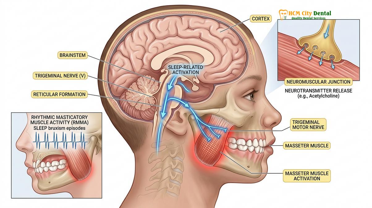
Psychological factors also contribute significantly to the prevalence of awake and sleep bruxism in the pediatric population. Children, much like adults, process emotional stress, anxiety, and environmental changes through somatic pathways. High-stress environments, academic pressures, family dynamics, or even excessive screen time before bed can elevate a child’s baseline anxiety levels, leading to increased muscle tension and nocturnal grinding. Furthermore, clinical studies have established a strong correlation between pediatric bruxism and neurodevelopmental conditions such as Attention Deficit Hyperactivity Disorder (ADHD). Children with hyperkinetic disorders frequently exhibit a higher incidence of parasomnias, including sleep talking, restlessness, and bruxism, suggesting a shared neurobiological vulnerability in motor control and sleep regulation.[2]
“Pediatric bruxism should no longer be viewed merely as a dental issue of tooth wear. It is a vital clinical biomarker—a neurological window into the child’s sleep quality, autonomic nervous system stability, and overall airway health.”
From a clinical perspective, Dr. Nguyen Van Cuong emphasizes that when parents report the unmistakable sound of child teeth grinding echoing from their child’s bedroom, the diagnostic focus must immediately broaden beyond the oral cavity. The sound itself—often described as a harsh, grating noise—is the mechanical result of the upper and lower teeth sliding against each other under immense muscular force. However, the root cause lies deep within the central nervous system’s attempt to regulate the child’s physiological state during sleep. Understanding this complex etiology is the first step in determining whether the bruxism is a transient developmental phase or a symptom of a more systemic health issue requiring intervention.
Airway Issues and Enlarged Tonsils: Primary Triggers in Toddlers
Enlarged tonsils and adenoids frequently obstruct a toddler’s upper airway during sleep, triggering micro-arousals and involuntary jaw movements as the brain attempts to reopen the respiratory passage.
One of the most critical and clinically actionable discoveries in pediatric dental medicine is the profound link between sleep-disordered breathing (SDB) and pediatric bruxism. In toddlers and young children, the anatomical structures of the upper airway—specifically the tonsils and adenoids (part of Waldeyer’s ring)—are highly active lymphoid tissues that frequently undergo hypertrophy (enlargement) in response to recurrent respiratory infections, allergies, or environmental irritants. When these tissues become excessively enlarged, they physically encroach upon the pharyngeal space, significantly increasing airway resistance and predisposing the child to obstructive sleep apnea (OSA) or upper airway resistance syndrome (UARS).[3]
The physiological cascade that links an obstructed airway to teeth grinding is a remarkable survival mechanism orchestrated by the central nervous system. When a child with adenotonsillar hypertrophy falls asleep, the loss of waking muscle tone causes the soft tissues of the throat to collapse further. As the airway narrows, the child experiences transient episodes of hypoxia (decreased oxygen) and hypercapnia (increased carbon dioxide). The brain detects this impending respiratory compromise and triggers an emergency micro-arousal. This arousal is accompanied by a surge of sympathetic adrenaline, which instantly activates the masticatory muscles. The resulting bruxism serves a mechanical purpose: by forcefully clenching and sliding the mandible forward, the child inadvertently pulls the tongue base away from the posterior pharyngeal wall, temporarily reopening the airway to allow a restorative breath.[4]
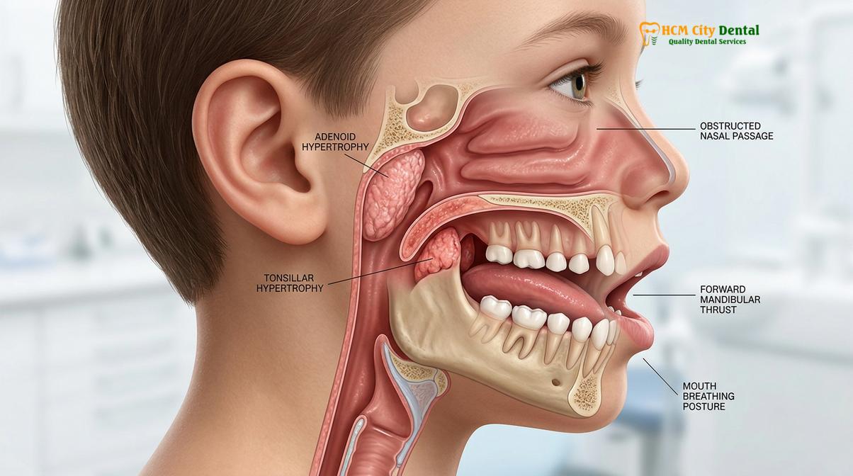
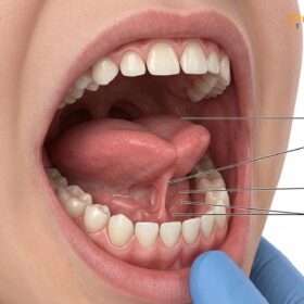
This repetitive cycle of obstruction, arousal, and grinding can occur dozens of times per hour, severely fragmenting the child’s sleep architecture. Consequently, children suffering from airway-induced bruxism rarely achieve the deep, restorative stages of sleep necessary for optimal cognitive and physical development. They often present with a constellation of secondary symptoms, including chronic mouth breathing, venous pooling under the eyes (allergic shiners), daytime hyperactivity, poor concentration, and morning headaches. Chronic mouth breathing, in particular, alters the resting posture of the tongue, dropping it to the floor of the mouth rather than resting against the palate. This lack of palatal support can lead to a narrow, V-shaped maxillary arch, further exacerbating the airway constriction and perpetuating the cycle of bruxism.[4]
| Clinical Indicator | Airway-Induced (Pathological) Bruxism | Physiological (Developmental) Bruxism |
|---|---|---|
| Breathing Pattern | Chronic mouth breathing, loud snoring, gasping | Quiet, consistent nasal breathing during sleep |
| Sleep Quality | Restless sleep, frequent waking, enuresis (bedwetting) | Deep, uninterrupted sleep cycles |
| Daytime Behavior | Hyperactivity, chronic fatigue, poor concentration | Normal energy levels, alert and focused |
| Anatomical Signs | Enlarged tonsils, narrow palate, venous pooling under eyes | Normal tonsillar size, broad developing dental arches |
| Duration | Persistent, worsens with respiratory illnesses | Transient, correlates with periods of tooth eruption |
Given this intricate relationship, a thorough pediatric airway screening is an indispensable component of the diagnostic process for any child presenting with severe nocturnal grinding. Dental professionals must look beyond the teeth to evaluate the patency of the nasal passages, the size of the tonsils, and the overall craniofacial development. In many cases, addressing the root cause through medical collaboration—such as an evaluation by an Ear, Nose, and Throat (ENT) specialist for potential adenotonsillectomy—can lead to a dramatic and spontaneous cessation of the bruxism, highlighting the importance of a multidisciplinary approach to pediatric health.
When is Pediatric Bruxism Considered Normal Development?
Pediatric bruxism is often considered a normal physiological phase during the eruption of primary and permanent teeth, helping children adapt to new bite relationships before naturally subsiding.
While the association between bruxism and airway pathology is critical, it is equally important for parents and clinicians to recognize that not all child teeth grinding is indicative of a disease process. In fact, a significant proportion of pediatric bruxism is classified as physiological or developmental. During the dynamic years of early childhood, the oral cavity undergoes continuous and rapid transformation. The eruption of the primary (deciduous) dentition, followed by the complex transition into the mixed dentition phase, creates a constantly shifting occlusal landscape. As new teeth emerge through the gingiva, they introduce novel proprioceptive feedback to the central nervous system via the periodontal ligaments.[5]
Physiological bruxism is widely theorized to be the neuromuscular system’s method of adapting to these changes. By grinding the newly erupted teeth against their antagonists, the child is essentially “test-driving” their developing bite, establishing stable occlusal contacts, and stimulating the growth of the alveolar bone. This developmental grinding is particularly common around the ages of three to four, when the primary dentition is complete, and again between the ages of six and nine, during the eruption of the first permanent molars and the exfoliation of the primary incisors. During these periods, the central nervous system is actively mapping the spatial relationship of the maxilla and mandible, and transient bruxism serves as a functional calibration tool.[5]
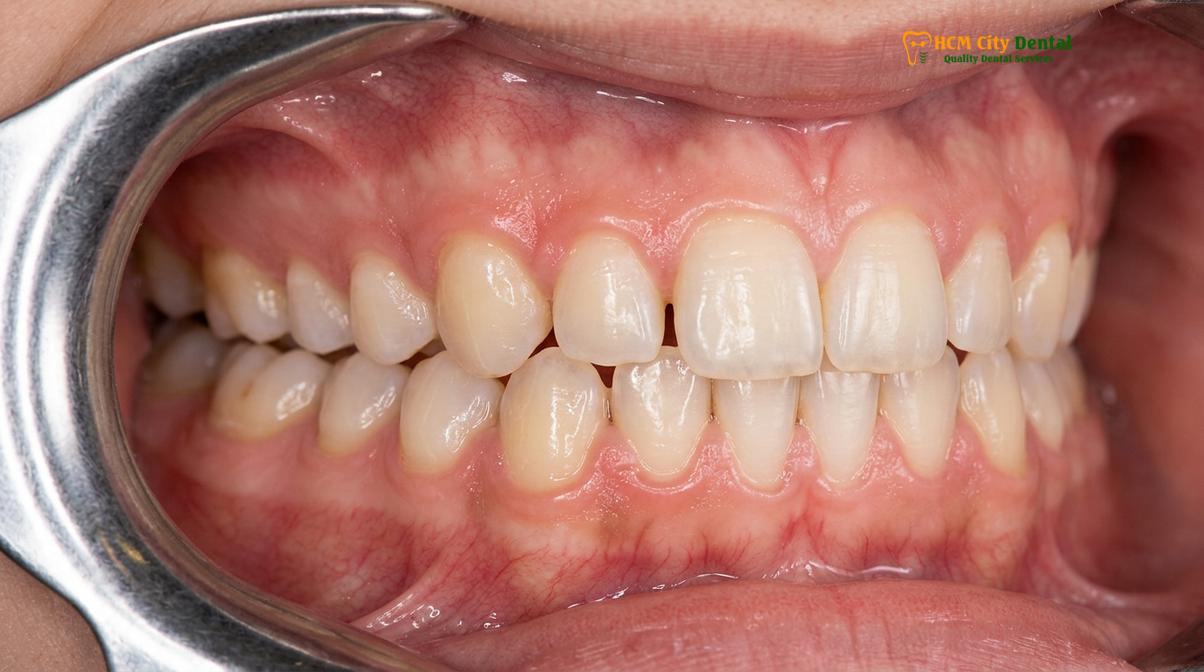
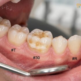
Clinically, the manifestation of physiological bruxism is often visible as mild to moderate wear facets on the incisal edges of the primary anterior teeth and the occlusal surfaces of the primary molars. Because the enamel of primary teeth is significantly thinner and less mineralized than that of permanent teeth, it wears down much more rapidly. While this flattening of the baby teeth can appear alarming to parents, it is generally a self-limiting process that does not require aggressive intervention. The primary teeth are designed to be temporary, and their gradual attrition rarely leads to pulpal exposure or significant dental pain in a healthy, developing child.
Clinical Case Review: Physiological Bruxism
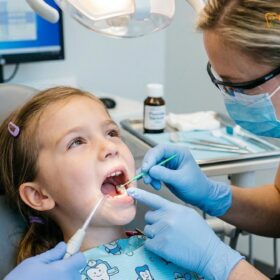
A 6-year-old patient visited HCMC Dental Clinic in Ho Chi Minh City accompanied by parents who were highly concerned about the audible grinding sounds occurring every night. The parents noted that the child’s front baby teeth appeared significantly flattened. Upon comprehensive examination, Dr. Nguyen Van Cuong confirmed that the child exhibited excellent nasal breathing, no signs of tonsillar hypertrophy, and normal sleep patterns. The wear facets were confined entirely to the primary dentition, which was actively preparing to exfoliate. The diagnosis was physiological bruxism related to the eruption of the first permanent molars. The clinical recommendation was active observation without the need for a protective splint. Within 18 months, as the permanent incisors and molars fully established occlusion, the nocturnal grinding spontaneously ceased, validating the conservative, evidence-based approach.
The critical clinical distinction lies in monitoring the transition from the mixed dentition to the permanent dentition. As the child approaches adolescence and the permanent bite stabilizes, the physiological triggers for bruxism should naturally diminish. If the grinding persists aggressively into the teenage years, affecting the newly erupted permanent teeth, the diagnosis shifts from a benign developmental phase to a potential parafunctional habit or secondary bruxism requiring targeted therapeutic management. Therefore, routine dental monitoring during these formative years is essential to ensure that normal developmental wear does not cross the threshold into pathological dental destruction.
Diagnostic Protocols and Safe Orthodontic Interventions
Clinical diagnosis relies on comprehensive pediatric airway screening and dental wear analysis, followed by conservative treatments like interceptive orthodontics or a soft pediatric mouthguard when medically indicated.
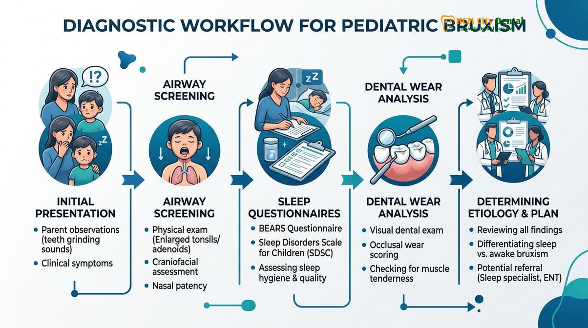
The clinical management of pediatric bruxism requires a highly nuanced and conservative approach, distinctly different from the protocols used for adult patients. Because a child’s craniofacial structures are in a state of active growth and development, aggressive or restrictive interventions can cause irreversible harm to the developing skeletal framework. The diagnostic journey begins with a meticulous medical and dental history, focusing heavily on sleep quality, respiratory health, and behavioral patterns. Clinicians utilize validated sleep questionnaires, such as the Pediatric Sleep Questionnaire (PSQ), to identify red flags for sleep-disordered breathing. This is followed by a comprehensive intraoral examination to grade the severity of dental attrition, assess the tone of the masticatory muscles, and evaluate the anatomical dimensions of the palate and airway.[2]
A cornerstone of this diagnostic process is the pediatric airway screening. By utilizing advanced imaging modalities, such as low-dose Cone Beam Computed Tomography (CBCT) when clinically justified, dentists can visualize the volumetric capacity of the nasal cavity and pharyngeal airway. If significant adenotonsillar hypertrophy or severe airway constriction is identified, the primary intervention is not dental, but medical. Referral to an otolaryngologist for potential surgical intervention (adenotonsillectomy) is often the most definitive treatment for airway-induced bruxism, frequently resulting in the complete resolution of the grinding behavior without the need for any intraoral appliances.[4]
When the etiology is linked to anatomical discrepancies such as a narrow maxilla or a retrognathic mandible, interceptive orthodontics becomes the treatment of choice. Procedures such as Rapid Maxillary Expansion (RME) utilize specialized appliances to gently widen the palatal suture. This not only creates adequate space for the erupting permanent teeth but, more importantly, increases the volume of the nasal floor, significantly reducing nasal airway resistance. By improving the child’s ability to breathe nasally during sleep, interceptive orthodontics addresses the root cause of the micro-arousals, thereby eliminating the neurological trigger for the bruxism. This proactive approach exemplifies the integration of comprehensive temporomandibular joint therapies with pediatric airway management.
Clinical Contraindication: Hard Acrylic Splints in Children
The use of rigid, hard acrylic stabilization splints (commonly prescribed for adults) is strictly contraindicated in growing children. These unyielding appliances can lock the maxilla and mandible into a fixed position, severely restricting normal transverse and sagittal craniofacial growth. Improper use of hard splints in pediatric patients can lead to permanent skeletal malocclusions, impacted teeth, and exacerbation of airway constriction.
In specific clinical scenarios where the bruxism is exceptionally severe—posing an immediate threat of pulpal exposure in the primary teeth or causing significant temporomandibular joint pain—a protective barrier may be temporarily required. In these carefully selected cases, a soft pediatric mouthguard may be fabricated. Unlike rigid adult splints, a soft pediatric mouthguard is constructed from pliable, resilient materials that provide a cushioning effect against occlusal trauma while still allowing for the natural lateral expansion of the dental arches. However, the use of these custom nightguards in children must be strictly monitored by a dental professional, with frequent adjustments or replacements to accommodate the rapid changes in the child’s dentition and jaw size. They are considered a temporary protective measure, not a definitive cure, and are always utilized in conjunction with therapies aimed at addressing the underlying etiology.
When to See a Doctor (Important Clinical Considerations)
While many cases of pediatric bruxism are benign and self-limiting, certain clinical presentations warrant immediate professional evaluation to prevent long-term dental, skeletal, or systemic complications. Parents should maintain vigilant observation of their child’s sleep habits and dental condition, acting promptly if specific red flags emerge. The distinction between normal developmental grinding and pathological bruxism often hinges on the presence of accompanying symptoms that indicate a deeper physiological struggle.

You should seek a comprehensive evaluation at a specialized facility, such as HCMC Dental Clinic, if your child exhibits any of the following clinical indicators:
- Severe Dental Attrition: If the wear on the teeth progresses beyond the enamel and exposes the underlying yellowish dentin, the child is at high risk for thermal sensitivity, dental decay, and potential nerve damage.
- Signs of Sleep Apnea: Loud, chronic snoring, witnessed pauses in breathing (apneas), gasping for air during sleep, or excessive daytime sleepiness and hyperactivity are strong indicators of sleep-disordered breathing requiring urgent airway assessment.
- Craniofacial Pain: Complaints of morning headaches, pain upon chewing, or tenderness in the jaw muscles and temporomandibular joints (TMJ) suggest that the bruxism is exceeding the physiological tolerance of the neuromuscular system.
- Persistence into Mixed Dentition: If aggressive grinding continues unabated as the permanent incisors and molars erupt, intervention may be necessary to protect the adult teeth from premature damage and to guide proper jaw joint management.
“Early intervention in pediatric bruxism is rarely about stopping the grinding directly; it is about identifying the silent struggles of the child’s airway and nervous system, ensuring they have the physiological foundation for healthy, restorative sleep.”
A thorough clinical examination will determine the most appropriate course of action, whether it be active observation, interceptive orthodontics, or the temporary application of protective dental splints. By addressing the underlying causes early, clinicians can guide the child toward optimal oral health and overall well-being, mitigating the risks associated with chronic parafunctional habits. For personalized diagnostics and advanced TMJ treatment protocols, consulting with experienced pediatric dental professionals is the most effective strategy for safeguarding your child’s developing smile.
Frequently Asked Questions
Should I be worried if my 5-year-old grinds teeth in sleep?
You should not be immediately alarmed, as mild sleep grinding is often a normal physiological phase during dental development. However, if the grinding is accompanied by loud snoring, daytime fatigue, complaints of jaw pain, or visible severe wear on the primary teeth, a clinical evaluation is highly recommended to rule out underlying sleep-disordered breathing or excessive occlusal trauma. Monitoring the transition as permanent teeth erupt is key to ensuring the habit remains benign.
Can enlarged adenoids cause teeth grinding in children?
Yes, enlarged adenoids are a primary trigger for sleep bruxism in young children. When hypertrophic adenoids obstruct the upper airway during sleep, the central nervous system initiates micro-arousals and involuntary jaw movements, pushing the lower jaw forward to mechanically open the respiratory passage and restore adequate oxygen flow. Addressing the adenotonsillar hypertrophy often leads to a complete cessation of the nocturnal grinding.
When do kids outgrow bruxism?
Most children naturally outgrow physiological bruxism between the ages of 9 and 12, coinciding with the complete eruption of their permanent teeth. As the mixed dentition phase concludes and the adult bite stabilizes, the neuromuscular triggers for grinding typically subside, provided there are no lingering airway obstructions or chronic psychological stressors. Persistent grinding beyond this age requires professional dental assessment.
Is a custom nightguard safe for a growing child?
A custom nightguard can be safe for a growing child only when specifically designed and closely monitored by a pediatric dental professional. Hard acrylic splints are generally contraindicated as they can restrict natural jaw growth; instead, a carefully calibrated soft pediatric mouthguard may be utilized temporarily to protect against severe attrition while allowing for normal skeletal development. Frequent adjustments are mandatory to accommodate erupting teeth.
How does pediatric bruxism affect permanent teeth?
While pediatric bruxism primarily affects baby teeth, persistent grinding into adolescence can lead to premature enamel wear, micro-fractures, and heightened thermal sensitivity in permanent teeth. If left unmanaged during the transition to adult dentition, chronic bruxism may also contribute to the early onset of temporomandibular joint dysfunction and altered facial muscle development, necessitating interceptive therapies to protect the permanent bite.
References
- Journal of Clinical Medicine. Global Prevalence of Sleep Bruxism and Awake Bruxism in Pediatric and Adult Populations: A Systematic Review. (2024).
- International Journal of Environmental Research and Public Health. Sleep Bruxism in Children: Etiology, Diagnosis, and Treatment. (2021).
- American Academy of Sleep Medicine. International Classification of Sleep Disorders, Third Edition. (2014).
- Journal of Dentistry for Children. The relationship between sleep-disordered breathing and pediatric bruxism. (2019).
- Pediatric Dentistry. Interceptive orthodontic management of airway-induced sleep bruxism. (2020).
For professional children’s dental care, preventive sealants, or to schedule a check-up, visit our Children & Pediatric Dentistry service page at HCMC Dental Clinic in Ho Chi Minh City.