Bleeding after tooth extraction is a normal physiological response that typically resolves within the first day of recovery. While mild oozing is expected as a blood clot forms, heavy, continuous bleeding that repeatedly soaks through gauze pads requires immediate clinical intervention to prevent complications and ensure proper socket healing.
Clinical Summary:
Post-extraction hemostasis relies on the successful formation and stabilization of a blood clot within the alveolar socket. Initial bleeding is managed through mechanical pressure using sterile gauze. If the socket bleeding won’t stop despite sustained pressure, it may indicate a disrupted clot, underlying coagulopathy, or localized tissue trauma. Dental professionals categorize bleeding into primary, reactionary, and secondary phases. Effective extraction aftercare guides emphasize avoiding negative oral pressure, strenuous physical activity, and chemical irritants like tobacco. Prompt evaluation by an emergency dentist is critical when active hemorrhage persists, accompanied by systemic symptoms such as dizziness or excessive swelling.
Key Takeaways:
- Mild oozing or pink-tinged saliva is a normal part of the healing process for the first 24 hours post-extraction.
- Firm, continuous pressure on a gauze pad for 30-45 minutes is the primary and most effective method to achieve hemostasis.
- A stable blood clot is essential to protect exposed bone and nerve endings, preventing the painful condition known as dry socket.
- Activities creating negative pressure, such as using straws, spitting, or smoking, can dislodge the clot and restart active bleeding.
- Continuous, bright red bleeding that fills the mouth rapidly is a dental emergency requiring immediate professional care.
Normal vs Abnormal Bleeding After Extraction
Differentiating between expected postoperative oozing and abnormal hemorrhage is crucial for patient safety. Normal healing involves minor blood-tinged saliva, whereas abnormal bleeding presents as a continuous, heavy flow of bright red blood that fails to clot.
When a tooth is removed from the alveolar bone, the body immediately initiates a complex physiological response to stop the bleeding and begin the healing process. This process, known as hemostasis, involves vascular spasms, the formation of a platelet plug, and a coagulation cascade that ultimately results in a stable fibrin clot[1]. Understanding the timeline and visual characteristics of this process is essential for patients to distinguish between normal recovery and potential complications.
In the immediate aftermath of the surgery, primary bleeding occurs. This is the active bleeding that happens while you are still in the dental chair. Your dentist will manage this by having you bite down on a sterile gauze pad before you leave the clinic. Once you return home, you enter the phase where reactionary bleeding may occur. As the local anesthetic wears off, the blood vessels in the surrounding tissues begin to dilate, which can cause a slight resurgence in bleeding.
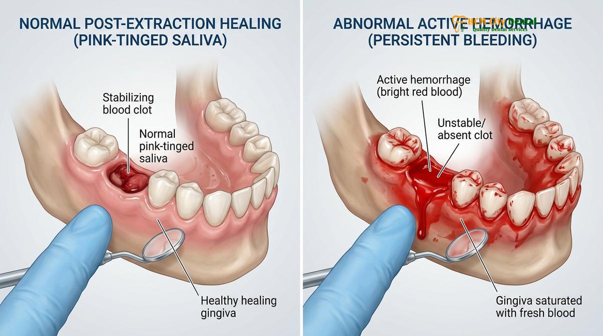
Normal post-extraction bleeding usually manifests as a slight oozing. Because blood mixes with saliva, it often looks much more dramatic than it actually is. A few drops of blood can turn a mouth full of saliva completely pink or light red. Patients frequently report a mild metallic taste in their mouth, which is also a standard part of the healing trajectory. By the end of the first day, this oozing should significantly diminish.
Conversely, abnormal bleeding presents a distinctly different clinical picture. If the blood is bright red, flows continuously, and rapidly pools in the mouth, the normal clotting mechanism has been compromised. In these cases, the gauze pad will become completely saturated within minutes. Another hallmark of abnormal bleeding is the formation of “liver clots”—large, dark, jelly-like masses of coagulated blood that sit over the socket but fail to seal the severed blood vessels beneath.
| Characteristic | Normal Oozing (Expected) | Abnormal Hemorrhage (Emergency) |
|---|---|---|
| Color and Consistency | Light pink to pale red; mostly saliva mixed with a small amount of blood. | Bright, vivid red; thick consistency; presence of large, jelly-like “liver clots.” |
| Flow Rate | Slow, intermittent oozing; gradually decreases over time. | Continuous, rapid pooling; fills the mouth quickly. |
| Response to Pressure | Stops or significantly slows down after 30 minutes of firm gauze pressure. | Continues heavily despite 45+ minutes of sustained, firm pressure. |
? Need urgent dental help in Ho Chi Minh City? Visit our Emergency Dentistry Hub for same-day appointments, 24/7 WhatsApp support, and comprehensive emergency dental services.
Immediate Steps to Control Post-Extraction Bleeding
The most effective initial intervention for post-extraction bleeding is the application of firm, sustained mechanical pressure directly over the surgical site. Proper gauze placement and patient positioning are fundamental to facilitating clot formation.
When you return home after a dental procedure, the responsibility for maintaining hemostasis transitions to you. The most critical tool at your disposal is mechanical pressure. Blood needs a static environment to coagulate; if blood is constantly flowing, the platelets cannot aggregate, and the fibrin network cannot form[2]. Therefore, applying the correct technique for pressure is paramount to stopping the bleeding efficiently.
If you notice that the extraction site is bleeding more than a mild ooze, the first step is to remain calm and wash your hands thoroughly. Take a piece of clean, sterile gauze and fold or roll it into a firm, thick pad. The size of the pad is crucial; it must be thick enough that when you bite down, the pressure is applied directly to the extraction socket, not just dispersed across the adjacent teeth.
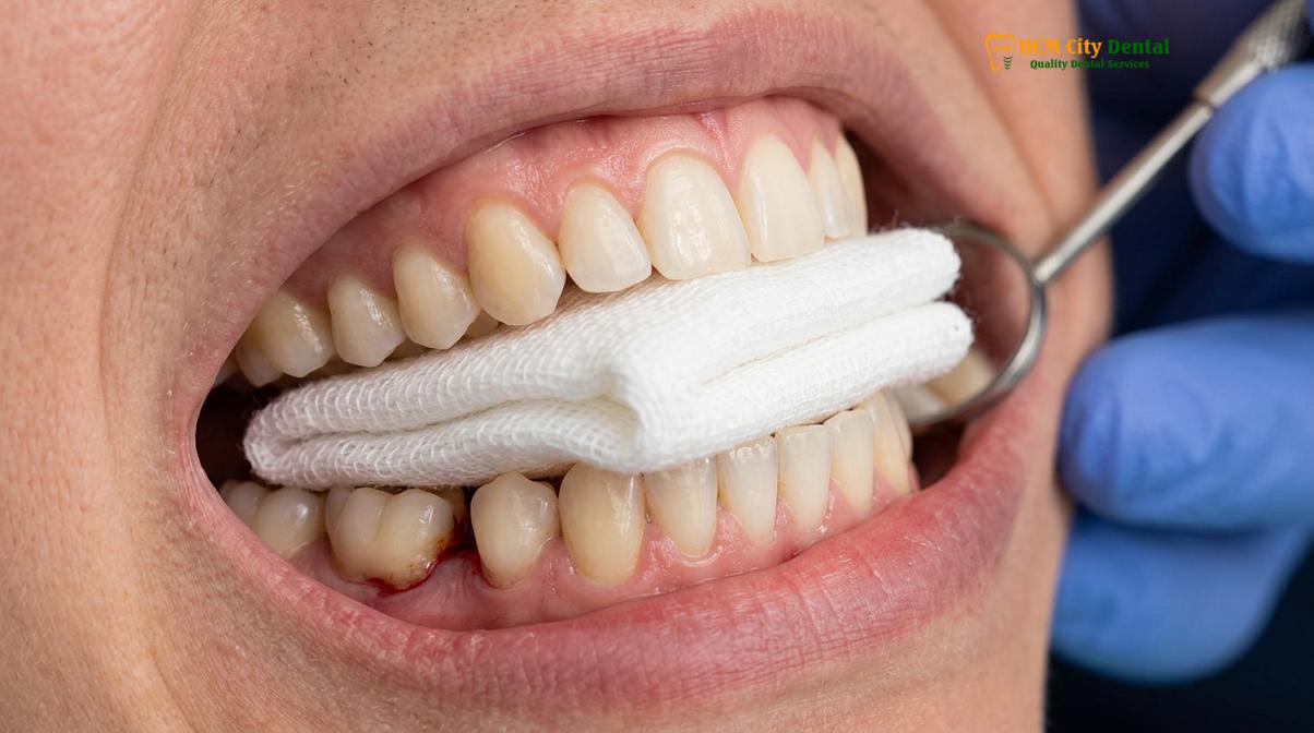
Place the folded gauze directly over the empty socket. Bite down firmly and continuously. One of the most common mistakes patients make is removing the gauze every five minutes to check if the bleeding has stopped. Every time the pressure is released, the fragile, newly forming clot is torn away, and the bleeding process restarts from zero.
“The success of post-extraction hemostasis relies entirely on the patient’s compliance with sustained mechanical pressure. Intermittent biting or frequent checking of the gauze disrupts the delicate fibrin matrix, transforming a routine recovery into a prolonged bleeding event.”
While applying pressure, your physical posture also plays a significant role in controlling bleeding. Lie back in a semi-reclined position or sit upright in a chair. Elevate your head above the level of your heart using pillows. This utilizes gravity to reduce the local blood pressure in your head and neck, thereby decreasing the hydrostatic pressure pushing blood out of the severed vessels in the socket.
Activities and Foods to Avoid Post-Extraction
Protecting the newly formed blood clot requires strict adherence to dietary and behavioral restrictions. Avoiding negative oral pressure and mechanical trauma is essential to prevent secondary bleeding episodes.
The stability of the blood clot is highly susceptible to environmental factors within the mouth. Any action that creates a vacuum or negative pressure can easily dislodge the clot from the socket walls. For this reason, patients must strictly avoid using drinking straws for at least several days following the procedure. The suction required to draw liquid through a straw generates enough force to pull the clot entirely out of the alveolar bone[3].
Similarly, forceful spitting and vigorous rinsing should be avoided. If you need to clear your mouth, let the liquid fall out gently into the sink rather than actively spitting. Smoking is particularly detrimental; not only does the inhalation create negative pressure, but the chemical toxins in tobacco smoke severely impair local blood circulation, delaying healing and increasing the risk of infection.
Dietary choices also play a major role in preventing post-operative bleeding. Hard, crunchy, or sharp foods—such as chips, nuts, and crusty bread—can physically scrape the surgical site and lacerate the healing tissues. Patients are advised to stick to a soft food diet, including items like yogurt, mashed potatoes, and lukewarm soups, ensuring that they chew on the opposite side of the mouth from the extraction site.
Why the Blood Clot is Critical for Healing
The blood clot serves as a biological dressing that protects the underlying alveolar bone and innervations while providing a scaffold for new tissue generation. Its preservation is the cornerstone of uncomplicated extraction recovery.
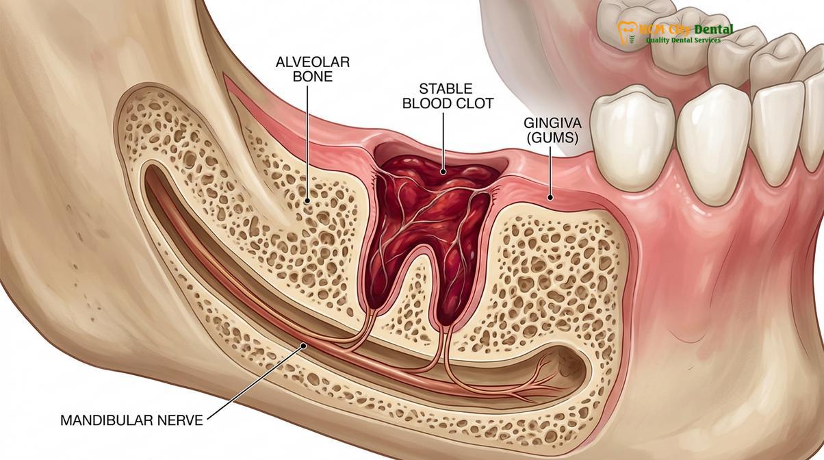
To truly understand the importance of managing bleeding, one must appreciate the profound biological role of the blood clot. When a tooth is removed, it leaves behind a deep defect in the alveolar bone, exposing the bone itself and the intricate network of nerve endings that once supplied the tooth. The body’s immediate response is to fill this void with blood, which rapidly coagulates to form a protective plug.
This blood clot is far more than just a stopper to prevent blood loss; it is a highly active biological matrix. In the initial recovery phase, the clot acts as a physical barrier, shielding the sensitive bone and nerves from the harsh environment of the oral cavity. Without this barrier, patients experience excruciating pain, a condition clinically known as alveolar osteitis, or “dry socket.”
Clinical Case Study: Managing Delayed Healing
A 34-year-old patient visited HCMC Dental Clinic in Ho Chi Minh City complaining of severe, radiating jaw pain three days after a molar extraction. Clinical examination revealed an empty, pale socket devoid of a blood clot—a classic presentation of alveolar osteitis. The patient admitted to vigorously rinsing with salt water on the first day, which had dislodged the initial clot. The clinical team gently irrigated the socket to remove debris and placed a specialized medicated dressing to soothe the exposed nerve endings. The patient experienced immediate pain relief and was monitored closely as secondary healing occurred over the following weeks.
Beyond protection, the clot serves as the foundational scaffold for tissue regeneration. It is rich in platelets, which release growth factors and cytokines that signal the body’s repair mechanisms. If the clot is lost prematurely, this entire regenerative cascade is halted, leading to delayed healing, increased risk of infection, and severe discomfort.
Professional Treatments for Uncontrolled Bleeding
When conservative home measures fail to achieve hemostasis, dental professionals utilize a variety of advanced clinical techniques and localized agents to stabilize the socket and halt active hemorrhage.
If you present to a dental clinic with uncontrolled post-extraction bleeding, the clinician will first clean the area to identify the exact source of the hemorrhage. Often, this involves removing unstable “liver clots” and irrigating the socket with sterile saline. Once the site is visible, the dentist may place specialized hemostatic agents directly into the alveolar void. Materials such as oxidized regenerated cellulose or gelatin sponges act as an artificial matrix, providing a physical structure that encourages platelets to aggregate and form a stable clot[4].
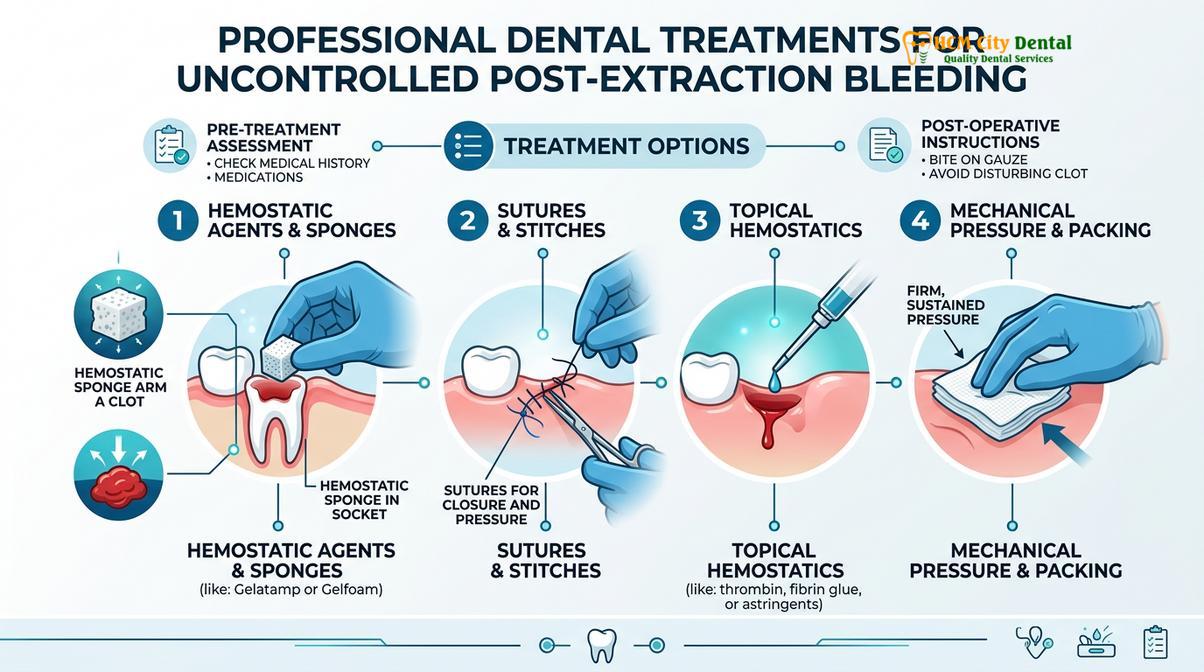
In cases where the bleeding originates from the gingival tissues rather than deep within the bone, the dentist may place sutures (stitches) to pull the gum margins tightly together. This mechanical closure applies direct pressure to the severed capillaries and significantly reduces the exposed surface area of the wound.
“Clinical intervention for persistent hemorrhage often requires a multi-faceted approach, combining mechanical suturing with localized hemostatic agents to ensure the socket is fully stabilized and protected against further disruption.”
For patients with underlying bleeding disorders or those on anticoagulant therapy, additional pharmacological interventions may be necessary. Dentists might use topical applications of tranexamic acid, an antifibrinolytic agent that prevents the premature breakdown of blood clots, ensuring that once hemostasis is achieved, the clot remains intact throughout the critical early stages of healing.
When Bleeding Gums/Socket is an Emergency
Recognizing the signs of a true dental emergency is vital. Continuous, heavy bleeding accompanied by systemic symptoms requires immediate medical or dental intervention to prevent severe complications.
While most post-extraction bleeding can be successfully managed at home with proper technique and patience, there are specific clinical scenarios where the bleeding crosses the threshold from a normal physiological response to a medical emergency. The primary indicator of an emergency is when the socket bleeding won’t stop despite your best efforts. If you have applied firm, continuous pressure with a sterile gauze pad for two consecutive 45-minute intervals, and the bleeding remains heavy, bright red, and rapidly fills your mouth, you must seek professional help.
Emergency Warning Signs: Seek immediate medical attention if post-extraction bleeding is accompanied by severe dizziness, confusion, extreme weakness, pale skin, or any difficulty breathing or swallowing. These symptoms may indicate significant blood loss or airway compromise.
According to general guidelines aligned with the Vietnam Odonto-Stomatology Association, patients experiencing uncontrolled hemorrhage should seek immediate emergency dental care rather than relying solely on home remedies. Systemic symptoms accompanying the bleeding are major red flags that dictate urgent evaluation at a hospital or specialized dental clinic[5].
Dr. Nguyen Van Cuong, a dedicated specialist at HCMC Dental Clinic, frequently guides patients through their recovery journey. He emphasizes that while a tooth extraction is a routine procedure, diligent aftercare is vital to prevent complications. Dr. Cuong advises patients to remain calm, avoid strenuous activities, and follow clinical instructions closely to ensure optimal healing.
If you are experiencing persistent bleeding or have concerns about your recovery in Ho Chi Minh City, contact HCMC Dental Clinic immediately for professional assistance. Our experienced team is equipped to handle post-operative complications and ensure your healing process remains on track.
References
- Journal of Oral and Maxillofacial Surgery. Management of post-extraction bleeding.
- American Dental Association. Clinical guidelines for dental extractions and hemostasis.
- International Journal of Oral Science. The role of the blood clot in alveolar healing.
- British Dental Journal. Efficacy of local hemostatic agents in dental surgery.
- Journal of Clinical Periodontology. Complications following third molar extractions.