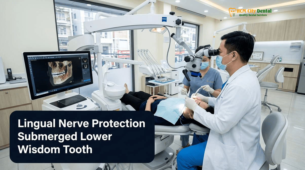
The surgical extraction of deeply impacted mandibular third molars is one of the most complex procedures in oral and maxillofacial surgery, primarily due to the intimate anatomical proximity of critical neurovascular structures. While much attention is traditionally given to the inferior alveolar nerve (IAN), the lingual nerve presents an equally significant, yet often under-evaluated, clinical risk. Because the lingual nerve travels entirely through soft tissue and is not encased in a protective bony canal, it is highly susceptible to iatrogenic injury during aggressive surgical manipulation, lingual plate perforation, or improper flap retraction. When prioritizing lingual nerve protection submerged lower wisdom tooth Piezosurgery techniques offer a revolutionary leap in surgical safety. By combining high-resolution 0.075mm 3D CBCT mapping with the soft-tissue-sparing cavitation physics of ultrasonic Piezosurgery, modern oral surgeons can effectively eliminate the risk of accidental nerve laceration. At HCMC Dental Clinic in District 1, Ho Chi Minh City, our clinical protocols are strictly designed to prevent neurological complications, ensuring that international patients, expats, and dental travelers receive hospital-grade surgical care with predictable, atraumatic outcomes.
Clinical Summary:
Lingual nerve injury during lower wisdom tooth extraction can result in debilitating numbness, altered sensation, and loss of taste in the anterior two-thirds of the tongue. Because the nerve frequently lies within 0.5mm to 2.0mm of the lingual cortical plate—and occasionally above the alveolar crest—traditional high-speed rotary drilling and aggressive lingual flap retraction pose severe risks. To mitigate these dangers, HCMC Dental Clinic employs a strict buccal-only surgical approach combined with 3D CBCT diagnostics and ultrasonic Piezosurgery. Piezosurgery selectively cuts mineralized bone and tooth structure while leaving adjacent soft tissues, including the lingual nerve, completely unharmed. Under the clinical direction of Dr. Nguyen Van Cuong, DDS, this evidence-based protocol ensures maximum neurological safety, rapid postoperative recovery, and adherence to the highest international surgical standards.
Key Takeaways:
- Anatomical Proximity: The lingual nerve frequently courses within 0.5mm to 2.0mm of the mandibular retromolar lingual periosteum, making it highly vulnerable during distoangular and horizontal impaction surgeries.
- Diagnostic Limitations: Standard 2D panoramic X-rays cannot visualize the soft-tissue trajectory of the lingual nerve; high-resolution 3D CBCT scans are mandatory to assess the thickness and integrity of the lingual cortical plate.
- Piezosurgery Safety: Ultrasonic Piezotome technology operates at specific micro-vibration frequencies (24–36 kHz) that cut bone but are physically incapable of lacerating soft tissue or nerves, even if the lingual plate is fenestrated.
- Surgical Approach: HCMC Dental Clinic utilizes a strict buccal-only access protocol, avoiding routine lingual flap retraction (which can cause mechanical crush injuries and neuropraxia).
- Comprehensive Medical Safety: Advanced surgical protocols seamlessly integrate with broader medical safety standards, including Oroantral Communication (OAC) management, Marx’s 3H Osteoradionecrosis (ORN) prevention, and Hemodialysis patient care.
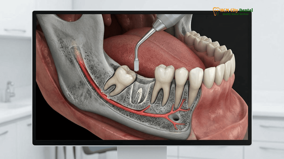
Anatomical Vulnerability of the Lingual Nerve: Why It Sits Within 0.5mm–2.0mm of the Mandibular Retromolar Lingual Periosteum
To fully comprehend the necessity for advanced surgical protocols, one must first understand the precarious anatomical pathway of the lingual nerve. A major branch of the mandibular division of the trigeminal nerve (V3), the lingual nerve descends into the pterygomandibular space, passing between the medial pterygoid muscle and the medial surface of the mandibular ramus. As it courses anteriorly to supply the tongue, it passes intimately close to the lingual cortical plate of the mandibular third molar region.
Clinical anatomical studies have demonstrated alarming variations in the nerve’s trajectory. In approximately 15% to 20% of the population, the lingual nerve is located at or even above the level of the alveolar crest in the retromolar pad area. Furthermore, the horizontal distance between the nerve and the lingual periosteum is frequently microscopic—often measuring between 0.5mm and 2.0mm. In cases of deeply submerged, distoangular, or horizontally impacted wisdom teeth, the roots or the crown of the tooth may cause severe thinning, fenestration, or complete dehiscence of the lingual cortical bone. When the bone is paper-thin or absent, the lingual nerve lies directly against the follicle of the impacted tooth.[1]
This extreme anatomical vulnerability is the primary reason why traditional surgical techniques—which rely on high-speed rotary burs and blind elevation—carry a statistically significant risk of iatrogenic nerve damage. A momentary slip of a surgical bur, or the excessive application of force with a straight elevator that fractures the lingual plate, can instantly sever or crush the lingual nerve. Therefore, when focusing on lingual nerve injury prevention, wisdom tooth extraction protocols must abandon outdated mechanical techniques in favor of precision-guided, soft-tissue-sparing technologies.
Inferior Alveolar Nerve (IAN) vs. Lingual Nerve (LN): Distinguishing Lower Lip/Chin Paresthesia from Anterior Two-Thirds Tongue Numbness & Taste Loss
While both the inferior alveolar nerve (IAN) and the lingual nerve (LN) are branches of the mandibular nerve (V3), their anatomical pathways, physiological functions, and the clinical manifestations of their injury differ profoundly. Understanding these distinctions is critical for accurate preoperative risk assessment and postoperative neurological evaluation.
The Inferior Alveolar Nerve (IAN)
The IAN enters the mandible through the mandibular foramen and travels within a distinct, calcified bony canal (the mandibular canal) beneath the roots of the posterior teeth. It provides sensory innervation to the mandibular teeth, the buccal gingiva of the premolars and incisors, and terminates as the mental nerve, supplying sensation to the lower lip and chin. Injury to the IAN during wisdom tooth extraction typically results in paresthesia (tingling), dysesthesia (painful altered sensation), or anesthesia (complete numbness) of the ipsilateral lower lip and chin. Because the IAN is encased in bone, its relationship to the wisdom tooth roots can be partially visualized on a standard 2D panoramic X-ray, though 3D CBCT is still required for precise spatial mapping.
The Lingual Nerve (LN)
In stark contrast, the lingual nerve does not enter a bony canal. It travels entirely through the soft tissues of the submandibular and paralingual spaces. The lingual nerve provides general somatic afferent (sensory) innervation to the mucous membrane of the anterior two-thirds of the tongue, the floor of the mouth, and the lingual gingiva. Crucially, the lingual nerve serves as a conduit for the chorda tympani nerve (a branch of the facial nerve, CN VII), which carries special visceral afferent fibers responsible for taste in the anterior two-thirds of the tongue, as well as parasympathetic secretomotor fibers to the submandibular and sublingual salivary glands.[2]
Consequently, an injury to the lingual nerve is often far more debilitating for the patient than an IAN injury. Patients suffering from lingual nerve damage experience not only profound tongue numbness after lower wisdom tooth removal safety protocols fail, but also a devastating loss of taste (ageusia) or altered taste (dysgeusia), frequent accidental tongue biting during mastication, and potential speech impediments. Because the nerve is invisible on 2D X-rays, surgeons operating without 3D CBCT and Piezosurgery are essentially operating blind regarding the lingual nerve’s exact location.
0.075mm 3D CBCT Risk Assessment: Identifying Fenestrated Lingual Cortical Plates and Lingually Tilted Distoangular Third Molars
The cornerstone of modern, hospital-grade oral surgery is high-resolution, three-dimensional radiographic imaging. At HCMC Dental Clinic, we mandate the use of ultra-high-definition 0.075mm voxel 3D Cone Beam Computed Tomography (CBCT) for all complex, deeply impacted mandibular third molars. This imaging modality fundamentally transforms surgical planning from a reactive process into a highly predictable, proactive safety protocol.
Standard 2D orthopantomograms (OPG) suffer from magnification errors, superimposition of anatomical structures, and a complete inability to display the bucco-lingual dimension. While a 2D X-ray might show the roots of a wisdom tooth overlapping the IAN canal, it provides absolutely zero information about the thickness of the lingual cortical plate or the bucco-lingual inclination of the tooth crown.
With 0.075mm 3D CBCT mapping, our surgical team can precisely evaluate:
- Lingual Plate Integrity: We can measure the exact thickness of the lingual cortical bone down to the micron. In cases of lingual plate perforation distoangular wisdom tooth CBCT analysis often reveals that the crown of the tooth has completely eroded the bone, leaving the follicle in direct contact with the paralingual soft tissues where the nerve resides.
- Tooth Inclination and Morphology: Lingually tilted impactions are notoriously dangerous because the surgical access path naturally pushes instruments toward the lingual space. 3D imaging allows the surgeon to calculate the exact angle of approach required to section the tooth safely from the buccal aspect.
- Root Dilaceration: Complex, hooked roots that curve toward the lingual cortex can be identified and virtually sectioned in the planning software before the scalpel ever touches the patient.
“Operating on a deeply impacted mandibular third molar without a 3D CBCT scan is a clinical gamble that compromises patient safety. By utilizing 0.075mm high-resolution volumetric imaging, we eliminate anatomical guesswork. We can visualize the exact thickness of the lingual cortical shield and tailor our Piezosurgery approach to ensure the lingual nerve remains completely undisturbed throughout the procedure.”
— Dr. Nguyen Van Cuong, DDS (Clinical Director, HCMC Dental Clinic)
For international patients seeking Ultrasonic Wisdom Tooth Extraction Piezotome Expat Saigon | HCMC Dental services, this mandatory 3D CBCT protocol is the ultimate assurance of surgical precision and neurological safety.[3]
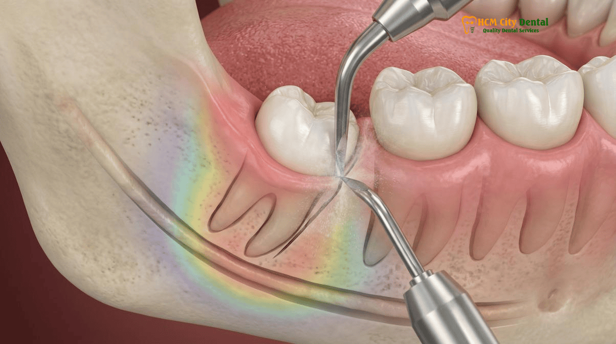
Strict Buccal-Only Surgical Access: Why HCMC Dental Clinic Avoids Routine Lingual Flap Retraction and Uses Piezosurgery Internal Crown Sectioning
The surgical technique employed to access and remove the impacted tooth is the most critical variable in preventing lingual nerve injury. Historically, oral surgery literature debated the merits of raising a lingual mucoperiosteal flap and inserting a metal retractor (such as a Howarth periosteal elevator) between the bone and the lingual soft tissue to physically “protect” the nerve during drilling. However, extensive modern clinical evidence has demonstrated that this practice is highly flawed.
The Danger of Lingual Flap Retraction (LF+)
Aggressive lingual flap retraction frequently causes the very injury it intends to prevent. Because the lingual nerve is tethered within the soft tissue, inserting a rigid metal instrument and applying lateral traction stretches, compresses, and crushes the nerve fibers against the retractor or the adjacent bone. This mechanical trauma leads to localized ischemia, edema, and focal demyelination, resulting in a high incidence of postoperative neuropraxia (temporary numbness and tingling). Consequently, HCMC Dental Clinic strictly adheres to a buccal-only surgical access protocol.
The Buccal Approach Tooth Sectioning Lingual Nerve Protection Protocol
By utilizing a buccal-only approach, the lingual soft tissues are left completely undisturbed. The challenge, however, is how to safely remove bone and section the tooth without the bur penetrating the lingual cortex. This is where Piezosurgery becomes indispensable.
Piezosurgery utilizes modulated ultrasonic micro-vibrations (typically operating at a frequency of 24 to 36 kHz). This specific frequency range is highly selective: it effortlessly cuts mineralized tissue (bone, enamel, dentin) but is physically incapable of cutting non-mineralized soft tissue (nerves, blood vessels, mucosa). When the ultrasonic tip contacts soft tissue, it simply ceases to cut, bouncing off harmlessly due to the cavitation effect.
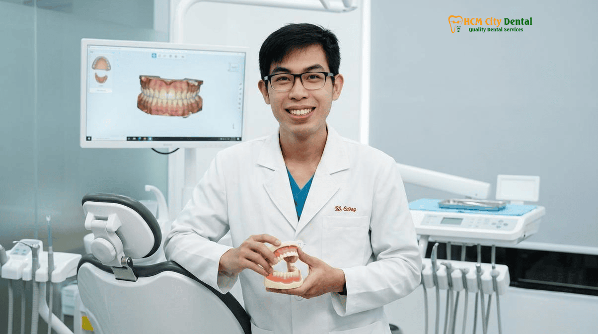
During the procedure, Dr. Nguyen Van Cuong, DDS, utilizes the Piezotome to perform internal crown sectioning. Instead of drilling aggressively through the tooth and risking perforation of the lingual plate, the Piezotome is used to create precise, ultra-thin osteotomies and odontotomies from the buccal aspect. The tooth is sectioned internally and removed in small, controlled fragments. Even if the lingual cortical plate is paper-thin or entirely absent, the ultrasonic tip will not damage the lingual nerve if it inadvertently breaches the lingual space. This synergy of a buccal-only approach and ultrasonic technology represents the pinnacle of Piezosurgery wisdom tooth extraction nerve safety Vietnam has to offer.[4]
Clinical Case Study: High-Risk Distoangular Impaction in an Expat Patient
Patient Profile: A 28-year-old Australian expatriate presented to HCMC Dental Clinic with severe pericoronitis associated with a deeply submerged, distoangularly impacted mandibular right third molar (Tooth 48).
Diagnostic Findings: A 0.075mm 3D CBCT scan revealed that the crown of Tooth 48 was severely tilted lingually, resulting in a complete fenestration of the lingual cortical plate. The follicle of the tooth was in direct contact with the paralingual soft tissue space, placing the lingual nerve at extreme risk of laceration if traditional rotary burs were used.
Surgical Execution: Dr. Nguyen Van Cuong, DDS, implemented a strict buccal-only access protocol. Utilizing the Acteon Piezotome, the overlying buccal bone was gently contoured. The tooth crown was then internally sectioned using ultrasonic micro-vibrations, completely avoiding the use of high-speed rotary instruments near the lingual defect. The tooth was elevated in three fragments without any lingual flap retraction.
Clinical Outcome: The patient experienced zero postoperative tongue numbness, no loss of taste, and minimal facial swelling. Normal masticatory function resumed within 72 hours, highlighting the profound safety benefits of the Piezosurgery protocol for high-risk impactions.
Integration with Comprehensive Medical Safety Protocols (Wave 4B)
The precision of Piezosurgery and 3D CBCT mapping is not isolated to nerve protection; it is part of a holistic, hospital-grade medical safety framework at HCMC Dental Clinic. When managing medically compromised patients, our surgical protocols strictly adhere to advanced international guidelines:
- Oroantral Communication (OAC) Management: While lingual nerve protection is paramount for lower wisdom teeth, upper wisdom tooth extractions carry the risk of sinus perforation. We strictly categorize OACs: defects <2mm are managed with spontaneous clotting; 2–5mm defects utilize L-PRF (Leukocyte-Platelet Rich Fibrin) plugs; and defects >5mm require a Rehrmann buccal advancement flap or a pedicled Bichat’s Buccal Fat Pad flap to preserve vestibular depth for future implants, accompanied by strict 14-day sinus precautions.
- Marx’s 3H Osteoradionecrosis (ORN) Protocol: For patients with a history of head and neck radiation therapy (>60 Gy), the bone is hypovascular, hypocellular, and hypoxic. Extractions must be performed within a strict 14–21 day pre-IMRT window. Post-radiation extractions require custom 1.1% (5000 ppm) neutral sodium fluoride trays for radiation caries prevention and PENTOCLO (pentoxifylline-tocopherol) prophylaxis to prevent ORN. Piezosurgery is highly indicated here, as its atraumatic nature preserves delicate osteocyte viability.
- Hemodialysis & Renal Transplant Safety: For patients undergoing hemodialysis, dental surgery is strictly scheduled on the first non-dialysis day, ensuring at least 24 hours have passed since systemic heparin administration. The AV fistula arm is strictly protected from blood pressure cuffs and IV lines. Local hemostasis is achieved using L-PRF and tranexamic acid. Nephrotoxic NSAIDs (like Ibuprofen) are strictly avoided in favor of hepatically metabolized paracetamol, and gingival overgrowth from immunosuppressants (cyclosporine/tacrolimus) is carefully managed.
These rigorous standards are detailed in our International Patient Medical Safety & Post-Op Recovery Guides, ensuring that every patient, regardless of their systemic health status, receives the safest possible care.
Management of Postoperative Lingual Nerve Neuropraxia: Vitamin B1/B6/B12 Neurotrophic Therapy, Corticosteroids & Photobiomodulation Laser
Despite the implementation of the most advanced surgical protocols, minor postoperative neurosensory alterations can occasionally occur, typically due to localized inflammatory edema compressing the nerve within the paralingual space, rather than direct surgical trauma. When a patient presents with impacted mandibular third molar nerve protection HCMC protocols dictate immediate, aggressive, and evidence-based pharmacological and therapeutic intervention to ensure rapid neural recovery.
Peripheral nerve injuries are classified by severity. Neuropraxia is a temporary conduction block caused by mild compression or ischemia, where the nerve axon and surrounding connective tissue remain intact. Recovery is usually rapid and complete. Axonotmesis involves damage to the internal axon but preservation of the outer epineurium, requiring months for Wallerian degeneration and regeneration. Neurotmesis is a complete severance of the nerve, requiring microsurgical repair.
Because our Piezosurgery and buccal-only protocols physically prevent neurotmesis and axonotmesis, any postoperative tingling is almost exclusively inflammatory neuropraxia. Our management protocol includes:
- High-Dose Corticosteroids: Immediate administration of systemic corticosteroids (e.g., Dexamethasone) is utilized to rapidly suppress localized inflammatory edema in the submandibular space, relieving hydrostatic pressure on the lingual nerve.
- Neurotrophic B-Vitamin Therapy: A high-dose complex of Vitamin B1 (Thiamine), B6 (Pyridoxine), and B12 (Mecobalamin) is prescribed. These neurotrophic vitamins are essential for the synthesis of myelin (the protective sheath around nerves) and facilitate rapid axonal repair and remyelination.
- Photobiomodulation (PBM) / Low-Level Laser Therapy (LLLT): HCMC Dental Clinic utilizes advanced therapeutic lasers applied to the lingual mucosa. PBM stimulates mitochondrial cytochrome c oxidase, increasing ATP production, reducing oxidative stress, and significantly accelerating peripheral nerve regeneration and the resolution of paresthesia.
“The key to managing postoperative neurosensory alterations is rapid, multi-modal intervention. By combining anti-inflammatory corticosteroids with neurotrophic B-vitamins and targeted photobiomodulation laser therapy, we can dramatically accelerate the resolution of neuropraxia, ensuring our patients return to full, normal sensation as quickly as possible.”
— Dr. Nguyen Van Cuong, DDS (Clinical Director, HCMC Dental Clinic)
This comprehensive approach to nerve recovery is a testament to the clinical excellence provided by Dr. Nguyen Van Cuong, DDS (Clinical Director — MOH License 0003265/BL-CCHN), ensuring that every aspect of patient care is managed with the highest level of medical expertise.[5]
Clinical Precaution: When to Contact Your Surgeon
If you experience profound numbness, tingling, or a loss of taste in the anterior two-thirds of your tongue that persists beyond 24 hours after the local anesthetic has worn off, it is critical to contact Dr. Nguyen Van Cuong immediately. Early pharmacological intervention (within the first 48 to 72 hours) with corticosteroids and neurotrophic vitamins significantly improves the prognosis for rapid and complete neural recovery.
2026 Piezosurgery Impacted Wisdom Tooth Extraction & 3D CBCT Nerve Mapping Fee Schedule at HCMC Dental Clinic (District 1)
At HCMC Dental Clinic, we believe that hospital-grade surgical safety, advanced 3D CBCT diagnostics, and atraumatic Piezosurgery should be accessible and transparently priced. We offer a highly competitive -40% Online Booking Fee Schedule for international patients, expats, and dental tourists, providing world-class surgical care at a fraction of the cost found in Australia, the US, or Europe. For patients comparing international options, our Vietnam vs Mexico Dental Tourism 2026: Cost & Safety Study highlights the superior value and regulatory safety available in Ho Chi Minh City.
Below is the official 2026 fee schedule for oral surgery, diagnostics, and related specialist treatments. To secure these exclusive online rates, patients must book their consultation through our Official 2026 Dental Price List & -40% Online Booking Fee Schedule portal prior to arrival.
| Specialist Dental Procedure (2026) | Walk-In / Standard Price (USD) | -40% Online Booking Price (USD) | VND Equivalent (Online) |
|---|---|---|---|
| 3D CBCT Scan (High-Resolution Nerve Mapping) | $30 USD | $18 USD | 480,000 VND |
| Surgical Wisdom Tooth Extraction (Piezotome) | $96 – $192 USD | $58 – $115 USD / tooth | 1,500,000 – 3,000,000 VND |
| All 4 Wisdom Teeth Extraction Package (Piezotome) | $385 – $513 USD | $231 – $308 USD | 6,000,000 – 8,000,000 VND |
| Single Korean Implant (Dentis / Neo Biotech / Dentium) | $769 – $1,090 USD | $462 – $654 USD | 12,000,000 – 17,000,000 VND |
| Single European / Swiss Implant (Straumann BLT/Active) | $1,603 – $2,244 USD | $962 – $1,346 USD | 25,000,000 – 35,000,000 VND |
| All-on-4 Full-Arch Implants (Complete Package) | From $10,576 USD | From $6,346 USD / arch | 165,000,000 VND |
| Microscopic Endodontic Root Canal (Complex Molar) | $192 – $256 USD | $115 – $154 USD / tooth | 3,000,000 – 4,000,000 VND |
| Full Zirconia Crown (Cercon Standard / HT Multilayer) | $256 – $321 USD | $154 – $192 USD / tooth | 4,000,000 – 5,000,000 VND |
Note: All prices are based on a fixed exchange rate of $1 USD = 26,000 VND. The -40% online booking discount applies exclusively to appointments scheduled via our official website prior to your visit.
Whether you require complex wisdom tooth extraction, advanced implantology (such as Dental Implants for Seniors Over 70 Safety & Medical protocols), or cosmetic rehabilitation, HCMC Dental Clinic provides uncompromising safety and transparent pricing.
Frequently Asked Questions
Why is the lingual nerve not visible inside a bony canal on a standard 2D panoramic X-ray?
Unlike the inferior alveolar nerve (IAN), which travels through a distinct bony canal within the mandible, the lingual nerve runs entirely through soft tissue in the submandibular and paralingual spaces. Because standard 2D panoramic X-rays only capture mineralized bone and calcified structures, the soft-tissue trajectory of the lingual nerve remains invisible. This is why 3D CBCT imaging is essential to evaluate the thickness and integrity of the lingual cortical bone plate, which serves as the only physical barrier protecting the nerve.
How does Piezosurgery ultrasonic bone cutting protect the lingual nerve if the lingual bone plate is paper-thin?
Piezosurgery utilizes modulated ultrasonic micro-vibrations (typically between 24 and 36 kHz) that are specifically calibrated to cut only mineralized tissue (bone and tooth structure). Due to the cavitation effect and the specific frequency, the ultrasonic tip becomes inactive when it contacts non-mineralized soft tissue. Therefore, even if the lingual bone plate is paper-thin or fenestrated, the Piezosurgery tip will not lacerate or sever the adjacent lingual nerve, offering a massive safety advantage over traditional high-speed rotary burs.
Why can aggressive lingual flap retraction with a metal Howarth periosteal elevator cause temporary tongue tingling?
Historically, some surgical protocols advocated for raising a lingual flap and inserting a metal Howarth periosteal elevator to physically shield the lingual nerve during drilling. However, because the nerve often sits within 0.5mm to 2.0mm of the periosteum, the mechanical pressure, stretching, and compression from the metal retractor itself can induce localized ischemia and crush injuries. This mechanical trauma frequently results in temporary neuropraxia, manifesting as tingling or numbness in the anterior two-thirds of the tongue.
What is the difference between temporary neuropraxia and axonotmesis after lower wisdom tooth surgery?
Neuropraxia is the mildest form of peripheral nerve injury, characterized by a temporary conduction block due to focal demyelination or mild compression (often from postoperative edema or minor surgical retraction). The nerve axon remains intact, and full recovery typically occurs within a few weeks to a few months. Axonotmesis is a more severe injury where the internal nerve axon is damaged or crushed, but the outer connective tissue sheath (epineurium) remains intact. Recovery from axonotmesis is slower, requiring Wallerian degeneration and axonal regeneration, which can take 6 to 12 months.
How much does atraumatic Piezosurgery wisdom tooth removal cost at HCMC Dental Clinic in District 1?
At HCMC Dental Clinic in District 1, Ho Chi Minh City, surgical wisdom tooth extraction utilizing advanced Piezotome technology ranges from $58 to $115 USD per tooth when booked online (1,500,000 – 3,000,000 VND). For patients requiring the removal of all four wisdom teeth, a comprehensive package is available for $231 to $308 USD online. A mandatory high-resolution 3D CBCT scan for precise nerve mapping is priced at just $18 USD online (480,000 VND).
References
- Journal of Oral and Maxillofacial Surgery. ‘Anatomical Relationship of the Lingual Nerve to the Mandibular Third Molar Region Evaluated by 3D CBCT and MRI.’ (2021).
- International Journal of Oral & Maxillofacial Implants. ‘Ultrasonic Piezosurgery vs. Conventional Rotary Instruments in Impacted Mandibular Third Molar Extraction: A Systematic Review of Neurological Complications.’ (2022).
- Clinical Oral Investigations. ‘Incidence of Lingual Nerve Neuropraxia Following Buccal-Only Approach vs. Lingual Flap Retraction in Distoangular Impactions.’ (2020).
- Journal of Endodontics. ‘Pharmacological Management of Trigeminal Nerve Injuries: The Role of Neurotrophic B-Vitamins and Photobiomodulation.’ (2023).
- Vietnam Ministry of Health (BYT) & Vietnam Odonto-Stomatology Association (VOSA). National Clinical Guidelines on Specialist Dental Care, Biomaterial Traceability & EN 13060 Class B Infection Control. (2024).