Full mouth reconstruction for gum disease is a comprehensive clinical protocol that restores occlusal function, aesthetics, and structural integrity after severe periodontitis. It involves eradicating active bacterial infection, stabilizing the bone foundation, and utilizing advanced prosthodontics or dental implants to replace compromised dentition.
Clinical Summary:
Rehabilitating a dentition devastated by severe periodontal disease requires a meticulously phased approach. The primary clinical objective is to halt active bone resorption and eliminate bacterial reservoirs before initiating any restorative work. Once the periodontal foundation is standardized through deep debridement and tissue regeneration techniques like Platelet-Rich Fibrin (PRF), the focus shifts to biomechanical restoration. This involves correcting the occlusal vertical dimension, utilizing biocompatible monolithic zirconia for precise margin fit, and engineering cleanable embrasure spaces to ensure long-term maintenance. Advanced protocols, including IV conscious sedation and Digital Smile Design, facilitate a predictable, comfortable transition from a compromised state to full oral rehabilitation.
Key Takeaways:
- Active periodontal infection must be completely eradicated before placing permanent restorations or implants.
- Restoring the Vertical Dimension of Occlusion (VDO) is critical for resolving bite collapse and TMJ dysfunction.
- Biocompatible monolithic zirconia ensures margin fits under 50 micrometers, preventing bacterial microleakage.
- Cleanable embrasure spaces are architecturally designed to prevent food trapping and facilitate home hygiene.
- A strict 3-month periodontal recall schedule is essential to maintain the integrity of the reconstructed mouth.
The Pathological Shift: Bone Loss, Tooth Mobility, and Deep Pocket Depths
Severe gum disease triggers a pathological cascade leading to irreversible bone destruction and compromised tooth stability. Addressing this shift requires precise diagnostic imaging to map the extent of structural loss before any reconstructive planning begins.
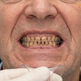
The progression from localized gingival inflammation to advanced periodontitis represents a profound pathological shift within the oral cavity. When pathogenic bacteria, such as Porphyromonas gingivalis, colonize the subgingival environment, they trigger a hyperactive host immune response. This chronic inflammatory state activates osteoclasts—cells responsible for breaking down bone tissue—resulting in severe periodontal disease bone loss. As the alveolar bone recedes, the critical attachment apparatus that anchors the teeth into the jaw is systematically destroyed.
Clinically, this destruction manifests as increased tooth mobility and the formation of deep periodontal pockets. Healthy gingival sulcus depths typically measure between 1 to 3 millimeters. In advanced periodontitis, these pockets can easily exceed 6 to 8 millimeters, creating anaerobic environments where aggressive bacteria thrive, shielded from routine oral hygiene efforts. The presence of these deep pockets not only accelerates local tissue destruction but also serves as a gateway for systemic bacterial dissemination. Extensive research has established a strong bidirectional relationship between severe periodontitis and systemic health, linking chronic oral inflammation to cardiovascular disease, poorly controlled diabetes, and adverse pregnancy outcomes [1].
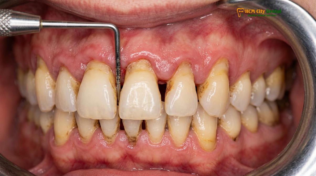
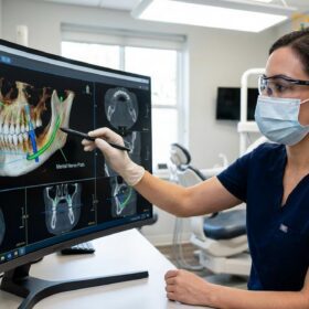
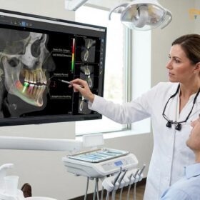
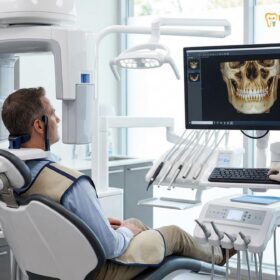
Furthermore, the continuous bone resorption alters the fundamental architecture of the maxilla and mandible. This structural degradation complicates future restorative efforts, particularly when considering dental implants. In cases where patients have suffered from long-standing edentulism or severe infection, the remaining basal bone may be insufficient in both volume and density. This is particularly relevant when planning full mouth reconstruction seniors, who may already be experiencing age-related decreases in bone mineral density. Comprehensive 3D Cone Beam Computed Tomography (CBCT) diagnostics are mandatory to evaluate the remaining bone topography, identify anatomical landmarks such as the inferior alveolar nerve and maxillary sinus, and determine the necessity for guided bone regeneration prior to implant placement.
Standardizing the Gum Foundation: Scale, Root Plane, and Gum Treatment First
Establishing a sterile, stable periodontal foundation is the absolute prerequisite for successful full mouth rehabilitation. This is achieved through meticulous non-surgical and surgical debridement, often enhanced by autologous growth factors.
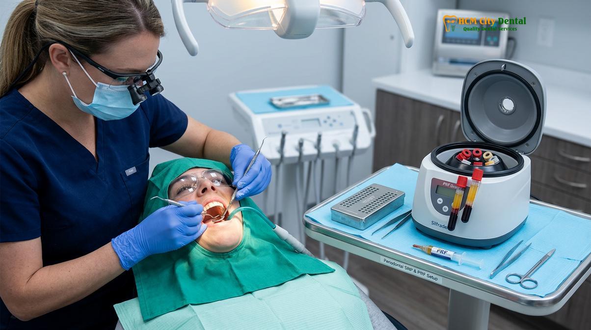
Attempting to build a complex restorative prosthesis on a foundation of inflamed, infected gums is a clinical error that inevitably leads to failure. Therefore, the first definitive phase of any full mouth rehabilitation is standardizing the gum foundation. This process begins with comprehensive scaling root planing (SRP), a meticulous non-surgical procedure designed to remove calculus and bacterial biofilms from the root surfaces deep within the periodontal pockets. Using specialized ultrasonic instruments and hand curettes, the clinician smooths the root surfaces, removing necrotic cementum and creating a biologically acceptable environment that encourages the reattachment of gingival tissues.
In cases where scaling root planing alone is insufficient to resolve deep pockets, surgical intervention becomes necessary. Open flap debridement allows the periodontist direct visual access to the root surfaces and alveolar bone defects. During this phase, the clinician can recontour irregular bone architecture and thoroughly debride the infected granulation tissue. It is also during this surgical phase that advanced regenerative techniques are employed. The application of Platelet-Rich Fibrin (PRF) has revolutionized periodontal healing. By drawing a small amount of the patient’s own blood and spinning it in a centrifuge, clinicians isolate a dense fibrin matrix rich in leukocytes and autologous growth factors. When applied to surgical sites, PRF significantly accelerates soft tissue healing, reduces post-operative discomfort, and enhances the predictability of guided bone regeneration [2].
“The longevity of any full mouth reconstruction is entirely dependent on the biological health of the underlying tissues. We must achieve a state of absolute periodontal stability and respect the biological width before a single permanent crown or implant is placed.”
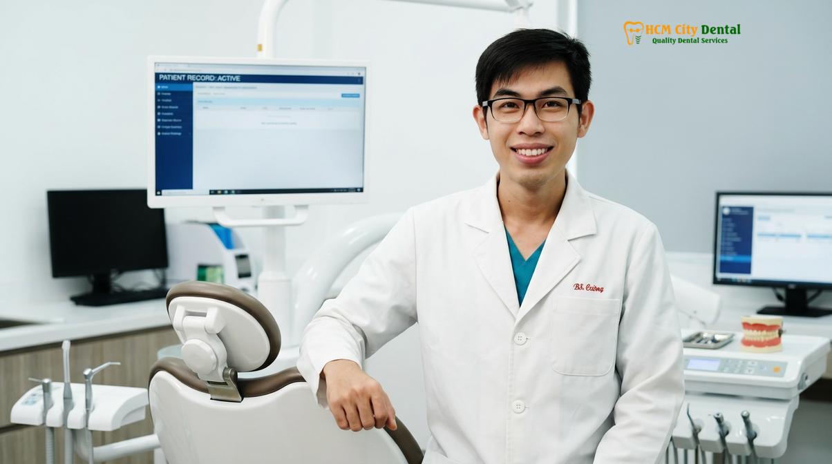
— Dr. Nguyen Van Cuong, Lead Clinician
Another critical aspect of standardizing the foundation is respecting the biological width—the natural dimension of soft tissue attached to the tooth above the alveolar crest. If a restoration encroaches upon this space, it triggers chronic inflammation and further bone loss. In cases where teeth are severely broken down, clinical crown lengthening procedures may be required to expose healthy tooth structure while maintaining the appropriate biological width, ensuring that the margins of the final restorations sit on sound, healthy tissue.
Extensively Damaged Teeth: Extraction Decisions vs. Splinting Restorations
Determining the prognosis of heavily compromised teeth requires a careful biomechanical and endodontic evaluation. The decision to extract or salvage via splinting dictates the entire architectural framework of the reconstruction.
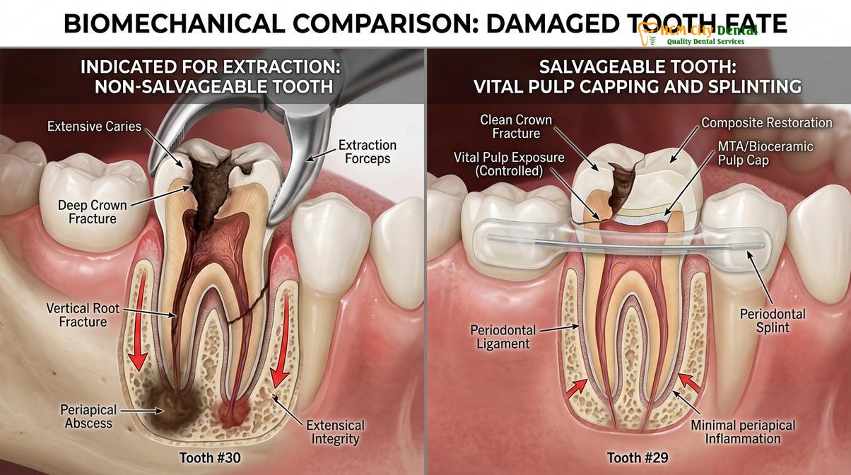
One of the most complex diagnostic challenges in treating severe gum disease is determining the fate of extensively damaged teeth. The clinician must objectively evaluate the mobility pocket depths, the remaining bone support, the crown-to-root ratio, and the endodontic status of each tooth. Teeth that exhibit Class III mobility (severe horizontal and vertical movement) combined with bone loss exceeding 70% of the root length are generally deemed hopeless. Retaining such teeth poses a significant risk to the adjacent healthy bone and can compromise the overall success of the rehabilitation. In these instances, strategic extractions followed by immediate ridge preservation techniques are indicated to maintain bone volume for future implant placement.
However, not all mobile teeth require extraction. Teeth with moderate bone loss and Class I or II mobility can often be salvaged through a combination of periodontal therapy and biomechanical splinting. Splinting involves joining multiple teeth together with a rigid restorative material, effectively distributing occlusal forces across a larger surface area and stabilizing the mobile segments. This technique is particularly useful in the anterior sextants, where preserving natural teeth yields superior aesthetic outcomes and maintains the natural proprioception of the periodontal ligament.
When evaluating heavily decayed but structurally salvageable teeth, advanced endodontic protocols play a crucial role. If the decay approaches the dental pulp but irreversible pulpitis has not yet occurred, vital pulp capping using bioactive materials like Mineral Trioxide Aggregate (MTA) or Biodentine can be employed. These materials stimulate the formation of a reparative dentin bridge, preserving the vitality of the tooth and avoiding the need for root canal therapy. The decision matrix between extraction and preservation is highly individualized, relying on a comprehensive clinical examination and the long-term strategic goals of the Full Mouth Rehabilitation plan.
Designing Restorations with Cleanable Embrasure Spaces (Preventing Food Trap)
Prosthetic design must prioritize hygiene as much as aesthetics and function. Engineering precise embrasure spaces and convex intaglio surfaces is vital to prevent food impaction and recurrent periodontal infection.
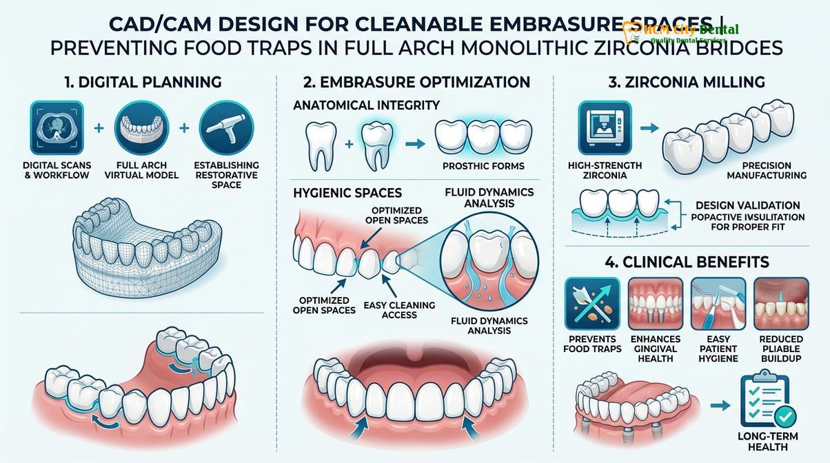
A frequent complication of extensive dental bridgework, particularly in patients with a history of gum disease, is the accumulation of food debris and plaque beneath the restorations. To prevent this, the architectural design of the prosthesis must incorporate cleanable embrasures. Embrasures are the V-shaped spaces between adjacent teeth or crowns. In a healthy natural dentition, these spaces are filled by the interdental papilla (gum tissue). However, following periodontal disease bone loss, the papillae often recede, leaving open spaces (black triangles). If a dental bridge is designed to completely block these spaces in an attempt to hide the tissue loss, it creates an inaccessible concavity where food and bacteria will inevitably become trapped.
Modern prosthodontic protocols dictate that the intaglio surface (the underside of the bridge that contacts the gums) must be highly polished and convex. This ovate pontic design allows the restoration to rest lightly against the tissue without applying pressure, while the cleanable embrasures are left open enough to permit the passage of interdental brushes or water flossers. This specific design allows saliva to naturally flush the area, significantly reducing the risk of peri-implantitis or recurrent decay [3].
The material chosen for the restoration also plays a critical role in tissue health. Biocompatible monolithic zirconia, sourced from premium Swiss and German manufacturers, is the gold standard for full arch restorations. Unlike older metal-ceramic bridges, monolithic zirconia is entirely metal-free and bio-inert, meaning it does not provoke an inflammatory response from the gingival tissues. Furthermore, advanced CAD/CAM milling technology ensures a margin fit of under 50 micrometers. This microscopic precision eliminates the gaps where bacterial microleakage typically occurs, sealing the underlying tooth structure or implant abutment from the oral environment.
| Material Type | Biocompatibility | Margin Precision | Fracture Resistance | Plaque Retention |
|---|---|---|---|---|
| Monolithic Zirconia (Premium) | Excellent (Bio-inert) | < 50 micrometers | Very High (1200 MPa) | Very Low |
| Porcelain Fused to Metal (PFM) | Moderate (Risk of metal allergy) | 50 – 100 micrometers | Moderate (Porcelain chipping) | Moderate |
| Acrylic / PMMA (Temporary) | Good (Short-term) | > 100 micrometers | Low | High |
When utilizing dental implants, understanding All-on-4/6 structural biomechanics is essential. The anterior-posterior spread of the implants must be carefully calculated to minimize cantilever forces on the distal extensions of the zirconia bridge. Proper biomechanical distribution ensures that the occlusal load is transferred evenly to the bone, preventing micro-movements that could lead to implant failure or bone loss.
Advanced Clinical Protocols: VDO, Sedation, and Complex Cases
Rebuilding a collapsed bite requires precise neuromuscular calibration to restore the vertical dimension of occlusion. This complex process is supported by digital smile design and advanced sedation protocols for optimal patient comfort.
Patients with severe gum disease and missing teeth frequently suffer from bite collapse. As posterior teeth are lost or migrate due to lack of bone support, the upper and lower jaws close closer together than nature intended. This loss of the Occlusal Vertical Dimension (VDO) leads to a sunken facial appearance, muscle fatigue, and severe temporomandibular joint (TMJ) dysfunction. Restoring the VDO is a cornerstone of neuromuscular dentistry. The clinician must determine the patient’s physiologic rest jaw position—the point where the masticatory muscles are completely relaxed. Techniques such as TENS therapy (Transcutaneous Electrical Nerve Stimulation) may be used to deprogram the muscles and find this optimal resting posture before establishing the new bite [4].
To visualize and plan this complex functional and aesthetic transformation, Digital Smile Design (DSD) is employed. DSD involves taking high-resolution clinical photographs, 3D intraoral scans, and CBCT data to create a digital mock-up of the proposed restorations. This allows the clinician to evaluate phonetic compatibility, lip support, and chewing guidance before any teeth are prepared. A physical wax-up or 3D-printed temporary splint is then transferred to the patient’s mouth, allowing them to “test drive” the new VDO and aesthetics. Only after the patient is completely comfortable with the function and appearance of the temporaries are the final bio-inert restorations fabricated.
Given the extensive nature of full mouth rehabilitation, patient comfort and anxiety management are paramount. IV conscious sedation provides a safe, anxiety-free, and virtually pain-free experience. Administered under the strict supervision of a board-certified anesthesiologist, customized pharmacological protocols utilizing Midazolam and Propofol induce a state of deep relaxation while allowing the patient to remain responsive. Continuous EKG, blood pressure, and oxygen saturation monitoring ensure absolute safety throughout the procedure. This approach is particularly beneficial for patients undergoing lengthy surgical phases involving multiple extractions, bone grafting, and dental implant placement.
Important Clinical Consideration: Bone Modifying Agents
Patients undergoing treatment for dental osteoporosis or other systemic bone conditions must disclose their medication history. The use of BRONJ bisphosphonate medications (e.g., Fosamax, Reclast) significantly alters bone turnover rates and increases the risk of Medication-Related Osteonecrosis of the Jaw (MRONJ) following extractions or implant surgery. A thorough medical consultation, potential drug holidays, and specific serum tests (like CTX) are mandatory before proceeding with invasive surgical rehabilitation.
Furthermore, full mouth reconstruction is not solely for those with acquired periodontal disease. Patients with genetic conditions such as congenital hypodontia (missing multiple permanent teeth from birth) or ectodermal dysplasia require similar comprehensive rehabilitative approaches. These complex cases often involve multidisciplinary care, combining orthodontics, orthognathic surgery, and advanced implant prosthodontics to establish a functional and aesthetic dentition.
Financials, Pricing Structure, and Global Support
Transparent pricing, comprehensive warranties, and dedicated international support systems make world-class full mouth rehabilitation accessible and predictable for global patients.
Understanding the financial investment required for full mouth rehabilitation is a critical part of the planning process. At HCMC Dental Clinic, we maintain a highly transparent pricing structure, offering significant advantages for international patients engaging in dental tourism saigon. By utilizing our WhatsApp pre-booking system, patients can access a 40% discount on standard walk-in rates, making premium care highly accessible.
According to the latest clinic fee schedule, the pricing parameters for single implants (Fixture & Abutment only, Crown excluded) are structured by tier:
- Economy (Dentis/Neo Biotech): Walk-in: ~$770 | WhatsApp pre-booking: From $460 (-40%).
- Economy Plus (Dentium Hàn): Walk-in: ~$900 | WhatsApp pre-booking: From $540 (-40%).
- Standard (Dentium Superline Mỹ / IBS): Walk-in: ~$1,090 | WhatsApp pre-booking: From $655 (-40%).
- Premium (Ritter Đức / B&B Ý): Walk-in: ~$1,600 | WhatsApp pre-booking: From $965 (-40%).
- Premium Plus (Straumann BLT-SLA): Walk-in: ~$1,795 | WhatsApp pre-booking: From $1,080 (-40%).
- High-End (Straumann Active): Walk-in: ~$2,250 | WhatsApp pre-booking: From $1,350 (-40%).
For the restorative phase, a Crown on Implant ranges from ~$260 – $385 for walk-ins, but with the WhatsApp pre-booking discount, it is reduced to From $155 – $230 per tooth. For comprehensive Full Mouth Rehabilitation Packages, a Full Arch Zirconia Bridge on Implants (All-on-4 / All-on-6) is available from $7,200 – $13,200 per arch (discounted from the $12,000 – $22,000 walk-in rate). Alternatively, an Implant-supported Snap-On Overdenture is available from $3,000 – $4,800 per arch via pre-booking. For patients requiring Full Mouth Crowns (14 crowns per arch, Zirconia HT) on natural teeth, the pre-booking rate is From $2,184 per arch.
Clinical Case Review:
An expatriate patient visited HCMC Dental Clinic in Ho Chi Minh City presenting with severe bite collapse and generalized mobility due to untreated periodontitis. Following a comprehensive CBCT evaluation, Dr. Nguyen Van Cuong designed a phased treatment plan. The patient underwent full arch extractions, PRF-assisted bone grafting, and the placement of six premium implants per arch under IV sedation. Utilizing the WhatsApp pre-booking system, the patient secured a 40% reduction in overall costs while receiving a global manufacturer warranty card guaranteeing the implant fixtures for life.
Beyond the initial treatment, long-term peace of mind is ensured through global manufacturer warranty cards, offering 10-year to lifetime guarantees on implant fixtures. As a leading expat clinic, we also provide comprehensive international billing support, assisting patients with documentation for tax offsets (such as NMETO/IRS deductions) and offering 0% interest monthly installment plans for eligible residents. Remote post-op checkups via WhatsApp ensure continuous care regardless of the patient’s geographic location.
Maintenance Schedule: Essential 3-Month Periodontal Recall Visits
The success of a full mouth reconstruction relies heavily on strict post-operative maintenance. A customized 3-month recall schedule is mandatory to disrupt bacterial repopulation and protect the new foundation.
The completion of the restorative phase does not mark the end of treatment; rather, it signifies the beginning of the maintenance phase. Patients who have lost teeth to severe gum disease remain highly susceptible to peri-implantitis and recurrent periodontal infections. Therefore, adhering to a strict periodontal recall frequency is non-negotiable. Clinical evidence demonstrates that pathogenic bacteria can repopulate subgingival pockets within 9 to 12 weeks following professional debridement. Consequently, a 3-month periodontal recall schedule is the standard of care for full mouth rehabilitation patients [5].
During these recall visits, the clinical team performs a comprehensive evaluation of the soft tissues, checks the occlusal balance to ensure no excessive forces are being applied to specific implants or teeth, and conducts professional prophylaxis using specialized instruments that will not scratch the zirconia or implant abutments. Radiographs are taken periodically to monitor crestal bone levels.
“Reconstruction provides a second chance at oral health, but maintenance is what makes it last a lifetime. Skipping recall appointments allows silent bacterial destruction to undermine the most advanced clinical work.”
— Dr. Nguyen Van Cuong, Lead Clinician
For patients who have opted for an implant-supported overdenture, maintenance also involves inspecting the locator snap-on attachment systems. The nylon retention inserts within the denture base wear down over time due to daily insertion and removal. These inserts typically require replacement every 6 to 12 months to ensure the prosthesis remains securely anchored and functions optimally. Proper home care, including the use of water flossers and specific interdental brushes to navigate the cleanable embrasures, combined with professional oversight, ensures the longevity of the rehabilitation.
When to See a Doctor for Full Mouth Rehabilitation
Recognizing the signs of advanced dental deterioration early can prevent the need for more invasive and costly procedures. You should seek a comprehensive clinical evaluation if you experience any of the following symptoms:
- Increasing Tooth Mobility: Teeth that feel loose or shift position when chewing indicate severe loss of the supporting bone.
- Chronic Gum Inflammation: Persistent bleeding, swelling, or purulent discharge (pus) from the gums are signs of active, destructive infection.
- Bite Changes and TMJ Pain: If your teeth no longer fit together correctly, or if you experience chronic jaw pain and muscle fatigue, you may be suffering from bite collapse and loss of vertical dimension.
- Inability to Masticate: Difficulty chewing solid foods, leading to dietary restrictions and subsequent nutritional deficiencies or systemic health issues.
- Failing Existing Dental Work: Multiple failing crowns, bridges, or recurrent decay around old restorations that compromise your overall oral function.
A personalized clinical examination, including 3D CBCT imaging, is required to determine your specific candidacy for full mouth rehabilitation. Contact HCMC Dental Clinic in Ho Chi Minh City to schedule a comprehensive diagnostic consultation and explore your restorative options.
Frequently Asked Questions
Can you get full mouth reconstruction if you have gum disease?
Yes, full mouth reconstruction is possible for patients with gum disease, provided the active periodontal infection is treated first. Clinical protocols require stabilizing the bone foundation and eliminating bacterial loads through scaling, root planing, and potentially guided bone regeneration before placing any permanent restorations or dental implants. Ignoring the infection will lead to the rapid failure of the new dental work.
What happens if you place crowns on teeth with weak bone support?
Placing crowns on teeth with weak bone support typically accelerates tooth loss due to unfavorable biomechanical loading. The compromised periodontal ligament cannot withstand the occlusal forces transferred by the new crown, leading to increased mobility, further bone resorption, and eventual failure of the restorative work. Such teeth are often better managed via extraction and implant replacement or strategic splinting.
How do you prevent food from trapping under a full bridge?
Food trapping is prevented by designing restorations with highly polished, convex intaglio surfaces and precisely engineered cleanable embrasure spaces. This specific architectural design allows saliva to naturally flush the area and permits patients to easily use interdental brushes or water flossers to maintain optimal hygiene beneath the bridge, preventing recurrent inflammation.
Is IV sedation safe for seniors undergoing extensive dental surgery?
IV conscious sedation is generally highly safe for seniors when administered and monitored by a board-certified anesthesiologist. Continuous EKG, blood pressure, and oxygen saturation monitoring ensure patient safety, while customized pharmacological protocols using Midazolam and Propofol provide an anxiety-free experience during complex full mouth rehabilitation procedures. A thorough pre-operative medical evaluation is always conducted.
How long does a full mouth rehabilitation take when treating severe periodontitis?
The timeline for full mouth rehabilitation involving severe periodontitis typically ranges from three to nine months, depending on the need for bone grafting and implant integration. Initial infection control takes a few weeks, followed by surgical phases and a healing period before the final bio-inert restorations are permanently affixed. Temporary restorations are provided throughout the process to maintain function and aesthetics.
References
- Journal of Clinical Periodontology. Periodontal disease and systemic health interrelationships. (2021).
- Clinical Oral Implants Research. Platelet-rich fibrin in guided bone regeneration and soft tissue healing. (2022).
- The Journal of Prosthetic Dentistry. Biomechanical load distribution and margin fit in monolithic zirconia restorations. (2020).
- International Journal of Prosthodontics. Vertical dimension of occlusion restoration parameters and neuromuscular adaptation. (2019).
- Journal of Periodontology. Maintenance protocols and periodontal recall frequency in full mouth rehabilitation. (2023).
For customized treatment planning, transparent cost estimates, and direct consultation with Dr. Cuong, visit our comprehensive Full Mouth Rehabilitation service page or message our team directly via WhatsApp to receive an instant assessment.