Yes, patients diagnosed with osteoporosis can safely and successfully receive dental implants. While reduced bone mineral density presents unique clinical challenges, advanced surgical modifications, precise 3D diagnostics, and specialized implant designs allow skilled oral surgeons to achieve excellent primary stability and long-term osseointegration without compromising systemic health.
Clinical Summary:
Receiving dental implants with osteoporosis requires a highly customized clinical approach due to altered bone microarchitecture and the potential risks associated with antiresorptive medications. Successful rehabilitation relies on comprehensive diagnostic imaging (CBCT and DEXA) to assess cortical thickness and trabecular density. To counteract the effects of porous bone, clinicians employ surgical modifications such as under-preparation drilling, osseodensification, and bicortical anchorage to maximize primary stability. For patients with severe maxillary resorption, alternative anchorage zones like zygomatic and pterygoid implants offer predictable solutions without the need for extensive bone grafting. Furthermore, strict pharmacological protocols must be observed to mitigate the risk of medication-related osteonecrosis of the jaw (MRONJ), ensuring a safe transition from surgery to final prosthetic restoration.
Key Takeaways:
- Osteoporosis is not an absolute contraindication for dental implants; high success rates are achievable with modified surgical protocols.
- Patients on bisphosphonates require careful medical clearance to prevent jaw osteonecrosis.
- CBCT 3D imaging is mandatory to evaluate the integrity of cortical and cancellous bone prior to surgery.
- Techniques like osseodensification and bicortical anchorage significantly improve implant stability in low-density bone.
- Zygomatic and pterygoid implants provide viable, graftless alternatives for severe upper jaw bone loss.
Bone Density and Dental Implants: Cortical vs. Cancellous Bone Integrity
Osteoporosis primarily degrades the inner cancellous bone, but the denser outer cortical bone often retains sufficient integrity to support dental implants when specialized surgical techniques are utilized.
To understand how osteoporosis impacts implant dentistry, it is essential to examine the fundamental architecture of the human jawbone. Bone tissue is broadly categorized into two types: cortical bone and cancellous (trabecular) bone. Cortical bone is the dense, hard outer layer that provides structural rigidity, while cancellous bone is the spongy, honeycomb-like inner matrix that houses blood vessels and bone marrow. Dental implants rely on both layers for stability; the cortical bone provides initial mechanical fixation (primary stability), and the cancellous bone facilitates the biological process of osseointegration, where living bone cells fuse to the titanium surface [1].
Osteoporosis is a systemic skeletal disorder characterized by a progressive decrease in bone mass and the deterioration of bone microarchitecture. This condition disrupts the natural remodeling cycle, where osteoclasts (cells that resorb bone) outpace osteoblasts (cells that form new bone). In the oral cavity, this systemic health issue manifests as dental osteoporosis, leading to increased porosity within the cancellous bone and a thinning of the cortical plates. The maxilla (upper jaw), which naturally consists of a higher proportion of cancellous bone, is typically more susceptible to osteoporotic changes than the denser mandible (lower jaw).
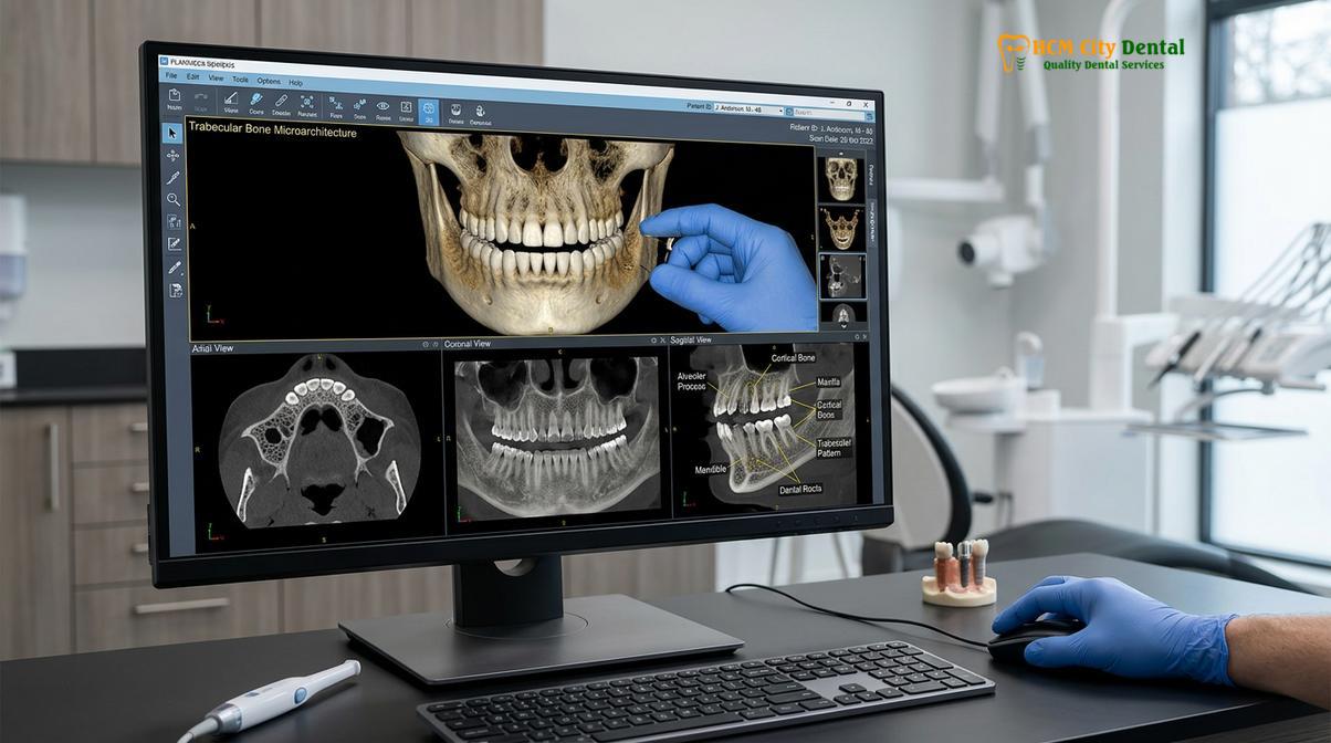
When a patient suffers from tooth loss, the jawbone undergoes disuse atrophy, a process known as bone resorption. Without the mechanical stimulation previously provided by natural tooth roots, the alveolar ridge begins to shrink in both height and width. In patients with osteoporosis, this resorptive process can be accelerated. Furthermore, if the patient has a history of periodontal disease bone loss, the cumulative effect can result in a severely compromised foundation for traditional implant placement. However, clinical evidence demonstrates that osteoporosis bone density alone does not dictate implant failure. The key to success lies in adapting the surgical approach to maximize contact with the remaining high-quality bone, particularly the basal bone, which is less affected by systemic demineralization than the alveolar bone.
Osteoporosis Medications: The Risk of BRONJ (Bisphosphonate-Related Osteonecrosis of the Jaw)
Patients taking antiresorptive drugs for osteoporosis must undergo strict pharmacological evaluation to mitigate the rare but severe risk of jaw osteonecrosis following invasive dental procedures.
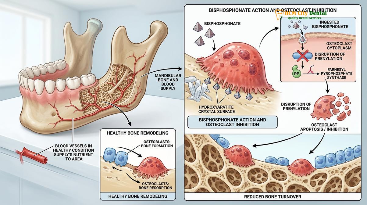
While osteoporosis itself poses biomechanical challenges, the medications used to treat the condition introduce significant biological considerations. The most commonly prescribed drugs for osteoporosis are antiresorptive agents, primarily bisphosphonates (e.g., Alendronate, Risedronate, Zoledronic acid) and RANKL inhibitors (e.g., Denosumab). These medications function by inhibiting osteoclast activity, thereby slowing down bone turnover and increasing overall bone density to prevent skeletal fractures. However, this suppression of bone remodeling can have adverse effects in the oral cavity, particularly following surgical trauma such as tooth extraction or implant placement [2].
The primary concern is a condition historically known as BRONJ bisphosphonate (Bisphosphonate-Related Osteonecrosis of the Jaw), now more broadly termed MRONJ (Medication-Related Osteonecrosis of the Jaw). Bisphosphonate jaw osteonecrosis occurs when the jawbone, having lost its ability to remodel and heal due to osteoclast suppression, becomes exposed to the oral environment and undergoes localized necrosis (tissue death). Because the jaws are subjected to constant micro-trauma from chewing and harbor a complex microbiome, they require a robust capacity for cellular turnover and angiogenesis (new blood vessel formation) to heal properly.
Clinical Warning: Never discontinue osteoporosis medications without consulting both your prescribing physician and your oral surgeon. A coordinated medical approach, potentially involving a C-terminal telopeptide (CTX) blood test to assess bone turnover rates, is essential before proceeding with implant surgery.
The risk of developing osteonecrosis depends heavily on the type of medication, the route of administration, and the duration of therapy. Patients receiving high-dose intravenous (IV) bisphosphonates for oncological conditions are at a significantly higher risk compared to those taking oral bisphosphonates for routine osteoporosis management. For patients on oral therapy for less than four years with no other risk factors (such as corticosteroid use or diabetes), the risk of MRONJ remains exceedingly low. Clinicians employ minimally invasive surgical techniques, prophylactic antibiotics, and meticulous soft tissue closure to further minimize this risk, ensuring that the benefits of restoring masticatory function far outweigh the potential complications.
Diagnostic Tools: Dual-Energy X-Ray Absorptiometry (DEXA) and CBCT Assessment
Accurate treatment planning for osteoporotic patients relies on combining systemic DEXA scores with localized 3D Cone Beam Computed Tomography (CBCT) to precisely map available bone volume and density.
In modern implantology, comprehensive diagnostics are the cornerstone of predictable outcomes, especially for patients with compromised bone quality. The evaluation begins with an assessment of the patient’s systemic skeletal health. A Dual-Energy X-Ray Absorptiometry (DEXA) scan is the gold standard for diagnosing osteoporosis. It measures bone mineral density, typically at the hip or lumbar spine, and provides a T-score. A T-score of -1.0 or above is considered normal, between -1.0 and -2.5 indicates osteopenia, and -2.5 or lower signifies osteoporosis. While a DEXA scan provides a systemic overview, it does not directly correlate with the localized bone density in the maxilla or mandible.
To evaluate the specific anatomical site intended for implant placement, clinicians utilize Cone Beam Computed Tomography (CBCT). Unlike traditional 2D panoramic X-rays, CBCT 3D bone diagnostics provide a highly detailed, cross-sectional view of the jawbone. This technology allows the surgeon to measure the exact height, width, and trajectory of the alveolar ridge, while also identifying critical anatomical landmarks such as the inferior alveolar nerve and the maxillary sinus [3].
More importantly for osteoporotic patients, CBCT software can estimate bone density using Hounsfield Units (HU). Bone is clinically classified into four types (D1 to D4). D1 is dense cortical bone (often found in the anterior mandible), while D4 is highly porous cancellous bone with a thin cortical layer (typical of the posterior maxilla in osteoporotic patients). By analyzing the HU values, the surgeon can anticipate the tactile feedback during drilling and select the appropriate implant macro-design—such as aggressive threading or tapered bodies—to maximize mechanical engagement in softer bone.
Surgical Modifications: Bicortical Anchorage and Under-Preparation Drilling Protocols
To achieve immediate stability in porous bone, surgeons utilize under-preparation techniques to compress the bone laterally and bicortical anchorage to engage the dense basal layers.
When placing implants in osteoporotic bone, the standard surgical protocols must be modified to compensate for the lack of dense trabecular support. The primary goal during surgery is to achieve high insertion torque and primary stability, which are critical prerequisites for successful osseointegration. If an implant is mobile at the time of placement, fibrous tissue will encapsulate it instead of bone, leading to failure.
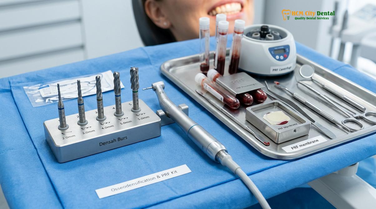
One of the most effective techniques is under-preparation of the osteotomy (the surgical hole). In dense bone, the osteotomy is drilled to a diameter nearly matching the implant. In osteoporotic bone, the surgeon intentionally uses drills that are significantly smaller than the implant diameter. As the wider implant is threaded into the narrower site, it compresses the surrounding soft cancellous bone outward, increasing the localized density—a concept known as bone compression or osseodensification. Specialized bur systems can even be run in reverse to compact bone laterally rather than excavating it, preserving precious bone volume [4].
Another crucial strategy is bicortical bone anchorage. Instead of relying solely on the crestal (top) cortical bone and the internal cancellous bone, the surgeon selects a longer implant designed to engage the opposing cortical plate (the basal bone). For example, in the upper jaw, the implant may be anchored into the dense floor of the nasal cavity or the sinus floor. This dual-cortical engagement acts like a structural pillar, providing exceptional rigidity even if the intervening cancellous bone is highly porous.
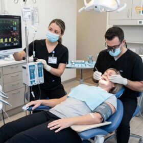
To enhance biological healing, clinicians frequently utilize Platelet-Rich Fibrin (PRF). This involves drawing a small amount of the patient’s blood and centrifuging it to isolate growth factors and healing cells. The resulting PRF membrane is placed into the surgical site to accelerate soft tissue closure and stimulate bone regeneration. Furthermore, to ensure a stress-free experience, complex procedures are often performed under IV conscious sedation. Administered by a board-certified anesthesiologist, this protocol uses medications like Midazolam and Propofol with continuous EKG monitoring, ensuring the patient remains completely comfortable and pain-free throughout the surgery.
Clinical Case Highlight: A 68-year-old female patient presented to HCMC Dental Clinic in Ho Chi Minh City with severe osteoporosis and failing dentition. Under the expert care of Dr. Nguyen Van Cuong, a customized treatment plan was developed utilizing CBCT diagnostics. By employing osseodensification protocols and PRF therapy under IV sedation, Dr. Cuong successfully placed multiple implants with excellent primary stability, entirely avoiding the need for invasive block grafting.
Alternative implant Placement Zones: Zygomatic and Pterygoid Implants
When severe maxillary bone loss precludes traditional implants, zygomatic and pterygoid implants offer a graftless solution by anchoring into dense, remote craniofacial structures.
In cases of advanced osteoporosis combined with long-term edentulism (toothlessness), the maxillary alveolar ridge may resorb to a point where traditional implants are impossible, even with bone grafting. This severe atrophy can also be seen in patients with genetic conditions affecting bone and tooth development, such as congenital hypodontia or ectodermal dysplasia. For these complex scenarios, alternative implant placement zones provide a highly predictable, graftless solution.
The zygomatic implant alternative involves using extra-long titanium implants (ranging from 30mm to 60mm) that bypass the compromised maxillary bone entirely and anchor directly into the body of the zygoma (the cheekbone). The zygomatic bone is dense, cortical basal bone that does not undergo the same resorptive processes as alveolar bone, nor is it significantly affected by osteoporosis bone density changes. This provides exceptional primary stability, often allowing for the immediate loading of a fixed provisional bridge on the same day as surgery [5].
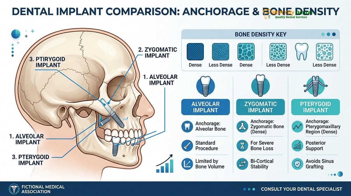
Similarly, pterygoid implants are placed in the posterior maxilla, angling backward to engage the dense pterygomaxillary pillar (the junction of the maxilla, palatine bone, and pterygoid process of the sphenoid bone). This region is highly resistant to resorption and provides a robust foundation for supporting the posterior segments of a full arch restoration. By combining anterior traditional implants with posterior zygomatic or pterygoid implants, surgeons can reconstruct an entire upper arch without the need for extensive sinus lifting or prolonged healing times associated with massive bone grafts.
Full Mouth Rehabilitation and Restorative Protocols
Restoring a full arch requires precise biomechanical planning, utilizing digital smile design and premium zirconia to ensure even load distribution and long-term stability.
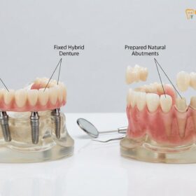
For patients requiring extensive treatment, such as full mouth reconstruction seniors, the surgical placement of implants is only the first phase. The restorative phase—designing and fabricating the prosthetic teeth—is equally critical, especially in osteoporotic patients where minimizing occlusal (bite) stress is paramount. A core component of Full Mouth Rehabilitation is the restoration of the Vertical Dimension of Occlusion (VDO). When teeth are lost, the bite collapses, leading to muscle fatigue, TMJ misalignment, and a sunken facial appearance. Clinicians use diagnostic temporary splints to establish a physiologic rest jaw position, transitioning to permanent restorations only when neuromuscular harmony is achieved.
The aesthetic and functional planning is guided by Digital Smile Design (DSD). This technology allows clinicians to create 3D digital mock-ups and tooth wax-ups, evaluating phonetic compatibility and chewing guidance before any final manufacturing occurs. For the final prosthesis, Biocompatible Monolithic Zirconia is the material of choice. Sourced from premium Swiss and German brands, this metal-free, bio-inert material offers immense flexural strength and achieves a margin fit under 50 micrometers, preventing bacterial microleakage and ensuring optimal soft tissue health.
Understanding the financial investment is crucial for patients seeking comprehensive care. HCMC Dental Clinic offers transparent pricing structures, including significant benefits for international patients utilizing dental tourism pathways. Below is an overview of the current pricing parameters, highlighting the exclusive 40% pre-arrival discount for WhatsApp bookings:
| Implant Tier / Package | Standard Walk-in Rate | WhatsApp Pre-booking Discount (-40%) |
|---|---|---|
| Economy (Dentis/Neo Biotech) – Fixture only | ~$770 (20M VND) | From $460 (12M VND) |
| Standard (Dentium Superline Mỹ / IBS) | ~$1,090 (28.3M VND) | From $655 (17M VND) |
| Premium Plus (Straumann BLT-SLA) | ~$1,795 (46.6M VND) | From $1,080 (28.0M VND) |
| Crown on Implant (Per Tooth) | ~$260 – $385 | From $155 – $230 |
| Full Arch Zirconia Bridge (All-on-4 / All-on-6) | ~$12,000 – $22,000 per arch | From $7,200 – $13,200 per arch |
| Snap-On Overdenture (Implant-supported) | ~$5,000 – $8,000 per arch | From $3,000 – $4,800 per arch |
To further support patients, the clinic provides global manufacturer warranty cards with 10-year to lifetime guarantees on implant fixtures, alongside remote post-op checkups via WhatsApp. Flexible financials, including 0% interest monthly installment plans and support for international tax offsets, ensure that world-class rehabilitation remains accessible.
When to See a Doctor
If you have been diagnosed with osteoporosis and are experiencing tooth loss, loose dentures, or difficulty chewing, it is imperative to seek a specialized clinical evaluation. Do not assume that your systemic condition disqualifies you from receiving implant therapy. However, because the intersection of bone density and antiresorptive medications requires expert management, you should consult an oral surgeon or a prosthodontist experienced in complex rehabilitations.
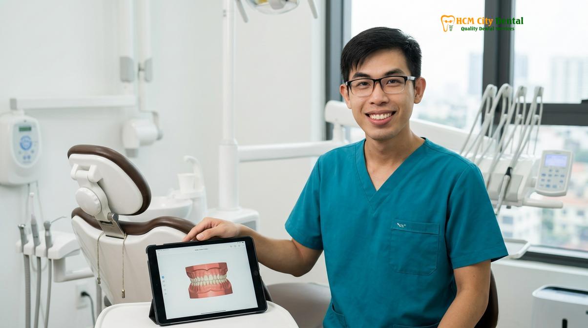
During your consultation, bring a complete list of your current medications, including the exact dosage and duration of any bisphosphonate therapy. Dr. Nguyen Van Cuong and the specialist team at HCMC Dental Clinic in Ho Chi Minh City utilize advanced 3D imaging to provide a definitive, personalized assessment of your bone volume and structural suitability. Early intervention not only prevents further bone resorption but also restores your ability to maintain a nutritious diet, which is vital for overall systemic health and managing osteoporosis.
Frequently Asked Questions
Can I get dental implants if I have osteoporosis?
Yes, patients with osteoporosis can successfully receive dental implants. While the condition reduces bone density, specialized surgical techniques like osseodensification and bicortical anchorage allow for secure implant placement with high long-term success rates. A thorough 3D CBCT scan is required to plan the exact trajectory and select the appropriate implant design to maximize stability in softer bone.
What is jaw osteonecrosis and how do you prevent it?
Jaw osteonecrosis is a rare condition where the jawbone fails to heal, often linked to antiresorptive medications. Prevention involves a thorough pharmacological review, potential drug holidays coordinated with your physician, and utilizing minimally invasive surgical protocols. Clinicians also use prophylactic antibiotics and advanced healing adjuncts like Platelet-Rich Fibrin (PRF) to ensure rapid soft tissue closure and protect the underlying bone.
How do pterygoid implants bypass bone grafts?
Pterygoid implants anchor into the dense pterygomaxillary region of the skull, which is highly resistant to resorption. This technique provides immediate structural support for upper jaw restorations without the need for extensive sinus lifting or bone grafting. By utilizing this dense basal bone, surgeons can provide fixed teeth for patients who have suffered severe alveolar bone loss.
Does osteoporosis bone density affect the healing time of dental implants?
Yes, reduced bone density typically requires an extended osseointegration period. Clinicians often recommend waiting four to six months before applying full occlusal loads to ensure the implant has completely fused with the surrounding trabecular bone. During this healing phase, patients are usually provided with a temporary prosthesis to maintain aesthetics and basic function without stressing the integrating implants.
Are zygomatic implants a safe alternative for severe bone loss?
Zygomatic implants are a highly effective and safe alternative for patients with severe maxillary bone loss. By anchoring directly into the cheekbone, they provide exceptional primary stability for full arch restorations, bypassing compromised alveolar bone. This advanced procedure requires specialized surgical training but offers a predictable, graftless solution for patients who were previously told they were not candidates for traditional implants.
References
- Journal of Oral Implantology. Impact of systemic osteoporosis on osseointegration and implant survival. (2021).
- Clinical Implant Dentistry and Related Research. Management of patients on antiresorptive therapy: BRONJ prevention protocols. (2020).
- International Journal of Oral & Maxillofacial Implants. Osseodensification and under-preparation drilling in low-density bone. (2019).
- Journal of Prosthetic Dentistry. Biomechanical considerations for full arch restorations in compromised bone. (2022).
- Clinical Oral Investigations. Zygomatic and pterygoid implant survival rates in severe maxillary atrophy. (2018).
For customized treatment planning, transparent cost estimates, and direct consultation with Dr. Cuong, visit our comprehensive Full Mouth Rehabilitation service page or message our team directly via WhatsApp to receive an instant assessment.